4976
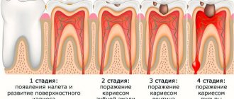
Increasingly, people turn to dental clinics with problems regarding the appearance of their teeth. Patients are concerned not only about changes in the shade of the enamel, but also about its deformation.
The main cause of such manifestations is considered to be dysplasia of enamel and dental tissues, which not only reduces the aesthetics of a smile, but can also lead to complete tooth destruction.
What it is?
Enamel dysplasia is a group of diseases of non-carious origin, characterized by abnormal development of dental tissue.
This pathology is congenital, developing during the intrauterine development of the fetus, or is provoked by genetic factors.
Abnormal tissue development begins during the formation of primordia. Symptoms of the disease are not age specific . They can appear both during the period of first teething and in adulthood.
Dysplasia can affect the entire dentition, but is most often localized in its anterior section. Violation of integrity with the same intensity is observed in all segments of the tooth: cement, dentin, enamel.
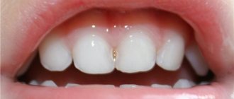
Photo: tooth enamel dysplasia in a child
How does it manifest?
This pathology has certain signs that allow it to be differentiated from other diseases affecting the enamel:
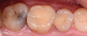
- change in the color of the enamel, which takes on a gray tint;
- thinning of the enamel, which results in increased tooth sensitivity;
- partial or complete absence of the enamel layer ;
- uncharacteristic shape of the cutting part, which includes many notches and chips;
- heterogeneity and surface deformation. Most often, longitudinal grooves and cracks are found on the enamel during visual inspection, and ribbing is detected during instrumental examination;
- the appearance of irregularly shaped white spots , which darken as the disease progresses. They first turn yellow and then turn brown;
- symmetrical arrangement of spots on the teeth of the same name in one jaw;
- surface uniformity in the spot area , here the enamel is dense and smooth, but over time it becomes dotted or grooved;
- the affected area is not stained with caries markers ;
- in the area of the pathological spot, tissue loss , leading to their gradual destruction;
- Pain syndrome may due to exposure to local irritating factors. Once the source is eliminated, the pain disappears.
Due to the disruption of dentin formation, the teeth take on an irregular shape: cone-shaped, barrel-shaped, pear-shaped.
Who is at risk?
Among the provoking factors are:
- late toxicosis or pathologies that caused a pronounced disturbance of metabolic processes during pregnancy;
- endocrine diseases (affecting metabolism) during the active growth of a child in the first years of his life;
- delayed reaction to taking certain medications;
- inappropriate living conditions;
- wrong approach to feeding a child.
Classification
When diagnosing pathology, the dentist relies on a certain classification, which distinguishes several types, depending on the area and nature of the localization.
There are:
- Root. The enamel surface is not damaged, but has a slight deviation in color.
Dentinal canals are present in small quantities, obliterated into the tooth cavity and have a crescent shape.The roots of the affected teeth are shortened and narrowed, which leads to their premature loss. The surface of the root part is dysplastic.
- Coronal .
It is characterized by a change in the color of the enamel, which gradually acquires a yellow tint. In the area of the coronal part, pathological abrasion of the enamel is observed, which begins to expose dentin. Most often, pathological areas are located in the central part of the tooth or closer to its cutting surface. - Odontodysplasia .
It is characterized by severe thinning of both enamel and dentin. Pathology can equally affect both temporary and permanent teeth. Most often, teeth are irregularly shaped and smaller in size. In some cases, denticles are found in the cavity.
Another 3 types are distinguished by the nature of enamel lesions:
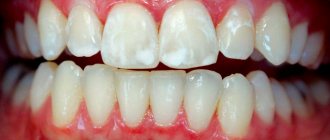
- Spotted .
It is expressed in the appearance of white round spots on any surface of the front and lateral incisors. The spots have clear boundaries and a shiny surface. Over time, it becomes rough. The thickness of the tissues of the affected area does not differ from the thickness of healthy enamel. In most cases, the spots do not change throughout life. - Erosive .
It is distinguished by cup-shaped or oval-shaped depressions formed as a result of damage to the enamel. The recesses are characterized by their overall width and depth. The bottom of the affected area is covered with thinned enamel, through which dentin is visible. Further development of the pathology leads to complete loss of enamel in the area of thinning. - Sulcata .
With this form of the disease, grooves are formed on the vestibular surfaces of the teeth. The groove is located parallel to the cutting part of the teeth. In the area of the furrow, the enamel is thinned or completely absent.
For those who should choose Pilot metal braces, read in a separate publication.
In this article we will talk about the causes of tooth enamel hypoplasia in children.
Here https://orto-info.ru/zubocheliustnye-anomalii/okklyuzii/perekrestnyiy-prikus.html we will discuss whether braces are effective for crossbite.
A 9 year old child has dysplasia on his teeth - Pain in legs
Dental dysplasia is a hereditary dental disease characterized by disruption of the formation of hard dental tissues.
According to statistics, this pathology is equally often diagnosed in both boys and girls in pediatric dentistry. In this case, dysplasia can affect the tissues of both milk and molar teeth.
Causes of dysplasia
The exact causes of enamel and dentin dysplasia have not yet been established. According to scientists, the development of the pathological process can be facilitated by anomalies of mesodermal cellular formations that arose during the period of embryonic development.
Mesodermal cell formation is the part of the germ layer from which blood vessels, bones, spine, kidneys and cartilage are formed.
Symptoms of dysplasia
The main manifestations of dentin and enamel dysplasia are observed in such dental diseases as:
- dentinogenesis imperfecta;
- Stanton-Capdepont syndrome;
- imperfect amelogenesis.
Stanton-Capdepont syndrome
Stanton-Capdepont disease or syndrome is a hereditary disorder of the formation of enamel and dentin tissue. The main symptoms of this pathology are:
- eruption of teeth having a natural shade and shape, and the subsequent change in the color of their enamel to gray;
- gradual destruction and chipping of tooth enamel;
- early, rapidly progressing abrasion of dentin;
- illumination of the contours of the dental cavity through dentin defects;
- decreased sensitivity of the pulp.
Molars with Stanton-Capdepont disease are subject to destruction and abrasion to a lesser extent than milk teeth. In some cases, the disease is completely asymptomatic, while the teeth are in excellent condition and look practically healthy.
Dentinogenesis imperfecta
Dentinogenesis imperfecta is a pathology characterized by a disruption in the formation of a layer of hard dental tissue located under the enamel (dentin). According to statistics, this disease is found in one out of 8,000 babies, with girls being diagnosed more often.
Depending on the symptoms, there are 4 forms of dentinogenesis imperfecta:
- Type I (the disease acts as one of the symptoms of a congenital disorder of bone formation, and its main signs, in addition to dental dysplasia, are bone fragility, changes in the shape of the middle ear and blue coloring of the eye sclera);
- Type II (the main signs of pathology are opalescence (translucency) of the teeth, a change in the natural color of the enamel to dull gray, increased abrasion of the teeth in the zone of closure of the dentition);
- root dysplasia (underdevelopment or complete blockage of dental roots, early loosening and loss of teeth, minimal difference in the shade of dental crowns from the natural one);
- coronal dysplasia (translucency of tooth enamel, frequent injury to the blood vessels of the pulp, accompanied by minor local bleeding, staining of teeth an amber or brownish-gray color by blood breakdown products).
Amelogenesis imperfecta
In turn, amelogenesis imperfecta is a disorder of the enamel formation process, leading to a change in shade and complete or partial loss of dental tissue.
Depending on the clinical picture, there are 4 subtypes of this pathology:
- type 1 (the enamel acquires a brown or yellow tint immediately after the teeth appear, unevenness of the dentin-enamel junction is observed);
- type 2 (1-3 years after the appearance of the first teeth, the enamel acquires a brown tint, becomes rough, matte, and becomes covered with cracks);
- type 3 (at the moment of teething, the teeth are covered with thin white enamel, which quickly disappears, exposing dentin);
- Type 4 (when teething, teeth are covered with matte, chalky enamel, which is easily separated from dentin under mechanical stress).
The sensitivity of the pulp with dental dysplasia is usually significantly reduced. Therefore, pain in people suffering from this disease is mainly caused not by increased sensitivity of the teeth, but by injury to the gums from the sharp edges of damaged crowns.
Source: www.adent.ru
Features of the period
By the age of 9, the child’s upper and lower incisors and first permanent molars erupt in the child’s dentition. The dentition still contains primary canines and molars. The permanent incisors are normally positioned evenly in the dentition without “protrusion” or “sagging” to the side.
By the age of 10, the process of formation of the roots of permanent teeth that have already erupted in the oral cavity (first molar, central and lateral incisors) is completed. These teeth are “stabilized” in the bone tissue.
At 9-10 years of age, the roots of the 4th milk tooth (1st primary molar) are reabsorbed and replaced with the 4th permanent tooth (1st premolar) first on the upper and then on the lower jaws.
From 10 to 12 years of age, the roots of the 5th primary tooth (2nd primary molar) and primary canine begin to be reabsorbed and replaced by the 2nd premolar and permanent canine, respectively.
The permanent canine is the last one to erupt.
By the age of 12–13 years, the replacement of baby teeth with permanent teeth is completed. There should be 24 teeth in the dentition: 12 on each jaw.
Typical problems.
NORMALLY, the permanent incisors are located “evenly” in the dentition without “protrusion” or “sagging” to the side. This indicates a harmoniously occurring process of physiological replacement of milk teeth with permanent ones.
If there has been early removal of baby teeth in children and, as a result, permanent chewing teeth are displaced forward, improper closure of the dentition occurs and crowding of teeth appears in the area of the frontal and chewing teeth.
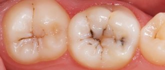
A common problem in the period from 9 to 12 years is poor oral hygiene, which is associated with the characteristics of the psychological development of children. As a result, caries develops in “young” permanent teeth. Most often, the 6th chewing teeth (1st molars) are affected in the area of fissures (natural depressions of the teeth located between the cusps of the tooth).
Fissure caries, remaining unnoticed, develops rapidly and is quickly complicated by inflammation of the nerve of the tooth (pulpitis).
Treatment and prevention
At the age of 9 - 12 years, “stabilization” and a decrease in the intensity of jaw growth occurs. Previously formed malocclusions are “fixed.” By the age of 12, when there are no baby teeth left, orthodontic treatment in most cases (but not always) is possible only with braces
IN CHILDREN WHO HAVE ORTHODONTIC TREATMENT STARTED AT AN EARLIER AGE AND WAS USING ORTHODONTIC PLATES AND TRAINERS TO CREATE SPACE IN THE TEETH, TREATMENT WITH BRACKETS PROCESSES QUICKLY AND WITHOUT REMOVAL OF PERMANENT TEETH .
BRACES CANNOT INCREASE THE SIZE OF THE JAW, SO IN ORDER TO “STRAIGHT” THE TEETH BY “PUSHING” THEM INTO THE AVAILABLE SPACE, IT IS OFTEN NECESSARY TO REMOVE THE PERMANENT TEETH.
THIS IS WHY ORTHODONTIC TREATMENT, IF NECESSARY, IS BETTER TO START AS EARLY AS POSSIBLE.
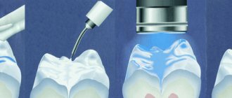
To prevent the development of caries of chewing teeth in the fissure area, a procedure called fissure sealing is performed - closing the fissures in order to prevent further plaque from getting stuck in them and the development of caries in them.
To prevent sports injuries to the upper incisors and canines, it is recommended to have an individual sports mouth guard made by a pediatric orthodontist.
Source: babysmiledent.ru
Causes
three main factors that cause enamel dysplasia :
- Diseases of a hereditary nature .
Most often, they lead to darkening of the enamel and its thinning. Basically, pathology begins to appear in children already on their baby teeth. As a result of the pathological process, it becomes thinner over the entire surface, leading to dentin exposure. In hereditary diseases, tooth enamel is yellow immediately after eruption. - General diseases, occurring in an acute form, leading to disruption of the metabolic processes of mineral components.
As a rule, due to such reasons, dysplasia can be focal in nature and affect single specimens.This pathology is most often complicated by carious processes, which are characterized by rapid progression.
- Pathologies of the skeletal system , leading to disruption of the process of tooth formation due to a lack of bone-forming elements. Such diseases include rickets, periosteal dystrophy, marble disease.
Causes of enamel dysplasia in primary teeth
Dysplasia is a lesion of bone tissue.
The disease is not inflammatory, but congenital. The disease is characterized by the fact that bone tissue forms and develops incorrectly. Affected teeth in children are destroyed very quickly, and as a result, the child loses his beautiful snow-white smile. But aesthetics is not the main problem with this disease. With dysplasia, the baby's diction is impaired, chronic pain appears, and the process of chewing food brings him discomfort and pain.
In most cases, dysplasia of dental tissues forms during fetal development or in infancy (at the stage of formation and development of teeth, and the period of their eruption). If dysplasia on baby teeth is not treated, the problem can also arise on permanent teeth.
Spotted dysplasia is the most common type
Why is the enamel of baby teeth subject to destruction? The exact reasons for this process are not fully known. There are several factors that may predispose to enamel dysplasia:
- heredity;
- disruption of the formation of dental tissue during the fetal development of the baby, including those associated with insufficient intake of minerals, especially calcium and phosphorus, into the mother’s body;
- use during pregnancy of antibacterial drugs, synthetic iron preparations with insufficient calcium intake;
- poor nutrition, consumption of poor-quality drinking water by a child.
The rapid manifestation of the disease already on the baby’s first teeth may indicate that the causes of enamel dysplasia most likely lie in the field of embryonic development or gestation.
Tooth decay in a child’s later life is evidence of problems with hygiene, receiving a qualitatively and quantitatively complete diet, and the supply of minerals and vitamins.
Consequences
Almost all types of dysplasia are characterized by complications and serious consequences.
If left untreated, dysplasia leads to at least an aesthetic defect . In more complex cases, dentin is exposed and infections occur.
Such teeth are most often affected by caries, which quickly reaches the pulp chamber, provoking purulent inflammation. In this case, not only hard tissues, but also adjacent soft tissues can be damaged.
Teeth affected by dysplasia are fragile and susceptible to easy chipping, which disrupts their integrity and shape.
In most cases, without appropriate therapy, the pathology leads to complete destruction of the tooth or its severe loosening, which is an indication for removal.
Treatment methods for enamel hypoplasia in children
If white spots on teeth are invisible to others, they are usually not removed. But the enamel, severely affected by the disease, ceases to protect the tooth tissue, resulting in complications - increased wear of teeth, destruction of dentin up to the loss of teeth, and the appearance of malocclusions. To prevent this, timely therapy is necessary.
Procedures for the treatment of enamel hypopalsia in children:
- remineralization (to prevent further destruction, the enamel is coated with varnishes containing calcium and fluorine);
- whitening (allows you to eliminate darkening of the enamel in mild forms of the disease);
- filling (indicated for underdeveloped enamel);
- prosthetics (this method is used to treat aplasia).
Remedies
Several types of methods are used to treat dysplasia. Some of them are corrective in nature, others are aimed at solving the problem by general strengthening of dental tissue .
Treatment methods are selected depending on the extent of the lesion and the intensity of the symptoms.
You will learn about the features of treatment of dentin dysplasia from our next review.
In the next article we will talk about the pros and cons of Clarity Sl self-ligating braces.
At this address https://orto-info.ru/zubocheliustnye-anomalii/zubov/formyi/konicheskie-i-shilovidnye.html you will find information about the sequence of treatment for spiky teeth.
Therapeutic
This group includes treatments aimed at visually correcting the appearance of enamel .
The following methods are used for this:
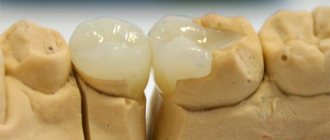
- Composite restoration. It is used for small foci of dysplasia. The procedure is performed under local anesthesia with preparation of damaged areas.
After preparing the cavities and the surface of the tooth, it is treated with an adhesive agent and a light-curing composite is applied. Most often, Consize and Evicrol are used for this.These materials allow you to accurately convey the appearance and structure of healthy enamel. They are highly durable and do not stain over time.
- Veneers. For extensive lesions, it is recommended to install veneers. They are thin ceramic plates that accurately replicate the relief of the vestibular and incisal parts of the teeth.
Before fixing the veneers, a small grinding is carried out, which is necessary to visually reduce the volume. The overlays are installed using special glue or conventional composite.In appearance, they are no different from the surface of real teeth and protect thinned enamel from the aggressive effects of external factors.
- Prosthetics with crowns .
Used in cases of volumetric destruction. Either single crowns or bridges are used, which are made from individual casts.
For one of the methods for eliminating enamel damage, watch the video:
General strengthening
General restorative treatment is indicated for minor manifestations of dysplasia in the spot stage, characterized by gradual modification.
Therapy consists of remineralization and includes the use of the following drugs :
- klamin _ Used orally, 1 tablet per day for a month. The drug must be taken 20 minutes before meals.
- calcium glycerophosphate . Just like the previous remedy, it is indicated for use for 1 month. The product should be taken no more than 1.5 g per day;
- multivitamin complex, for example, Kvadevit or Complivit. Take no more than 2 tablets per day;
Also prescribed:
- electrophoresis with a solution of calcium glycerophosphate . Conduct at least 10 sessions 3 times a year;
- enamel treatment with sodium fluoride.