535
Odontogenic cysts develop more often than other tumor-like neoplasms and are diagnosed at any age.
On the upper jaw, neoplasms appear three times more often than on the mobile jaw arch.
A cyst is a serious enough disease that you should consult a doctor at the first suspicion of it.
Concept of pathology
A dental cyst is a benign neoplasm measuring from 0.5 to 3 cm. It is a capsule with a connective tissue membrane, which is filled with pasty or liquid, purulent or serous contents, consisting of living and dead bacteria, dead leukocytes, decomposed cells, etc.
The contents of the capsule are under pressure of 30-95 cm of water. Art. The neoplasm gradually increases in size. There is still no agreement among experts about what exactly causes its growth - internal pressure, cell vegetation, or both at the same time.
New growths can form fistulas - canals connecting their internal cavity with the vestibule and oral cavity.
One of the main negative consequences is the growth of the tumor and pressure on adjacent tissues, which leads to jaw atrophy and displacement of tooth roots. There are other, more serious consequences of odontogenic cysts, which can only be avoided by promptly consulting a doctor.
What difficulties may arise during the treatment process?
Sometimes during the treatment process the doctor encounters some difficulties:
- If a pin is located in the canals, its removal may result in perforation or root fracture. The result is tooth extraction.
- In order to re-seal the canals, it is necessary to unseal them. This is not an easy and time-consuming process, which is often accompanied by perforation.
- After the canals have been unsealed, they can be filled again only after anti-inflammatory therapy, which often takes 2-3 months.
Read also: There is a balloon on the gum, what to do?
Reasons for development
Cysts grow from granulomas - small (up to 5 mm) nodules that occur in certain diseases and abnormalities in the development of teeth and oral mucosa. Granuloma is distinguished not only by its small size, but also by the absence of a membrane.
Granulomas can be caused by infection, trauma, or a combination of both. The infectious process occurs in the tooth or gum itself, or is transmitted through the bloodstream from other organs.
Causal infections localized to the gum or tooth include:
- pulpitis;
- caries;
- periodontitis;
- periodontitis;
- an infection resulting from insufficient sterilization of the canal during its treatment.
The cyst can be provoked by infections that have penetrated into the periodontium through the vascular bed from other organs: tonsillitis, sinusitis, otitis media, sinusitis.
The development of pathology can be a consequence of injury:
- when eating too solid food;
- due to mechanical impact (sports or other circumstances);
- with improper prosthetics (overload of the supporting tooth);
- errors during treatment, for example, perforation of the bottom of the maxillary sinus, breakage of an instrument in the bone, etc.
Combined causes - from trauma and infection include severe eruption of third molars and other impacted teeth , pathologies of jaw development, etc.
Risk factors are:
- weak immunity;
- hypothermia;
- stress;
- excessively intense mental and physical work.
Description and causes of pathology
A dental cyst is a rather dangerous disease, and without timely treatment it can lead to serious complications. In most cases, such a formation under the crown is formed due to unqualified specialists or insufficient oral hygiene. This tumor forms in the part of the bone tissue or gum where bacteria penetrate.
Among all cystic lesions in dentistry, the formation under the crown is the most difficult. This is explained by the fact that pathology is most often detected at a late stage, when most of the tooth is already affected.
The dental cyst itself is a rather dense formation that contains fluid with epithelial cells and dead bacteria. Outwardly, it resembles a capsule. Initially, its size may be about a few millimeters.
Pathology occurs for the following reasons:
- penetration of infection into the root canals;
- chronic periodontitis;
- advanced form of caries;
- poor-quality filling;
- complications after sinusitis;
- chronic inflammation under the crown;
- insufficient oral hygiene;
- weak immunity.
Despite the fact that the main cause of the pathology is associated with poor-quality prosthetics and treatment, the disease does not develop immediately after the completion of dental procedures. The cyst can take years to develop. Sometimes it is almost impossible to determine the exact cause of a cystic lesion.
First signs
At the beginning of the growth of the tumor, there are no pronounced symptoms. Usually the first signs of development are:
- mild pain when pressing on the tooth or chewing;
- slight swelling and redness of the gums (initially bright red, then bluish);
- unpleasant taste and odor from the mouth;
- change in enamel color (blackening or greying), which may indicate a lack of minerals due to pathology;
- sensitivity to sour, sweet, cold, hot;
- feeling of gums bursting from the inside;
- numbness of the lower lip and chin (Vincent's symptom - inflammation of the mandibular nerve).
Frequent inflammation of the sinuses can also be a consequence of the appearance of a neoplasm.
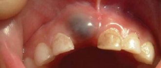
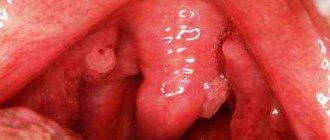
When the pathology reaches an advanced stage, its symptoms are difficult to ignore. The pathology begins to be observed visually - in the form of a protrusion on the gum or palate.
The following symptoms appear:
- Severe swelling and redness of the gums, swelling of the cheek.
- Aching, intractable pain.
- A headache is possible if the inflamed capsule is located near the nasal sinuses.
- Fever, general malaise.
- The appearance of a fistula with leaking exudate.
- Enlarged cervical lymph nodes.
Is tooth depulpation necessary before prosthetics and how is the procedure performed?
Come here to understand when dental crown replacement is required.
At this address https://zubovv.ru/protezirovanie/nesemnyie-p/koronki-np/kogda-opravdano-kombinirovannyimi.html we will talk about the positive properties of a cast combined crown.
Symptoms
Typically, a cyst does not manifest itself at an early stage of formation. As a rule, the disease is discovered accidentally - during the examination of other ailments or already in advanced stages. Although there are signs by which you can determine that it is time to seek help from a dentist:
- Painful sensations in the root part of the tooth. The pain syndrome at an early stage is insignificant; during palpation examination it may increase slightly.
- Due to the cystic tumor in the root area, the tooth darkens and may move slightly to the side.
- As the cyst increases in size, the symptoms intensify. There is an increase in temperature, the appearance of lethargy, headaches, and swelling of the gum tissue. Chewing food becomes painful. Flux may develop.
The main sign indicating the formation of a large cystic tumor under the crown is the presence of a fistula on the gums.
Types of neoplasms
There are more than five types of odontogenic cysts. Their pathogenesis, symptoms and treatment methods are largely similar, but each type has its own characteristics.
Let's look at commonly diagnosed types in more detail.
Paradental
Occurs with pathologies of teething, most often third-party painters. They account for 3% of all cases. They manifest themselves as a small bluish swelling located above the crown of the erupting unit.
Characterized by:
- the presence of blood in the capsule (because of this it acquires a bluish tint).
- severe swelling;
- possibility of capsule rupture.
Treated with surgery (facilitate tooth eruption).
Radicular
The root type develops from granulomas formed by inflamed and dead pulp in the apical third of the tooth. It can grow up to 3 cm. It often grows asymptomatically, without destroying bone tissue or affecting teeth.
The root enters the cavity of the capsule and usually grows towards the vestibule of the mouth. Characterized by slow growth over months or even years. However, the patient may not be aware of it. But gradually the bone plate becomes thinner and begins to bulge from the gums.
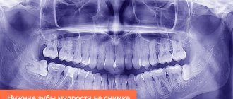
It is clearly visible on an x-ray in the form of a rounded area of rarefaction with clear boundaries. It is often treated endodontically through the root canal. But surgery or enucleation (enucleation) may be required.
Residual
A residual cyst grows in the socket left by tooth extraction. Often due to improper extirpation (the apex was not removed, or the wound became infected), it is clearly visible on an x-ray. Can grow up to several centimeters. It is treated surgically.
Types of disease
There are different types of cysts depending on the causes and location. Based on location, the following types of disease are distinguished:
- radicular (located on the root) is most often a complication of chronic periodontitis, when there is no quality treatment,
- paradental in most cases develops due to an inflammatory process in the area of wisdom tooth growth during its problematic eruption,
- a follicular cyst develops after infection of the tooth buds,
- a primary cyst occurs as a result of pathologies in the development and eruption of teeth,
- eruption cyst develops in children during the appearance of permanent teeth,
- residual cyst in the root is a formation that remains in the bone tissue after tooth extraction.
The formation can be located in the maxillary sinuses, in the area of the front teeth, or in the area of eights.
Consequences of refusing treatment
Delay in treatment is fraught with complications that can be very serious, even life-threatening. The most common consequence is bone resorption due to compression of the neoplasm. The capsule grows, “conquering” space from adjacent tissues and putting pressure on them.
It is mainly the jaw bones that undergo atrophy, not the teeth themselves. The roots are practically not resorbed; they can only change their location under the pressure of the cyst. But the alveolar bone surrounding them is resorbed, so with an advanced pathological process, the teeth may simply fall out.
Thinning of the jaw bones can lead to jaw deformation and fracture. A cyst growing in the area of the bottom of the maxillary sinuses leads to its thinning and destruction.
In addition, untreated formations can lead to abscesses and phlegmon, the development of periodontitis, periostitis (flux) and chronic sinusitis.
The most dangerous complications are malignancy (malignancy) and sepsis.
What is a dental cyst?
A dental cyst is a cavity in the bone tissue that contains pus and is lined with a dense fibrous membrane. This formation is most often a complication of chronic periodontitis. The disease is dangerous because it is almost impossible to diagnose it in the early stages, since it does not make itself felt in any way and nothing bothers the patient. In the absence of proper treatment, the disease can lead to very serious and life-threatening consequences.
Expert opinion. Dentist Petrov I.O. : “Due to the absence of symptoms, patients in most cases turn to the dentist in the later stages of the disease, when they feel obvious pain and a significant deterioration in their general condition. A cyst can only be detected using an x-ray."
Diagnostics
The most informative diagnostic method is radiography and tomography. Today, tomography is recognized as the most accurate method. Spiral tomographs make it possible to obtain a three-dimensional model and sections of cysts of any etiology and location.
On radiographs and tomograms, the capsule manifests itself as a rarefied bone of a round shape with clearly distinguishable boundaries.
If it is directed towards the palate, the palatal plate is destroyed. A severely thinned bone bends upon palpation, producing a crunching sound (Dupuytren's syndrome). If the bone has completely resorbed, the surface fluctuates.
As a rule, clinical and radiological studies can accurately determine the presence of a cyst and its type. If in doubt, a cytological examination of the contents is performed.
Electroodontodiagnostics (EDD) allows you to determine the condition of the pulp of teeth located in the area of pathological formation.
Advantages of metal-free crowns for front teeth and their characteristics.
In this publication, we will look at the pros and cons of metal crowns on coated teeth.
Here https://zubovv.ru/protezirovanie/nesemnyie-p/koronki-np/nepriyatnyiy-zapah-iz-pod.html read what to do if there is a smell from under the crown of a tooth.
What can cause a cyst to form under the crown and how to get rid of it?
Dental patients are often concerned about a cyst under the crown. What to do in this case, how to carry out the treatment and how long will it last? Is it painful and what consequences will appear if you do not pay attention to the symptoms of malaise?
Having installed a crown, we expect that the tooth will no longer be disturbed and that it is reliably protected from any infections, bacteria, or diseases. But unfortunately, people often experience recurring cavities and other damage. One of the most dangerous and unpleasant is the appearance of a cyst in the root part of the tooth.
Therapy methods
Therapy is carried out depending on their type, location and stage of development of the pathological phenomenon. The main method of treatment is surgery. But there are cases when a cyst can be dealt with using therapeutic (conservative) methods.
Conservative treatment
Conservative treatment provides access to the tumor through the root canal of the tooth. That is, in essence, this is endodontic treatment. Unfortunately, it is impossible to cure the formation with one dose of medication.
Small radicular cysts are treated conservatively. To provide access to them, it is necessary to remove the artificial crown.
Treatment involves the following:
- Local anesthesia.
- Removing the crown.
- Opening the tooth canal, gaining access to the tumor.
- Opening the wall, removing the contents, antiseptic and antibacterial treatment of the cavity.
- Filling the cavity with self-absorbable osteoplastic material.
- Temporary filling of the canal and tooth, prescribing antibiotics if necessary.
- Radiography.
- Pause 3-6 months.
- X-ray – to assess the changes that have occurred. If the cyst has decreased in size and there is no inflammation, the canal is permanently filled and a crown is installed.
The effectiveness of conservative treatment is 70%. Its advantage is low invasiveness and tooth preservation. The disadvantage is the additional stage and the duration of the procedure.
Therapeutic treatment is carried out only for certain types of tumors. In most cases, surgical treatment is necessary - access to the formation through the gum wall with detachment of the flap and trepanation of the bone at the site of its projection.
If the condition of the roots allows them to be used further, treatment (cleaning, disinfection, filling) is usually carried out before the main operation. Before this, the crown is removed. However, in some cases, retrograde filling can be performed through the apex of the root during the main operation to remove the cyst.
Root apex resection
If the root has grown into a radicular type, its apex is removed along with the capsule. The operation is performed under local anesthesia:
- A flap is removed from the side of the gum in the projection of the cyst.
- A section of bone no smaller than the size of the capsule is cut out.
- The root is cut off at the level of the surface of the new growth.
- The capsule shell is separated from the adjacent tissues and removed along with the root apex.
- The remaining part of the root is examined and, if necessary (there is no cement in the canal), it is filled retrogradely.
Hemisection
Hemisection is the operation of removing one root of a multi-rooted tooth (premolar or malar).
If, when removing a formation, it is not possible to preserve the root that has grown into it, it is removed along with part of the crown located above it.
After the stump is formed, an artificial crown is made and installed on the tooth.
Cystectomy
Cystectomy is the complete removal of the cystic capsule. Radical surgery provides treatment effectiveness close to 100%.
Indications for cystectomy are:
- a problem resulting from improper development of odontogenic epithelium;
- a small neoplasm, in the area of which there are 1-2 intact teeth (cystectomy in this case is often combined with resection of the root apex);
- a large mandibular tumor with no roots in its area and maintaining a bone thickness of at least 5-10 mm, which excludes a fracture;
- a large maxillary formation that does not have teeth in its area and has not destroyed the bottom of the maxillary sinus.
The disadvantage of cystectomy is its relative complexity.
Cystotomy
The essence of cystotomy is that only the anterior wall of the cyst and its contents are removed. As a result of the operation, the chamber cavity is connected to the oral cavity or its vestibule, the pressure disappears, and the cavity begins to gradually grow from the inside with new bone.
Cystotomy is indicated in the following cases:
- Formation involving 3 or more intact teeth.
- The radiograph shows the absence of a periodontal fissure. This suggests that the alveolar bone surrounding the root has resorbed.
- Large maxillary formations that destroyed the palatine plate and the floor of the maxillary sinus.
- Large mandibular neoplasms that have significantly thinned the jaw bone (to a thickness of less than 5 mm). In this case, the preserved shell strengthens the jaw and prevents its fracture.
Cystotomy is less traumatic than cystectomy , but requires long-term monitoring and hygienic wound care. This is its disadvantage.
Possible complications
A cyst formed under the crown must be treated. If it increases to a large size, a fracture of the jaw is possible, and not only diseased teeth, but also healthy ones begin to fall out.
A cyst that is not treated in a timely manner can become an impetus for the development of other formations. The danger lies in the fact that in this case the nature of their origin may be malignant. In an advanced form of the disease, an abscess may develop; along with the blood flow, pus will begin to spread throughout the human body.
Complications of a dental cyst without treatment
Reviews
Surgical treatment of a cyst is a serious enough procedure to leave a mark on the memory.
If you have had a cystectomy, cystotomy, or hemisection, please share your experience with this procedure.
How long did it last, what was the pain, how quickly did it heal? You can leave your comment at the bottom of this page.
If you find an error, please select a piece of text and press Ctrl+Enter.
Tags dental crowns cyst
Did you like the article? stay tuned
No comments yet
Tooth cyst under the crown: is treatment possible?
Crowns reliably protect teeth from mechanical stress and temperature influences. In addition, thanks to dentures, it is difficult for bacteria to enter under the crown. For a crown to truly perform these functions, it must be perfectly tailored to the surface of the tooth and gum. Crown installation is not always successful, which can lead to unpleasant consequences.
Installation of the prosthesis has several stages. First, the tooth is prepared - ground and depulped (the nerve is removed). This way there is less chance that the tooth will start to hurt. However, pulpless teeth live less, so dentists are increasingly avoiding this procedure. However, the tooth under the denture may hurt in both cases.
If the denture was not fitted successfully, then food debris begins to accumulate between the crown and gum. Conventional hygiene procedures do not cope with this problem, so bacteria begin to multiply in the leftover food, the result of which is a tooth cyst under the crown . It is dangerous because it goes unnoticed for a long time. Only when the cyst grows strongly and begins to fester can it be detected directly.
Places of dental cyst formation
Is it possible to treat a tooth cyst under a crown? First of all, the patient must make sure that it is a dental cyst. This diagnosis can be made by a dentist.