13042
In dental practice, there is a special classification of carious lesions, the founder of which was the famous American specialist Green Vardimar Black. Based on this scale developed by the scientist, dentists assign the degree of development of the disease to a certain class, which serves as the basis for choosing the optimal treatment method.
The essence of the system
Black's classification of carious lesions is a system for dividing the disease into certain classes depending on the location of the destroyed area of hard tissue and the coverage of certain elements of the jaw row.
Despite the fact that this scale was developed more than a hundred years ago and does not include secondary and root caries, its use is widespread in modern dental practice.
Dr. Black identified 5 main classes of caries, to which another degree of development of the disease was later added.
The purpose of creating this classification was to select the optimal method of therapy - selection of a suitable material for filling and a method of preparing the affected surface.
The topography of types of cavities according to Black is well demonstrated in the following diagram:
Features of treatment of caries of different classes
The point of distributing different forms of the disease into groups is not only to facilitate the doctor’s task when making a diagnosis. Black's classes are very important in dentistry, as they are a “guide” to treatment. Depending on the location and severity of the damage to the tooth tissue, the doctor chooses the method of preparing the cavity and the method of installing the filling material.
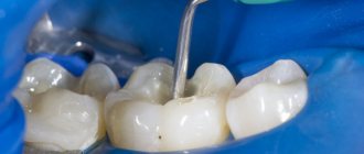
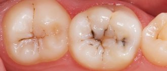
I class
Incorrectly performed treatment of group I caries can cause the filling to fall out of the cavity of the diseased tooth when chewing ; the risk is determined by the location of the lesion. Therefore, when treating caries, which is included in group 1 of the Black classification, the dentist uses several techniques to prevent such consequences:
- reduces the bevel of tooth enamel;
- applies the composition parallel to the base of the carious cavity (when working with composites);
- lays light-hardening mixtures at an angle in several layers (to change the direction of shrinkage);
- produces the final reflection of the filling through the side walls of the tooth.
There are many ways to reduce the risk of a filling falling out. For the treatment of first class dental caries, special algorithms have been developed that take into account the peculiarities of working with different materials. All of them are reflected in curricula and specialized instructions for dentists.
II class
This class of caries has its own characteristics. The main task of the dentist when treating teeth affected by the disease of the second group is to prevent the edge of the filling from overhanging and to ensure its tight fit to the bottom of the filled cavity.
The process can be complicated by teeth that are too widely or too closely spaced, so one of the stages of treatment may be bringing the contact surfaces closer or apart with the help of wooden wedges and holders.
All procedures, including preparation of the tooth cavity and spreading of contact surfaces, are performed after adequate anesthesia.
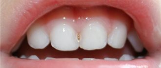
III and IV classes
Features of the preparation of carious cavities of the third and fourth categories according to Black do not play such an important role; the competent selection of filling material comes to the fore in the treatment of this type of dental caries. Since darkened areas of enamel are in visible places, it is necessary to use a filling that matches the color.
To do this, the prepared tooth is filled with a composite of not one, but two shades :
- white or milky for dentin restoration;
- almost transparent for restoration of tooth enamel.
The main difficulty in treating carious cavities on visible contact surfaces is to correctly assess the transparency of the tooth. There are no exact criteria for this; the dentist is forced to rely on his own feelings. Therefore, it will be better if an experienced specialist undertakes the treatment of caries of class IV and III according to Black.
V class
According to Black's classification of carious cavities, class five lesions are located in close proximity to the gums. This is the main difficulty in their treatment. If the patient is worried about gingival bleeding and noticeable discomfort in the cervical area, the doctor may suspect a deep location of the cavity affected by decay.
In this case, dental care is provided in several stages:
- Removing plaque from the surface of a diseased tooth.
- Determining the shade of the future filling.
- Anesthesia.
- Opening the cavity, cleaning out softened tissues.
- Correction of the gum edge.
- Treatment, placing a filling in the cavity treated with the drug.
- Polishing.
If the clinic adheres to the Black classification, the doctor will be recommended to use a composite ionomer composition. Due to its properties, this material is ideal for filling large cavities.
Dr. Greene Vardiman Black
Dr. Greene Vardimar Black is a well-known personality who is at the forefront of the development of dental science in the United States of America. He was born in 1836 in the city of Winchester.
At the age of 17, the young man became interested in medicine and worked for several years as an assistant to dentist D.S. Spira, while simultaneously gaining theoretical knowledge on this topic.
After completing his education, Greene Vardimar Black opened his own dental office in Jacksonville. In addition to providing services to the public, Dr. Black never stopped studying science and improving himself.
In 1870, a specialist invented a mechanical drill equipped with a foot drive. The gold amalgam composition developed by Dr. Black is also used in modern dentistry.
In addition, the specialist brought the terminological base to the standard, and also developed a classification of carious cavities and cutting dental instruments.
Dr. Black compiled several books that described methods for preparing the tooth surface, touched on the features of therapeutic dentistry, and also described some pathologies. In addition, Mr. Black taught dental science at the College of Chicago and also served as dean of the Northwestern University School of Dentistry.
Taxonomy according to the degree of caries activity
https://www.youtube.com/watch?v=ytcreatorsru
Vinogradova identified three degrees of caries activity, based on many years of experience in clinical analysis of the development of the disease in children. The classification takes into account the number of affected teeth and carious cavities, their location and dynamics.
Compensated
This form of pathology was detected in half of the children. The enamel is white, dense with a healthy shine. Damage to the chewing surface of molars is typical. Starting from the age of 14, premolars are involved in the pathological process. Caries progresses slowly, and the lesion itself is isolated. Children belonging to this group only need to undergo an oral examination once every 12 months.
Important! This type does not cause unpleasant sensations, such as pain, increased sensitivity to cold and hot irritants, and is easily treatable if you consult a dentist in the initial stages.
Subcompensated
The frequency of second degree activity ranges from 25%. The enamel is still resistant to caries, retains its shine and has a grayish color. Typical localization is on the chewing surfaces of the first and second molars and incisors.
The first signs of damage are single foci of dull enamel. The furrows are rough, the surfaces are covered with plaque. The growth of the carious process is carried out in width; the presence of deep caries is not observed.
Subjectively, the pathology does not give children any sensations. The subcompensated form requires a preventive examination and sanitation of the oral cavity every six months.
Decompensated
This variety affects the teeth of 11% of children. The enamel has the following features:
- Lack of shine.
- Chalky or matte enamel shade.
- Roughness in physiological grooves.
The speed of development is high. After 3-4 months complications are observed: pulpitis, periodontitis. The fissures are pigmented, and after opening, a carious cavity is found in them in 100% of cases. To prevent further progression of the disease, examinations and sanitation every 4 months are recommended.
Important! The main feature of the decompensated type is its intensive development and acute course. Pathology quickly leads to loss of ability to work.
What other systems exist?
Black's classification is topographical; dentistry uses several more ways to divide types of caries into features:
Universal classification ICD 10
ICD 10 is a generally accepted and unified classification of diseases that applies to all human organs, including teeth. The classification of caries according to this system is described in detail here.
Histological
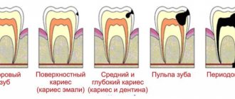
Involves sorting according to histological characteristics, i.e. the conclusion is made based on what tooth tissue is affected: enamel, dentin or cement. The classification includes 3 corresponding varieties:
- Enamel caries.
- Dentin caries
- Cement caries.
According to the clinical course
Using diagnostic methods and analysis of patient complaints, the doctor determines the nature of the disease:
- Spicy.
- Chronic.
According to the depth of the lesion
The main method that helps to choose an approach to treatment. For example, medium caries lesions of the contact surface of the tooth (type 2 according to Black). There are 4 types:
- At the spot stage.
- Surface.
- Average.
- Deep.
In relation to the condition of the pulp
A conclusion is made about the involvement of the pulp in the process of tooth destruction.
- Simple.
- Complicated.
By number of teeth affected
How many teeth are affected by caries at the time of the patient’s visit to the dentist.
- Single.
- Multiple.
- Generalized.
If you find an error, please select a piece of text and press Ctrl+Enter.
Tags caries treatment
Did you like the article? stay tuned
Previous article
Pathological causes of burning tongue and proper treatment
Next article
Gentle and effective treatment of periodontitis with a smart device Vector
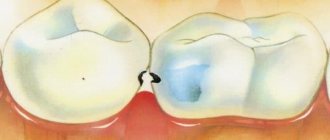
Prevention of approximal caries
The development of interproximal caries can be prevented. To do this, you need to thoroughly clean the interdental spaces using an irrigator or dental floss. In the dentist's office, you can have your teeth professionally cleaned once every six months, and the doctor can treat the dental spaces using special strips.
The use of fluoride-containing toothpastes significantly reduces the risk of caries formation by 30-50%. It is also worth paying attention to the diet: a large amount of carbohydrates in food leads to prolonged acidification of saliva and faster destruction of tooth enamel.

