A bacterial infection such as caries is distinguished by the fact that its activity continues even after treatment, at least until the pathogenic microflora is destroyed. The resumption of this disease is called secondary caries or it is also called caries under a filling.
The difficulty here is that the development of the disease in the secondary form may not manifest itself in any way, the disease affects deeper and deeper layers, and the patient does not feel any discomfort and the problem becomes obvious only during the treatment process, when the old filling is removed. It also happens that the disease affects tooth enamel, which has not previously experienced its harmful effects.
Soreness
After a visit to the dentist, patients often experience pain, this happens for the following reasons:
- increased sensitivity as a result of intervention in the oral cavity from the outside;
- the appearance of a cyst;
- development of periodontitis;
- allergic reaction to the filling material used;
- inflammatory process in tissues and nerves;
- relapse of the disease.
Causes of secondary caries
The main reasons are a violation of the technology for preparing the tooth surface before filling, shrinkage of the filling or a loose joint between the enamel and the filling mass. The larger the size of the filling, the higher the risk of complications, since the affected area increases.
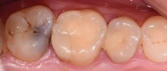
Secondary caries forms around the filling - at the border of the filling material and hard dental tissues.
The pathogenesis of the disease includes 3 stages:
- First phase.
Formation of a gap (microcrack) between the contact surfaces of the composite and the walls of the dental cavity.
- Second phase.
Saliva, along with bacteria, enzymes and other components, penetrates into the eaten away microcrack.
- Third phase.
The bacteria Streptococcus mutans multiply and colonize. Microorganisms produce organic acids that destroy enamel and the integrity of the filling material. Rejection of composites from the walls of the dental cavity occurs.
The reason for the need to change old fillings
The question of the need to replace an old filling with a new one often arises before patients; many of them doubt whether this should be done, since the tooth does not cause any concern. However, it should be said that the appearance of caries under the crown is quite possible and this can be caused by very specific reasons, in particular, there may be a medical error, for example, incomplete removal of infected tissue. In addition, the filling does not last forever, its service life is on average about 5 years, and after that a variety of situations are possible.
Thus, we can say that replacement of the filling must be performed in the following cases:
- violation of the tightness of the seal due to which pathogenic organisms penetrate under it;
- severe wear of the seal as a result of its long service life;
- violation of anatomical proportions during the installation of a filling;
- aesthetic unattractiveness of the installed product.
It is worth remembering that, despite the importance of the aesthetic component, health issues must be put at the forefront, therefore, if problems arise with installed fillings, you need to take care of their timely replacement.
Treatment of secondary caries under a filling
If a small carious lesion occurs on the chewing surface, there is no need to remove the entire filling. The doctor eliminates only part of it and fills this place with the same filling material.
In cases where the tooth is completely affected by caries, re-filling is prescribed according to the standard scheme.
Stages of filling
- Drilling out an old filling with a drill.
- Cleaning necrotic (decayed) dental tissues.
- Antiseptic treatment with a solution of chlorhexidine bigluconate or hydrogen peroxide.
- Installation of an insulating medical gasket at the bottom of the cavity.
- Tooth restoration using layer-by-layer application of a photopolymer composite.
- “Adjusting” the filling to the bite.
- Surface grinding and polishing.
Installing a tab
A ceramic inlay is used if, after the removal of an old filling, a wide cavity has formed. An inlay is a microprosthesis made in a dental laboratory using dental impressions. It is stronger and more stable than any filling.
The individual inlay firmly adheres to the walls of the dental cavity and is securely fixed with adhesive cement. The cost of such a microprosthesis is 2-3 times higher than that of a standard filling.
Ceramic inlay
Causes of secondary disease
Secondary caries can manifest itself for various reasons, the most common include the following:
- incorrect initial installation of a filling, when there is a difference between the tooth and the surface of the material used, which serves as a place for the accumulation of harmful microorganisms;
- insufficient level of enamel preparation for the installation procedure;
- incomplete removal of tissues affected by caries;
- Caries under a filling can develop if the first filling was too large.
Important! Secondary caries under a filling in a patient who visits the dentist in a timely manner is a very unpleasant moment. Often the disease develops due to errors made during the initial treatment. But experienced doctors believe that this can be avoided if all manipulations are carried out under the control of a dental microscope. A high-precision device allows you to clearly visualize the entire course of treatment, carry out work with pinpoint precision, and even take pictures (photos and videos) at all stages.
Treatment of the disease
There is only one way out of the situation - re-installation of a new filling after complete removal of the old one
Treatment of this disease follows a standard template, in all cases without exception:
- Complete removal of the old filling.
- Removal of necrotic carious dentin under a filling.
- Antiseptic treatment of the formed cavity.
- Placement of a new filling.
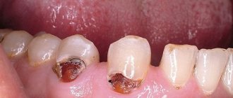
If you neglect the first point and leave part of the filling in place, secondary caries may occur again. In a situation where the filling occupies a large part of the tooth, the doctor may suggest orthopedic treatment - placing a crown.
And this will be quite competent and justified, since after removing the filling and tissues affected by caries, almost nothing will be left of the tooth, and making a “patchwork quilt” from old and new fillings is very short-lived and can lead to further destruction.
Symptoms
As mentioned above, it is not easy to recognize secondary caries; however, there are certain symptoms, the appearance of which serves as an alarming signal and a reason for an early visit to the dentist. These include the following factors:
- The appearance of pain in individual teeth.
- Inflammation of the gums, their swelling.
- Manifestation of bleeding gums.
- The appearance of an unpleasant odor in the mouth.
You can also pay attention to some external signs, in particular, the filling will change its color either completely or along the edges, in addition, cracks or chips may appear in the enamel.
Cost of treatment
The cost of treatment depends on many factors.
The pricing of dental services is influenced by several factors:
- regional peculiarity;
- clinic status;
- level of qualification of dentists;
- materials and technologies used.
Treatment of secondary caries is more expensive, in contrast to the initial visit to the doctor.
- Repeated filling includes unsealing the canal, cleaning it, and medicinal treatment. This is approximately 2-2,500 rubles. Plus, re-installation of the filling - from 900 rubles and above. The amount may increase significantly when complications are identified and treated.
- For adhesive tooth restoration you will have to pay from 6,000 to 47,000 rubles. Such a wide range of prices is due to the use of different materials and methods of restoring the missing tooth fragment.
- The installation of a crown also depends on the material from which it is made. The price for the service starts from 10,000 rubles.
- Microprosthetics involves the installation of a ceramic inlay. The cost of the service ranges from 5,000 rubles and above.
Risk factors and diagnosis
There are several external factors, the action of which contributes to the patient developing caries under the filling;
- prolonged exposure of the tooth to food that is too cold or hot;
- prolonged consumption of excessively hard foods or the habit of gnawing on something;
- excess friction as a result of malocclusion;
- insufficient attention to oral hygiene.
A specialist can diagnose the disease during a routine examination, however, this is not always enough and here X-ray and visiography data come to the aid of the dentist.
Nature of pain
The nature of pain in a tooth under a filling can vary.
This can range from a completely insignificant reaction to cold to acute and almost unbearable pain.
Based on the nature of the pain, you can easily determine the cause of its occurrence:
- Sensitivity during chewing. Increased sensitivity of the tooth usually indicates that the doctor overdried the crown during its installation. In this case, there is no point in worrying too much. Over time, the nerve endings will return to normal and stop reacting painfully to stimuli.
- Sensitivity to sweet, sour, hot and cold. Such problems indicate the low quality of the material of the installed filling. The easiest way to get rid of the pain is to have it refilled by a more experienced specialist.
- It's a dull pain. If the pain is aching in nature, and the gums are red and bleeding, most likely we are talking about a common allergic reaction. Any antihistamines will help cope with it.
- Severe and throbbing pain. Such symptoms indicate the development of acute pulpitis in the patient.
- Mild associated pain. Such pain may mean nothing, or may indicate the presence of chronic pulpitis.
If you suspect pulpitis, it is strongly recommended to seek help from a dentist regarding pulp removal as soon as possible. The specialist will remove the old filling, remove the pulp, fill the affected canals and re-place the filling.
If treatment is started in a timely manner, partial preservation of the pulp inside the dental canals is possible. The last point is especially important, since teeth from which soft tissue has been removed are more susceptible to destructive processes.
Secondary VS recurrent – let’s understand the terminology
When they talk about secondary caries, they mean that under the filling placed on the tooth, new foci of the disease are formed. The reason for their appearance is microorganisms that have penetrated through cracks under the filling. In addition, secondary caries also affects the damage to tooth enamel, which is located near the installed filling.
The concept of recurrent caries includes the resumption of the disease in the place where previously treatment was carried out. In most situations, this occurs through the fault of the doctor, since either there was poor quality treatment of the affected area, or due attention was not paid to the use of antiseptics and the infection was not completely destroyed.
Prevention
The best prevention is competent and qualified treatment. If the doctor did everything correctly, carefully removed all diseased tooth tissue, and adjusted the filling perfectly, the risk of recurrent caries is extremely low. If the patient follows all oral hygiene standards, relapse may not occur.
You should definitely visit the dentist twice a year; in this case, secondary caries can be detected at an early stage of development. In some cases, if the edges of the filling have become rough, it is enough to simply polish the filling and the place where it joins the tooth enamel to prevent it. Sometimes this precaution is enough to prevent the development of recurrent caries.
Development mechanism
A secondary disease that affects healthy teeth develops in three stages:
- Microscopic gaps appear between the material used in treatment and the tooth.
- Pathogenic microorganisms begin to penetrate into these cracks and crevices.
- The proliferation of pathogenic organisms and the release of toxins that have a destructive effect on tooth enamel and installed fillings.
As a result, rejection of the material used begins.
Providing dental care
Treatment depends on the extent and severity of secondary caries. After a complete diagnosis, the dentist chooses a specific method of therapy, or completely removes the tooth if its deep layers along with the root were affected.
Re-filling
The re-sealing procedure is carried out in several stages:
- drill out the damaged part of the tooth;
- remove damaged or dead dental tissue and filling remains;
- clean enamel and dentin;
- treat the tooth cavity with antiseptics;
- lay an insulating gasket;
- install a new filling.
To ensure the effectiveness of the procedure, carious areas are stained with a special solution. Based on the results of the reaction, it becomes clear whether all damaged tissue was removed.
Adhesive restoration
This treatment method is an alternative to installing a dental crown. The main advantage of adhesive restoration is the minimal impact on tooth enamel, while achieving high integrity of the tooth itself.
The procedure is as simple as possible. The prepared tooth is treated with an adhesive polymer, the main task of which is to restore damaged enamel and reduce its sensitivity to various temperature effects. The treatment method is reliable and durable, and as a result the patient receives an attractive smile.
Microprosthetics
Today, microprosthetics is considered to be a progressive and effective method of treating teeth affected by caries, even in young patients.
The technology involves the installation of special ceramic (or other materials) prosthetic inlays. A cast of the tooth affected by caries is made, its edges are processed and sent to the laboratory to create suitable inlays.
The design fully matches the shape, size and color of the tooth, which makes its presence invisible to others. In addition, thanks to the tight fixation, such an insert does not feel like a foreign body in the mouth. If the procedure is not possible, a re-installation of the crown is used to restore the tooth.
Crown installation
A crown is installed if it is impossible to carry out fillings, extensions and other treatment methods. However, crown installation is possible if there is no damage to the tooth root.
For the manufacture of crowns, alloys from medical steel are used (due to their unaesthetic appearance, a similar method is used in the treatment of distant teeth), metal-ceramics (they are durable and attractive in appearance) and ceramics (most similar to natural teeth, but unable to cope with heavy loads ).
Preparation for the procedure includes removing the dead tooth or grinding it down and then polishing it. After this, a plaster cast is made, the basis of which will serve to create a future crown. If the crown is being made for a long time, the patient is given an auxiliary plastic structure that will protect the ground tooth.
Initially, the crown is installed on a temporary cement mortar so that the behavior of the diseased tooth in relation to the adjacent healthy teeth can be observed. If no negative effects were noticed, the product is fixed with permanent cement.
Possible consequences
If you ignore the emerging symptoms of the return of caries, the patient risks getting a number of problems and complications, namely:
- deep damage to bone tissue infection;
- caries damage to adjacent healthy teeth;
- the process of destruction of the root and dental canal;
- tooth loss.
The main danger of secondary caries is that an inflammatory process develops in the pulp, which over time will lead to tissue death.
Diagnostics
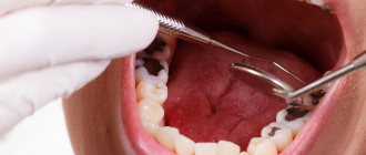
The dentist diagnoses the disease using basic and additional research methods. The first step is to examine the diseased tooth. If a cavity forms near the filling or a carious process is visible through the enamel, these signs will be sufficient to determine the disease. The specialist also collects an anamnesis of the disease and asks about primary treatment.
If the dentist sees that the filling has changed color, but cannot immediately identify caries, he will order an x-ray. An X-ray will also be needed if secondary caries has occurred in a pulpless tooth.
Using X-rays, you can assess the depth and localization of carious destruction. In the picture, the filling will look like a white spot, and the caries will look like a dark spot.
Tooth lesions due to hidden caries
Usually, hidden caries is not difficult to notice even with the naked eye; the extent of the damage received and its nature can be determined due to the duration of the process and its localization on the tooth damaged by the infection. Localizations of hidden caries include:
- on the back and adjacent walls of the tooth, which is extremely difficult to notice during a visual inspection;
- under crowns or fillings installed with errors and inaccuracies;
- in those areas of the tooth that are located under the gum, since it is difficult to clean out food debris with a toothbrush;
- in the natural pits of the so-called “back teeth”, which are also not easy to notice during examination.
Usually, the diagnosis of hidden caries becomes possible in the later stages of the disease, less often in the middle stages and almost never in the early stages. That is why the dentist begins treatment when the peripulpal tissues and dentin already have extensive damage.
Why does a tooth bleed?
The situation when a tooth bleeds is familiar to everyone. According to WHO, 100% of adults have caries, and 15-20% are diagnosed with severe periodontal diseases. The cause is not always pathological, but it is impossible to identify it on your own. If your gums are inflamed or your teeth are bleeding, the dentist will determine the treatment. Doctors at Dr. Granov’s clinic not only relieve tooth pain and bleeding, but also eliminate its cause.
Reasons why a tooth bleeds
Droplets of blood appear when brushing with a brush that is too hard, when injured with sharp objects, even while taking certain medications, such as aspirin. But these reasons are related to the gum, and not the tooth itself. Often, already at the appointment, the patient complains of tooth bleeding, but the doctor sees that the source of concern is the gums.
But in some cases, the cause is actually inside the tooth, which is bleeding due to:
- granulations - loose bleeding tissue that fills the carious cavity and gum pocket;
- inflamed pulp - connective tissue inside the tooth, penetrated by nerves and blood vessels.
Diseases that cause teeth to bleed
Bleeding manifests itself as a complication of caries. It accompanies the following pathologies:
- periodontitis - inflammation of the tissues surrounding the tooth and holding it in the socket, accompanied by the formation of granulations;
- chronic pulpitis - inflammation of the pulp of tissues in which blood vessels and nerves pass;
- dental abscess - the formation of a purulent cavity in it, which destroys the vessels of the pulp.
Timely treatment of inflamed tissues prevents their further destruction.
If overgrown bleeding areas of the pulp or periodontal tissue are left unattended, the inflammation spreads to the bone or even provokes the development of a tumor.
Leave your phone number. The clinic administrator will call you back.
By leaving a request on the site, you consent to the processing of personal data
Make an appointment
Periodontist consultation
500 rub.
Teeth are bleeding: what to do
It is impossible to independently determine the cause of bleeding. All the more dangerous are attempts at self-medication. Trauma or cauterization of inflamed areas sometimes becomes an impetus for the formation of a tumor. Therefore, when a tooth or gum bleeds, all manipulations must be carried out by a dentist.
At Dr. Granov’s clinic, doctors use instrumental and hardware methods to diagnose any pathologies of the teeth and periodontium - the tissues that support it in the socket.
Treatment and prevention
Treatment of caries of an already pulpless tooth is based on the removal of a previously placed filling. Next, the preparation procedure is carried out and it is extremely important that it is carried out at the highest level of quality, otherwise the potential threat of the return of caries will arise again. Then a new filling is installed.
In the fight against caries, prevention plays a vital role, since preventing a disease is always easier than treating it later, especially in the late and advanced stages. Therefore, it is necessary to regularly and promptly visit the dentist for preventive examinations. If any damage, even the most minor, occurs, you should immediately visit a specialist to fix this problem.