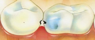
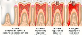
Deep caries is a disease characterized by severe destruction of hard tissue. The structure of the tooth includes enamel, dentin and pulp - the inner, soft structure. The first stage of a carious lesion is the appearance of a white spot on the enamel - the demineralized area manifests itself as a change in color. Subsequent destruction of the enamel, the appearance of a dark spot, is characteristic of medium caries. In deep caries, it affects not only the enamel, but also the dentin located near the pulp (nerve of the tooth). The caries cavity is separated from the neurovascular bundle by a thin strip of dentin, so it is very important to stop the pathological process and prevent pulpitis.
Deep dental caries can be primary or secondary. In the first case, it develops on a previously healthy tooth and is a consequence of the progression of secondary caries and the failure to provide timely dental care. In the second case, deep caries develops in a previously treated tooth under a filling. In addition, the disease can occur in two forms: acute and chronic. In acute flow, a narrow inlet leads into the cavity.
Chronic deep caries is characterized by a funnel-shaped wide entrance to the cavity, the bottom of which, on the contrary, is narrower.
Classification
According to the international classification of diseases ICD-10, dental caries is included in a separate section. Deep caries (dentine caries) is assigned code/code K02.1. Depending on the degree of compensation (since caries can worsen and acquire different activity), the dentist can use codes K. 02.0-02.9 to encrypt the diagnosis.
Acute form
Patient complaints:
- The occurrence of acute pain when exposed to mechanical, thermal and chemical stimuli, while the pain disappears immediately after the cause of the irritation disappears.
- Change in tooth color.
- A carious cavity that is constantly increasing in size.
- Food getting stuck in a carious cavity.
During a dental examination:
- A deep carious cavity, with the entrance hole often being smaller than the width of the cavity.
- Chalky color of dentin/enamel.
Chronic form
Patient complaints:
- Mild pain.
- The presence of a cavity into which food enters.
- Change in tooth color.
During a dental examination:
- Wide entrance hole into a deep carious cavity.
- The bottom and walls of the cavity are filled with pigmented plaque.
- Damage to peripulpal dentin.
Deep caries under filling
Dentists distinguish two types of carious lesions under fillings.
- Secondary caries (when a carious lesion develops due to the formation of microcracks between the filling and hard tissue and the penetration of microbes into them).
- Recurrence of caries (if during the previous treatment the affected tissue was not completely removed).
Complaints:
- Sensitivity to temperature stimuli.
- Darkening of tooth tissues (gray dentin tissue is visible through the enamel).
- Mobility of the filling
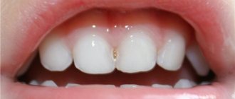
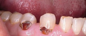
In the photo: 3 teeth destroyed by deep caries
Causes
There are many theories of the occurrence of carious processes. Their graphic symbol became the “Case trefoil” (circles overlapping in the center). According to him, caries develops when three conditions coincide: low enamel resistance, cariogenic flora and easily digestible carbohydrates. Modern dentists supplement the “trefoil” with another circle and, accordingly, a condition - the duration of the effect.
In addition, deep caries can develop in a previously treated tooth - due to treatment defects, under a filling, or when a filling is chipped.
Reasons for development:
- General (insufficient fluoridation of drinking water and consumption of foods with insufficient minerals and vitamins; predisposition to caries at the gene level and malfunctions in the mother’s body during the period of intrauterine formation of tooth germs; ecology).
- Local (food residue remaining on the teeth after eating - dental plaque begins to form within 2 hours after brushing; reduced secretion of saliva in certain diseases and conditions; dental plaque).
Diagnostic methods in dentistry
- Clinical examination. Using a probe and a special dental mirror, the doctor examines the appearance and assesses the condition of the teeth and gums. When examined with a light microscope in deep caries, 3 zones are clearly visible: decay and demineralization, transparent and intact dentin, replacement dentin and changes in the pulp.
- Thermometry. Study of tissue response to temperature. With deep caries, the tooth reacts to temperatures below 18-20 and above 45 degrees. To determine the reaction, dentists use cold water irrigation, chloroethyl and pieces of heated gutta-percha.
- X-ray examination. A photograph of one, two or several adjacent teeth can be taken, a panoramic photograph in which two rows of teeth will be captured. The image allows you to evaluate the quality of the fillings and identify a carious cavity. Significant disadvantages of the method: radiation exposure and the possibility of detecting occlusal caries in large cavities.
- Transillumination. Transillumination of dental tissue with a photopolymerization lamp. Carious cavities and cracks appear as dark spots. This method is not very informative if there is a filling on the tooth.
- Luminescent method. Fluorescent stomatoscopy is performed in a darkened room. A beam of ultraviolet rays is directed onto the enamel. When it comes into contact with healthy areas of the tooth, a blue glow is observed. With caries, the glow goes out and dark spots become clearly visible.
- Caries markers. Using caries markers, the boundaries of the carious cavity are determined. To stain demineralized tissues in dental practice, preparations based on fuchsin or 2% methylene blue are used. Their operating principle is simple: healthy dentin has small pores, and the dye does not penetrate into them, while infected tissues with large pores acquire a certain color.
- Laser diagnostics. A highly accurate diagnostic method that can be used even in childhood. Diagnostics is based on fluorescent radiation. The radiation spectrum of healthy tissues differs from the spectrum of infected tissues. The laser beam, hitting the infected area, is reflected from the carious cavity, and the diagnostic device emits a special sound signal.
- Electroodontometry (EDO) . A healthy tooth responds to a current strength of 2-6 μA. Caries reduces the insulating properties, and the electrical resistance decreases to 15-20 μA. A decrease in excitability to 60 μA indicates an inflammatory process of the coronal pulp, up to 100 μA - of the root pulp, more than 100 μA - with the death of the pulp.
Symptoms and self-diagnosis
- The presence of a carious cavity (a dark spot on the enamel).
- Pain (from hot/cold, sweet/sour, when pieces of food get into the cavity).
- Bad breath (especially noticeable with multiple dental lesions).
- Violation of the filling (its loss, mobility, chipping).
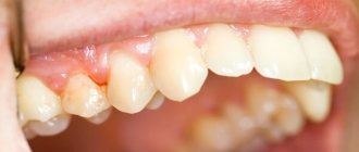
In the photo: advanced deep caries on the left tooth
With chronic deep caries, the asymptomatic period can last several years. Only when the destruction of dentin reaches the bottom of the tooth does pain occur when pressed.
Complaints
Patients with acute deep caries complain of pain that occurs as a result of the action of thermal, mechanical, and chemical stimuli and disappears immediately after their elimination. Thus, inserting a tampon soaked in hot (no more than 50°C) or cold water, as well as ether, into a carious cavity is usually accompanied by a sharp pain reaction. A carious cavity is detected in the tooth within the peripulpal dentin with overhanging edges of the enamel. The enamel around the entrance hole of the cavity is brittle and chalky in color. The carious cavity is made of softened dentin of a whitish or gray-yellow color. When probing, pain is noted in the area of the enamel-dentin junction, as well as (less pronounced) at the bottom of the cavity at the points of the thinnest dentin above the pulp. Often these are the projection sites of the pulp horns, which directly respond to stimuli. There is no connection between the carious cavity and the tooth cavity. In case of acute deep caries, probing the bottom of the carious cavity must be carried out very carefully. At the projection points of the pulp horns, the arch of the tooth cavity is very thin, the dentin is softened, and it is easy to pierce it with a probe and injure the pulp. This is accompanied by sharp pain and the appearance of a drop of blood in the carious cavity.
With chronic deep caries, there may be no complaints of pain or there may be minor short-term pain after exposure to thermal, chemical and mechanical stimuli. The defect of hard tissues within the boundaries of the peripulpal dentin occupies a significant part of the tooth crown and is open to the outside (the fragile overhanging edges of the enamel break off). The transverse dimensions of the cavity exceed its depth. The carious cavity is made of rather dense, but without sclerotic shine, pigmented dentin. The pigmentation of its walls and bottom has a wide range - from yellow-brown to brown, or even almost black. Probing the walls and bottom of the cavity is painless, since zones of transparent and secondary dentin are well defined underneath them. The surface of carious dentin is rough when probing, in some cases it yields to the pressure of the probe and is quite difficult to excavate. Such a cavity can take years to form.
Treatment
General stages of treatment:
- Anesthesia (local anesthesia with lidocaine drugs)
- Opening the carious cavity (the dentist removes the overhanging edges of the enamel with a bur).
- Necrectomy (removal of softened dentin).
- Formation of a cavity using air-water cooling (the walls should meet the bottom of the cavity at a right angle; in deep carious cavities, the bottom is stepped or roller-shaped).
- Finishing the cavity (smoothing the edges of the enamel and removing damaged areas; necessary for a tight fit of the filling and to prevent the development of secondary caries).
- Antiseptic treatment and drying of prepared hard tissues (warm solutions of furatsilin, chlorhexidine, dimexide, etonium are used; drying is carried out with sterile cotton swabs).
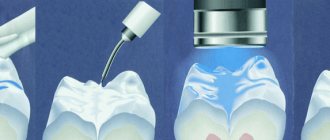
- Etching the enamel.
- Washing and drying the surface.
- Application of primer and adhesive (necessary for better fixation of the filling to the dentin).
- Adding filling material (glass ionomer cement or composite material).
- Polymerization of the filling (treatment with a polymerization halogen lamp).
- Polishing.
- Post-bonding (grinding roughness).
- Application of fluoride preparations.
The duration of one procedure can be from 40 minutes to one and a half hours.
Tools and materials
Medical pad
Even gentle tissue preparation injures the processes of odontoblasts and negatively affects the condition of the pulp. The therapeutic pad has a healing effect on the pulp, stops the inflammatory process, stimulates reparative processes, relieves pain, and withstands pressure after hardening.
In case of deep caries, the dentist usually applies a therapeutic pad with an antiseptic and odontotropic effect. These can be pads based on calcium hydroxide (“Calme-cin”, “Dycal”, “Alkaliner”, “Reocap-E”, etc.), zinc-eugenol cement (the main component is eugenol - an antiseptic of plant origin), combined medicinal pastes.
A warm therapeutic pad is applied with a thickness of no more than 0.5 mm only to the bottom of the prepared carious cavity.
Temporary filling
With deep caries, the layer of healthy dentin is so thin that cariogenic microbes penetrate into the pulp. If during a visual examination the dentist has this kind of suspicion, then a medicinal tab is installed and a temporary filling is placed.
The following one-component materials are often used: dentin paste, water-based or artificial dentin, cements, polymer materials. A temporary filling can also be diagnostic. It is installed in order to see how the pulp behaves after preparation. If replacement dentin begins to be produced, the patient will no longer be bothered by pain. A tooth with an infected pulp requires endodontic treatment.
Temporary filling
Insulating gaskets
The purpose of the insulating gasket is to protect dentin and pulp from chemical and thermal influences. In addition, it is placed between the bottom of the carious cavity and the permanent filling to prevent increased sensitivity after drilling.
Functions of insulating pad:
- Improving the fixation of permanent fillings.
- Dentin sealing to prevent microbial invasion.
- Remineralizing effect on underlying dentin.
Depending on the condition of the cavity, the dentist may use a base or thin-layer lining. If the thin-layer only protects the pulp from chemical attack, then the basic one creates the optimal geometry of the carious cavity, protects the pulp from thermal irritants and allows the use of a smaller amount of filling material.
Materials for creating insulating gaskets: insulating varnishes, glass ionomer cements, polycarboxylate and zinc phosphate cements (the last two are practically not used).
Medicinal paste
Therapeutic pastes are prepared immediately before being introduced into the carious cavity. The medicinal substances included in their composition may be different. Their choice depends on the clinical situation and the individual preferences of the dentist. Their main disadvantage is the rapid loss of activity and insufficient mechanical strength, so pastes are used during the period of “active” treatment for a temporary filling, followed by replacement with some type of cement.
List of medicinal substances used for the preparation of combined pastes:
- Anti-inflammatory substances (prednisolone, salicylates, hydrocortisone, indomethacin).
- Odontotropic substances (Algipor, calcium hydroxide, dentinal sawdust, fluorides, calcium glycerophosphate, collagen).
- Antimicrobial agents (ethonium paste, chlorhexidine, lysozyme, metronidazole).
- Proteolytic enzymes (imozymase, profezim).
- Other products (natural oils - sea buckthorn, clove), dimexide.
Preparations:
- Pulpomixine;
- Pulpanest;
- Pulperyl;
- Contrasil;
- Calcipulpe;
- Septomixine forte.
Treatment of deep caries under the gum
The pathological process developing under the gum often does not involve areas visible to the eye, and therefore is detected in a deep form. Often in such cases, pulpitis develops and treatment is impossible without removing the nerve.
Peculiarities:
- Subgingival carious processes require gum correction (coagulation, excision). It is necessary for high-quality dental treatment and isolation of the working area from saliva and blood.
- If there is significant destruction of the walls, it is necessary to install an inlay (or an inlay with a pin).
- If the crown of the tooth is completely infected and the root is intact, a crown is placed on the living tooth.
Tooth extraction is carried out if the deep parts of the roots become infected.
Features of treatment in children
Treatment of deep carious lesions in children is associated with a number of difficulties. Children get tired quickly and often close their mouths. Treatment of a carious cavity is complicated by abundant salivation and mobility of the head and tongue.
Preschool children
Carious cavities at this age have wide entrance holes, they are flat and small. Dentin is easily removed. During treatment, the pulp horn may be exposed, so the dentist must be very careful.
Mechanical treatment begins at the 2-3rd visit. It is preceded by coating the teeth with fluoride varnish. The doctor gently treats the carious surface with a spherical bur, removing softened dentin, treats the cavity with non-irritating antiseptics and fills the tooth with polycarboxylate or glass ionomer cements.
In childhood, delayed treatment is possible (without anesthesia and a drill). The purpose of the method is to prevent tooth decay and wait until the child gets used to the dental environment.
Stages of treatment:
- Treatment of carious cavity with hand tools.
- Temporary tooth restoration with glass ionomer cement.
This material is temporary, it tends to be stained with dyes, chip, and sag, so the minimum service life of such a filling is 6-12 months. After this time, the tooth must be restored with permanent material.
Pupils
To restore carious defects in fillings at this age, dentists use silver amalgam, chemically and light-curing composite materials. When treating teeth with deep caries and immature roots, preference is also given to silver amalgam. An invasive fissure sealing method can also be used.
How to treat during pregnancy
A carious lesion can cause intrauterine infection of the fetus, delay its development, provoke miscarriage and premature birth. In addition, acute pain during caries causes the release of specific hormones that negatively affect the embryo (they disrupt the nutrition and breathing of the fetus). Therefore, at the first symptoms of deep caries, it is necessary to contact a dentist, who, taking into account the duration of pregnancy, will select a safe treatment regimen. The best time for treatment is from 13 to 32 weeks of pregnancy.
- Anesthesia. Modern drugs used for anesthesia penetrate the systemic circulation, but do not cross the placental barrier. Therefore, if the dentist offers local anesthesia, you should not refuse.
- Radiography. Images during pregnancy are taken with a radiovisiograph - a local device (for an area of 2-3 cm) with an extremely low level of radiation. The abdomen is protected with a special apron with lead plates that do not transmit x-rays.
- Filling. After treating the carious cavity, hypoallergenic light-curing fillings are installed. Photopolymer lamps are used to illuminate them. Their blue glow is not an ultraviolet cure, but a stream of light passing through a filter.
Anesthetics used: articaine (considered the least toxic, but not used in case of anemia in a pregnant woman), ultracaine, brilocaine, primacaine.
How is the treatment carried out?
You can begin dental treatment after pain relief . First, the carious dental cavity is cleaned from softened dentin, then the doctor begins filling the tooth. There are also more severe cases when the disease affects the pulp tissue (damage to the dental nerve occurs). In this case, the attending physician performs depulpation of the tooth (removal of the soft tissue of the pulp, including the removal of the dental nerve).
In addition to the fact that the doctor will use filling material, he can also use medicinal calcium-containing pads, various antiseptics, insulating fluoride-containing sealants.
If deep caries is treated skillfully and in a timely manner, then even with deep destruction of dentin, it is possible to save a living tooth , restore it and return all its functions to it. If treatment is delayed, the problematic tooth will have to be removed.
During treatment it is important:
- maintain the vitality of the dental pulp
- avoid the development and recurrence of caries
- stabilize the remineralization of predentin and dentin
- activate the functions of the pulp to accelerate the formation of renewed replacement dentin.
It is important to approach the treatment of deep caries with extreme caution; it is especially important to carefully treat the bottom of the affected cavity. For processing, it is best to use only a spherical bur, while the bottom of the cavity is not leveled.
The use of irritating medications for treatment (ether, alcohol) is unacceptable. The choice of lining material will depend on the general condition of the dentin at the bottom of the cavity.
Folk remedies
Treatment of deep caries with folk remedies is impossible, because... requires the intervention of a qualified dentist. At home, resorting to traditional medicine recipes can only temporarily relieve the severity of pain and signs of inflammation.
Relieve pain:
- Alcohol tincture of propolis, vodka, Japanese Sophora, onion peel tincture, fir oil.
Relieve inflammation:
- Gargling with decoctions of doom bark, chamomile, and sage.
What not to do:
- Use honey or milk powder. The carbohydrates they contain activate the growth of bacteria and contribute to the intensive growth of carious lesions.
- Use hydrogen peroxide (possible burn to the mouth).
Deep caries of wisdom tooth
The reason for the development of deep caries of wisdom teeth is the rapid accumulation of plaque and an inconvenient location, which interferes with hygiene procedures. It happens that the “eight” erupts from the gums already with deep caries, and the person is not aware of its development until the pain begins to bother him. Another option for the unnoticed course of the disease is interdental caries, which, in addition to the wisdom tooth, also affects the adjacent molar. It is almost impossible to notice such caries at the spot stage.
Most dentists advocate the removal of carious wisdom teeth, because... It is sometimes simply impossible to completely remove infected tissue. Even in the case of high-quality filling, there is a high risk of developing secondary caries due to inaccessibility to a toothbrush.
At the same time, the G8 may be required in the future. For example, as a support for a prosthesis.
Considering all of the above, the question of treating or removing tooth decay remains open.
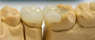
In the photo: deep caries of a wisdom tooth
Treatment of deep caries with pulp damage
If, after installing a temporary filling or even at the diagnostic stage, the doctor determines that the pulp is infected, a more complex multi-stage treatment is planned.
Most often, the pulp is removed, and the root canals are cleaned and processed. However, if the inflammatory process occurs at the initial stage, the doctor may recommend complete or partial preservation of the pulp - antibacterial therapy will be required.
If there are indications for removal of the neurovascular bundle, further tactics are determined taking into account the individual characteristics of the course of the disease and the extent of destruction of the coronal part. The doctor can fill the canals, and then the coronal part, or install a stump inlay or a crown if your own is badly damaged. The last option requires contacting an orthopedic dentist.
After treatment for deep caries, pain may persist for some time. As a rule, we are talking about a time of no more than 1–2 days, and the pain is moderate, observed only when chewing. If the pain does not subside or intensifies, it is important to return to the dentist - especially if the pulp is intact.
Consequences and complications
Post-filling sensitivity, which manifests itself as pain when pressure is placed on the filling and a feeling of discomfort, normally goes away within 1-2 weeks. If noticeable pain persists longer, this is one or another complication that arose due to a dentist’s mistake.
- Change in color of the filling (due to eating food containing dyes on the first day).
- Depressurization of the space between the tooth and the filling (due to a violation of the filling technique; fraught with the development of secondary caries and tissue infection).
- Postoperative sensitivity (due to overdrying of dentin).
- Filling fracture (during processing, placement or thinning).
- Filling loss (violation of the filling insertion technology and cavity formation).
- Papillitis (after irrational filling; manifested by swelling and redness of the gingival papilla).
- Inflammation and necrosis of the pulp (due to overheating of dentin, traumatic treatment of the cavity; endocanal treatment is required).
Deep caries filled - tooth hurts and aches
A slight aching pain is the norm after treatment of deep caries. The fact is that the bottom of the carious cavity is located close to the nerve, and the filling material transfers pressure to the pulp. Within a month, a protective layer of dentin will develop, and the tooth will stop hurting. If you are experiencing acute pain or fever, you should consult a dentist. After the x-ray, he will determine a further treatment plan.
It hurts to chew after filling
Pain when biting and chewing food after filling can occur when the surface tension of the filling on the tooth surface is high. In most cases, refilling the tooth is sufficient. There is no need to treat the canals or remove the nerve.
Pulpitis after treatment of deep caries
If after treatment you are bothered by increasing pain, then pulpitis has probably developed. You should immediately contact your dentist to remove the infected pulp and fill the root canals. With timely treatment, there is a chance to leave the pulp alive, i.e. do not remove the nerve. The doctor will remove only the coronal part of the pulp, using the so-called biological treatment method (after removing the tissues affected by caries, a therapeutic calcium-containing pad is placed in the cavity and a temporary filling is installed).
Gums hurt after treatment
A slight inflammation of the gums after treatment is possible. It usually goes away on its own. If after treatment the gums become inflamed, then after eating your mouth should be rinsed with solutions of medications (furacilin, chlorhexidine), decoctions of chamomile, sage, calendula tincture, etc. If the inflammation does not subside after 2-3 days, you should see a doctor (to rule out chronic gingivitis and other complications).
Tooth sensitivity after treatment of deep caries
The cause of sensitivity may be a violation of the tightness of the filling material to the tooth surface due to overdrying/underdrying of the dental cavity. When overdrying occurs, nerve endings are damaged and even die. If the cavity is not dried enough, drops of water remain on the walls of the cavity, which prevent the penetration of the adhesive into the dentinal layers. Only a doctor can clarify the origin of sensitivity, so if the dynamics are negative, be sure to contact him for advice. The problem may be solved by refilling the tooth.
After treatment, the tooth reacts to cold, hot or sweet
Increased sensitivity is a normal reaction of the pulp to intervention, but if the tooth reacts to irritants with increased sensitivity for more than 5-7 days, you should consult a dentist. Perhaps the tooth was not completely cured, and the dental canal became infected.
Can there be throbbing pain after treatment?
If deep caries with penetration of infection into the tooth tissue has not been completely treated, then aching or paroxysmal pain may occur. They can occur both during eating and at rest (particularly at night). These are symptoms of developing pulpitis. A characteristic sign of such toothache is irradiation to the temple or ear. At home, you can only relieve pain (take Ibuprofen, Analgin). Then you need to contact your dentist to clarify the diagnosis.
Diagnostics
Acute caries is most dangerous if its treatment is ignored, although, unlike other types, this disease is very easy to detect. It is enough to pay attention to such symptoms as:
- change in enamel color, darkening;
- the appearance of prolonged acute pain;
- exacerbation of the reaction to temperature changes and various external stimuli.
As soon as any of these signs begin to appear, you should immediately seek help from a doctor. Even if there are few signs of carious lesions or they are expressed to a small extent, you still need to visit a specialist, since a small spot on the tooth surface may hide a large affected area.
It will not be difficult for any competent dentist to make the correct diagnosis of decompensated caries, since it has well-manifesting signs:
- large cavity width;
- narrow entrance;
- softened dentin in large quantities;
- acute pain on probing.
Conventional diagnosis can be carried out using a mirror and a probe, but if the probing area is visually poorly visible, then in this case you need to take an x-ray or use the technique of highlighting carious cavities (transillumination). Sometimes a diagnostic technique such as fissurotomy is also used, when caries hidden from the eye are examined using preventive excision of depressions and pits of the enamel.