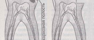
Features of caries between teeth
The oral cavity needs daily cleansing. One of the most difficult places for hygiene procedures is the space between the teeth. If you do not use floss and other modern means, a large number of bacteria accumulate in this place, which destroy the natural structure of the enamel through organic acid.
It is quite difficult to detect interdental caries on your own, so you need to visit your dentist regularly.
Due to the daily destruction of tooth cells, caries is formed. The location in the interdental space is complicated by the fact that infection quickly spreads to healthy tissue, affecting an increasingly larger area.
Initial caries can rarely be seen with the naked eye. As a rule, a doctor is consulted when caries has reached the root of the tooth, causing acute pain.
Depending on the location, there are several types of caries between teeth:
- chewing teeth . The most common type of disease, since it is between the chewing molars that the most food particles accumulate. Due to the location of these teeth, the disease is almost impossible to notice in the early stages. Moreover, caries can often form under the gum, which leads to damage to the tooth root;
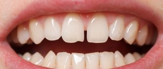
- anterior incisors . It occurs somewhat less frequently, especially if you follow the rules for cleaning the oral cavity. The defect can be noticed at the initial stage due to the formation of characteristic darkening;
- between baby teeth . In itself, it is not a complex disease, but if not treated in a timely manner, the pathology can spread to the root part, creating unfavorable conditions for the development of permanent teeth;
- partial caries between teeth . Only the side of one tooth is affected, when the disease has not yet spread to the neighboring ones. If detected, urgent treatment is required, as caries progresses quickly.
It is recommended to treat the disease as early as possible. According to statistics, deterioration of caries is observed after 1-2 weeks after the transition to the acute carious stage.
Treatment of interdental caries of chewing teeth
Despite the fact that molars are located in the lateral part of the jaw and are not too noticeable when smiling, interdental caries of molars is very dangerous, and at an advanced stage it requires multi-stage and expensive treatment. The disease develops more slowly on chewing teeth, but is more difficult to detect. As a rule, a patient comes to the doctor when interdental caries has affected the dentin layer on one or two teeth. Often, caries between chewing teeth affects the pulp of two teeth at once: this is the most unpleasant situation, since endodontic treatment of the canals of chewing teeth is a long and very expensive process. If you let the situation take its course, the patient risks acquiring a whole bunch of complications (pulpitis, periodontitis, and so on).
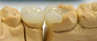
To treat interdental caries of chewing teeth, special wedges and matrices are used that highlight the working surface, help the doctor form the shape and restore the anatomy of the tooth (cusps, fissures). When filling caries between molars, the emphasis is on strength, reliability and durability, and not on aesthetics, as is the case with the smile area. Glass ionomer fillings are most often used to restore molars.
Causes
The main factor provoking the onset of the disease is improper oral hygiene. This type of caries is especially common since most people only use a brush, but for complete cleaning you should use floss, regular professional cleaning, and also use a mouthwash.
The course of the disease can be aggravated due to weak immunity, as well as in the absence of important microelements in the menu. This is why it is important to take vitamins and also maintain a balanced diet.
Reasons for development
The etiology of damage to hard tissues in this case is similar to the factors causing classic damage. The pathological process develops due to the accumulation of food particles and the active reproduction of pathogenic microorganisms in an environment favorable to them. The disease can spread very quickly, affecting not only the enamel, but also dentin, reaching the roots and causing other diseases. If you do not consult a doctor promptly, you may subsequently need to remove the nerve or install a prosthesis.
Where does interdental caries appear?
It is observed in places of close contact of dental units. It often occurs between the posterior chewing molars, which are actively involved in the process of chewing food. The front incisors and canines are affected much less frequently, since they are used only for biting food.
It is especially unpleasant when the disease affects “eights”. The distant location, as well as uneven roots, complicate filling and depulpation. Most often, experts recommend complete removal without subsequent prosthetics.
Caries between the gum and tooth is not considered interdental. A carious cavity should form precisely in the areas between adjacent units located in the vicinity.
Who gets the disease?
This is one of the most common dental problems. The disease occurs in children of all ages and adults.
It is often found on primary incisors and premolars in children 4 years of age and older. This feature is due to the fact that at this time the child begins to eat all the food from the parent’s table and at the same time needs regular high-quality oral hygiene.
Some parents mistakenly believe that if their son or daughter’s first teeth are damaged, they don’t need to be filled, since they will soon fall out anyway. Caries between teeth is treated both in the presence of temporary units and in the development of permanent ones. The pathological process can spread further, including to the rudiments of the molars and molars. Infection will lead to serious problems later in life.
But you should not be afraid that the child will be in pain during treatment. In the Dentika clinic, in case of deep lesions, the need for depulpation and canal cleaning, both the usual method of pain relief and sedation are used, that is, when performing medical manipulations, the baby sleeps and does not feel pain.
Why does pathology appear?
The main reason for the appearance of caries between teeth in adults and children is the accumulation of food debris in the interdental space and the active proliferation of pathogenic bacteria. With regular brushing, it is almost impossible to completely clean the contact points between the units. It is necessary to use additional devices (irrigator, floss).
The disease develops as follows:
- a person does not eat properly or does not regularly cleanse, or provides poor quality care;
- mucous membranes and enamel are covered with a dense coating;
- biological residues produce acids;
- the resulting substances corrode the outer layer and after some time affect the dentin.
The problem occurs in people with malocclusion, dystopic figure eights and other physiological characteristics. This is due to the difficulty of performing hygienic procedures and detecting carious lesions during a visual examination without x-rays and other precise techniques.
There is a list of factors that can lead to caries between two teeth:
- smoking, alcohol abuse;
- poor food quality;
- unfavorable environmental situation in the locality;
- reduced immunity;
- a history of chronic diseases affecting the condition of hard tissues;
- abuse of sweets and other carbohydrate foods.
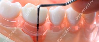
Diagnostic methods
Due to the fact that caries between teeth is poorly diagnosed by visual examination, additional hardware methods are required. Modern clinics use the following diagnostic methods:
- radiograph . The most accessible and accurate way. In the picture, the affected area has a characteristic dark tint, in contrast to the light healthy enamel and dentin tissue;
- Diagnodent device . In recent years, most clinics have used this diagnostic method. During the procedure, the affected tissue is captured by light waves, which is completely safe for the patient’s health;
- visual dental examination . Effective only in the middle and late stages of the disease. Diagnostics are carried out using special mirrors and probes, which make it possible to detect defects even in the most inaccessible places. Read more about modern methods for diagnosing caries here.
The choice of diagnostic method is determined by the doctor in each specific case. With interdental caries, it is especially important to detect the defect as accurately as possible, since bacteria often destroy the surface of several adjacent elements of the dentition at once, which may not be noticeable at the first examination.
How to independently diagnose interdental caries
To independently determine carious lesions, the patient can observe the following symptoms:
- In the first case, you will need a mirror and good lighting. Taking a closer look at the teeth, you will notice that the structure of the tooth is different in color. Healthy areas have the same shade and are transparent. The damaged area will be visible through the healthy enamel as a dark spot.
- The second factor in developing caries can be minor pain. It usually occurs under the influence of chemical irritants (sweet, sour and salty foods), as well as thermal irritants (eating food at different temperatures). In both cases, the pain quickly stops. Immediately after the stimulus ceases to act. When visually examining a tooth, its destruction may not be noticed, but the frequency of pain that occurs should serve as a signal to contact a dentist.
- The presence of carious damage can result in bad breath and altered taste, which spread from a certain area of the dental system. A carious defect is an excellent place for the accumulation of bacteria and food debris, and the vital activity of organisms and the rotting of products is accompanied by unpleasant odors.
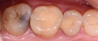
Treatment
Interdental lesions are treated with the same methods as other types. The main difference is the need for dental therapy on several teeth at once, which always affects the cost. Treatment of deep caries between teeth is a rare practice, so standard filling is used to eliminate the defect.
When interdental caries appears, prepare for the fact that you will have to treat two teeth at once.
Before installing fillings, a complete sanitation of the oral cavity is carried out and the affected tooth and neighboring objects are cleaned. In some cases, it is recommended to pre-clean professional enamel with ultrasound. After this, a hole is drilled without reaching the nerve. This stage is carried out both under local anesthesia and without it. Note that not only a standard drill, but also laser treatment of caries can be used to remove affected tissue.
Only after this can you proceed to installing the filling material. As a rule, light seals are used, which are easy to install and have a long service life. After these manipulations, it is important to carry out two necessary procedures:
- completely recreate the natural surface of the tooth , which has tubercles and fissures. This step allows you to preserve chewing function;
- reconstruct the dense arrangement of teeth to avoid recurrent caries in this area. For this purpose, special dental matrices and wedges are used.
When you first contact the dentist, you should monitor the treatment process at the stage of restoring the shape of the tooth. Some doctors neglect these steps, which ultimately affects the patient's chewing function.
Treatment of the disease
Caries between teeth is treated in the same way as regular one, with the only difference being that two units have to be “fixed” at once. In some special cases, in order to get to the affected part, the dentist is obliged to get rid of healthy tooth tissue by drilling. First, the specialist must prepare the cavity and then treat it.
It is important to be careful about the process so as not to touch or even damage the nerve. After which the doctor installs a plate that separates the teeth and inserts a filling. At the end of the process, it is put in order by grinding and polishing.
Prevention
To avoid secondary infection, it is important to follow simple preventive measures, which boil down to the following points:
- professional cleaning . This is an important procedure that can be used to eliminate enamel contamination and remove tartar. It is recommended to undergo cleaning at least once a year. In addition, after the procedure, a special paste is usually applied that restores the amount of calcium and fluoride in the structure of tooth enamel (for example, Fluoride varnish). For patients with thin enamel, mineralization of teeth is recommended;
- proper oral hygiene . For high-quality cleaning, you should use not only a brush, but floss. With its help you can remove food debris in the most difficult to reach places. It is recommended to floss after every meal;
- avoiding snacking . Dentists recommend avoiding frequent snacking between meals, as this leads to an increase in the number of small food particles between the teeth, which are subsequently not cleaned by flossing or brushing. Read more about caries prevention here.
With good oral hygiene and regular dental checkups, tooth decay can be completely avoided.
Treatment methods
To treat caries of the front teeth, different methods are used than for crowns of the invisible zone of the oral cavity. The most effective for this is considered to be standard filling, but only with the use of special materials, as well as restoration using special overlays.
Each of these methods has its own characteristics, advantages and disadvantages.
Seals
Filling is used as an independent method of restoring the crown of a tooth if no more than 1/3 of its surface is destroyed.
For this procedure, special materials are used that meet the maximum aesthetic requirements. These include light-curing and silicate composites.
They have low shrinkage rates, but allow you to accurately recreate the shade of a natural crown. The filling procedure follows the standard procedure:
- The dentist, under local anesthesia, prepares the carious cavity , after which he rinses and dries it.
- Then a rubber dam is applied and the crown is isolated with metal plates.
- After this, the surface of the cavity is treated with acid , washed and an adhesive substance is applied .
- Finally, the crown is restored with composite , applying it in layers. Each layer is cured with a special lamp, which is applied to the crown for 15 to 40 seconds.
This method is affordable and allows you to correct a dental defect in one visit. But it is worth noting that over time, the composite will begin to differ from the natural part of the crown. In addition, the junction of the filling with the tooth may lose its tightness .
What does caries look like in baby teeth? Photos of stages, description of symptoms, treatment methods.
How does a wisdom tooth cyst manifest itself? All symptoms are listed here.
Here https://zubovv.ru/lechenie/zubyi/lz-mudrosti/rastet-v-shheku-pochemu-i-chto-delat.html recommendations on what to do if a wisdom tooth grows and your cheek hurts.
Veneers
This technique involves the restoration of the crown part of the tooth by applying special ceramic plates . They provide a thin coating on the front side of the tooth and its cutting surface. The thickness of such a plate is about 1 mm.
It is made from impressions of the patient’s dentition, taking into account all anatomical features and shade of enamel. Before installing veneers, it is necessary to carry out minor crown preparation .
This is done to ensure that veneers do not increase the size and thickness of the teeth. The preparation is so gentle that it does not require the mandatory use of anesthetics. The overlays are installed using special glue .
The advantage of this system is that it allows you to restore the crown even if it is extensively damaged. As a disadvantage of the method, one can note its cost and the inability to observe the processes occurring under the overlay.
Lumineers
At their core, lumineers are the same overlays as veneers, only they are thinner. The cross-section of the plate is almost 2 times narrower than that of veneers. This allows them to be fixed without turning .
To create better adhesion, the surface is sanded and slightly leveled. The lumineer is applied to the prepared vestibular plane using a thin layer of glue.
Lumineers are more expensive than veneers, but otherwise they are in no way inferior to them.
Which fillings are preferable for front teeth?
Each filling has its own advantages and disadvantages; to understand this, you should familiarize yourself with each of the options.
Silicate fillings
Silicate or silicophosphate materials are often used for restoration, but they have some disadvantages:
- yellowish tint, which does not always match the natural color of the enamel;
- the complexity of the technology for its installation: in order for the filling on the front tooth to withstand long-term use, the dentist must be an experienced and competent specialist;
- such material cannot be polished;
- The filling has a slightly sour taste.
Silicate is most in demand in pediatric dentistry, due to the low percentage of toxic substances.
Plastic fillings
Plastic material is most often preferred by older specialists, however, such fillings also have their drawbacks:
- long installation process;
- difficult surface treatment;
- high rate of mass settlement after hardening;
- high risk of darkening over time;
- Such a filling on the front teeth has a short service life.
Photopolymer fillings
Photopolymer is a modern solution. The material is highly durable, looks aesthetically pleasing, and lasts for eight years. In addition, the available color palette can satisfy the requirements of any patient.
The shade is selected as close as possible to the enamel; it is even possible to make a translucent restoration.
Among the advantages of the material:
- complete reconstruction of the anatomical shape of the incisor;
- high resistance to temperature changes;
- uniform hardening of the working mass;
- fillings for the front teeth made of photopolymer are absolutely safe;
- the presence of proper hygiene procedures extends the life of the material to ten years.
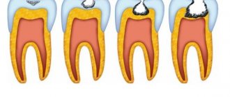
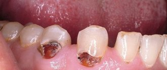
Stages of the carious process
Regardless of where the destruction is localized on the tooth, its classification is always the same.
It is customary to distinguish 4 types of caries according to the depth of the lesion:
- Spotted stage (macula cariosa) . Due to the loss of various microelements by the enamel, especially calcium salts, it becomes matte, losing its natural shine. This is a demineralization process. The spots may be chalky or dark brown. The enamel structure begins to slowly deteriorate, and the spot develops into a full-fledged cavity defect. When caries between teeth is diagnosed, treatment of the spotted form consists of improving the quality of oral hygiene and the use of remineralizing therapy.
- Superficial dental caries (superficialis caries) . When food irritants come into contact with the area of enamel destruction, significant sensitivity appears, which quickly passes. The tissue around the cavity is practically unchanged, which makes it impossible to independently determine it. That is why only a doctor can identify the lesion after a thorough examination.
- Medium caries (mediocris caries) . This is a deeper defect that develops due to the lack of treatment for superficial lesions. It affects not only the enamel, but also the upper layers of dentin.
- Deep caries (profunda caries) . At this stage, dentin is almost completely damaged. Complaints about thermal and chemical irritants, which gradually disappear after their removal.
Why does it occur
The reasons for the appearance of caries between teeth are the same as for classic ones. This means:
- poor oral hygiene;
- hereditary predisposition;
- high activity of pathogenic microorganisms affecting enamel;
- the presence of large gaps in the interdental spaces, in which food debris often accumulates.
The difficulty is that very often the problem is detected in the later stages. While a person can notice a dark spot on the enamel on their own, the darkening between the incisors or molars is difficult to see without a dental mirror. Most often, the dentist discovers enamel destruction during a routine examination or during the treatment of other units.
The disease can be deceptive: a tiny carious cavity, upon detailed examination, can turn out to be deep and voluminous, affecting both dentin and pulp. In this case, the patient has to undergo endodontic treatment of two decaying teeth at once.