5683
The destruction of hard tooth tissue, which is known as caries, is quite common. This pathology causes especially many problems if it is localized between the front incisors. The destruction processes in this place occur quickly, which can lead to the loss of teeth in the most visible place.
What is the difference?
The front incisors are often very close to each other. Food gets stuck in between them, and it is very difficult to clean it out using conventional methods. This leads to the formation of plaque and the development of caries.
It is the front teeth that most often come into contact with food and experience heavy loads when biting, so such processes in this place are a fairly common occurrence. A peculiarity of the development of caries between the front incisors is that their destruction occurs very quickly .
This is where the dentin layer is thinnest, so decay spreads inward and quickly reaches the root. The danger of this process is that it is often noticed when the tooth is almost lost. And in this case it is quite difficult to restore it.
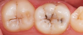
Photo: carious cavities between the front teeth
In addition to stopping the destruction process, the dentist must provide an aesthetic appearance after treatment. And filling cavities here is very difficult, since the pulp is located close to the enamel, and access to the damaged area is difficult.
In addition, if treatment is carried out incorrectly or untimely, complications often develop on the front teeth .
- If bacteria have penetrated the pulp and begun their destructive action, pulpitis . Inflammation develops over several hours and leads to severe pain.
- Periodontitis is also a frequent accompaniment of caries between the front teeth.
- There may be a change in the bite , a shift in the dentition, and even a deterioration in diction.
- In advanced cases, gumboil or abscess .
- It is also worth mentioning the occurrence of psychological discomfort and complexes due to an unaesthetic appearance.
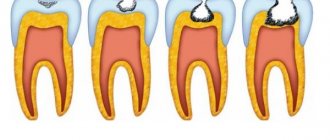
Stages of destruction
Between the incisors, the destruction process goes through the same stages as on other teeth, only it goes faster due to the structural features.
- The initial stage is characterized only by the thinning of the top layer of enamel, which is why white spots are visible in the spaces between the teeth.
- At the next stage, the enamel is gradually destroyed, forming depressions and the process moves into the stage of superficial caries, which can be identified by dark spots between the teeth.
- Medium caries is characterized by darkening of a large area and the appearance of sensitivity to cold or hot.
- If the tooth begins to decay, then it is deep caries . In this case, cavities and large gaps are formed between adjacent incisors. Pain is felt, and the sharp edges of the enamel can be felt with the tongue.
Types and forms of caries
Cervical caries
There are several types of caries in dental practice:
- Cervical caries forms in the area of the gum where plaque appears.
- Fissure carious deposits are located between uneven areas of the oral cavity.
- Root caries will bother the patient when the destruction has reached the very root.
Carious lesions can take the following forms:
- Initial form. At this stage, the destruction will look like a stain.
- In a superficial form, the carious spot will be rough.
- The middle stage of caries is a form in which the destruction process is located at the border between enamel and dentin.
- Deep caries is located deep in the dentin.
Causes
Caries between the front teeth does not always occur due to improper oral care.
Other factors may also trigger this process.:
- cracks, chips of enamel as a result of injuries or biting off hard food;
- often eating too acidic foods and sweets also destroys the top layer of enamel;
- braces and staples used to correct bites in children and adolescents injure dentin and make oral care difficult;
- in some cases, a person produces too little saliva, which causes bacteria to multiply more;
- Suffer from this form of caries mainly children, old people and women during pregnancy, which is associated with hormonal imbalances.
Most often, the processes of destruction of the front teeth occur in people with reduced immunity, endocrine diseases and metabolic disorders.
Excessive consumption of carbohydrates and sweets contributes to the development of bacterial microflora in the oral cavity. And a lack of calcium and fluoride makes tissues more susceptible to destruction.
Causes
Caries occurs under the influence of many factors, among which are:
- weakened immune system;
- genetic predisposition;
- poor oral hygiene
- lack of vitamins and minerals in the body;
- tooth injury
- active consumption of foods rich in carbohydrates;
- infection of the body with streptococcal infection (such microorganisms actively corrode tooth enamel);
- constant wearing of dentures, which damage the teeth and make it difficult to carry out hygiene measures;
- problems with saliva production.
The above factors create a favorable environment for the development of pathogenic microorganisms. Regular and excessive consumption of sweet, salty and sour foods accelerates the process of destruction of dental tissue.
Symptoms
In order not to lose teeth in the most visible place, caries must be treated on time, in its first stages.
To do this, you need to know what symptoms accompany this disease and how to determine where the destruction began:
- Teeth react with pain to cold, hot, sweet or sour.
- The sensitivity of the enamel becomes aggravated during hygiene procedures.
- Bad breath occurs due to decay of plaque and the development of bacterial microflora.
- White or dark spots appear on the enamel.
- In the later stages of the process, severe pain and carious cavities appear, which can be felt with the tongue.
Read the following publication to see what measures are recommended for the prevention of cervical caries.
In a separate article we will discuss the dangers of rotten teeth during pregnancy.
Here https://www.vash-dentist.ru/hirurgiya/rezektsiya/verhushki-kornya-zuba-zachem-nuzhna-operatsiya-i-kak-ona-provoditsya.html you will find a video of how the root apex resection operation is performed.
Diagnostics
Caries between the front teeth can sometimes be noticed independently. It is expressed in the form of white spots, dark specks, discoloration of the enamel or indentations.
But it is best to start treating the disease while these signs are not yet noticeable, and the carious process is diagnosed during a routine examination at the dentist:
- In the early stages, the disease can be detected, for example, by using special dyes or dental floss . Fluorescent irradiation allows you to see dark spots against the background of luminous enamel.
- When performing electroodontometry, the excitability of the pulp is measured.
- Many dental clinics use the capabilities of the Diagnodent device, which irradiates the enamel with light waves and analyzes the reflected light. When the mineral composition of tissues is disturbed, which happens with caries, it gives a signal.
Caries, which develops only on the lateral surfaces of the teeth and is not visually noticeable, is diagnosed after an x-ray. The images immediately show areas affected by caries.
They stand out as black spots on the light background of dentin. Some X-ray machines can detect affected areas even at the initial stage.
Signs of caries in front teeth
- Pigmentation.
When enamel begins to deteriorate under the influence of acids, its color first turns white. This spot stands out because it is lighter than the surface of the tooth. Over time, when the infection penetrates deeper, the white spot begins to change its color and darken. - Carious cavity.
Acids contained in food and released by bacteria destroy enamel. First it becomes loose, and then a cavity appears in it. The greater the volume of tooth tissue is destroyed, the deeper and more noticeable it is. - Unpleasant sensations.
At the initial stage, caries is asymptomatic, since there are no nerve endings in the infected enamel. But as soon as bacteria penetrate deeper into the dentin and pulp, unpleasant sensations may occur. They usually appear in response to irritants - sour, sweet, hot or cold foods. - Food particles.
Food particles begin to get stuck between the infected and healthy teeth, although this did not happen before.
The symptoms of caries in the front teeth are exactly the same as in the rest. But we notice any changes in the smile area faster, so you have every chance to save the tooth from destruction. To do this, you just need to seek help from a dentist when one of these signs appears.
Treatment methods
In the first stages of the disease, before the formation of a carious cavity, the process can be stopped . For this we use:
- special products for oral hygiene;
- remineralizing therapy using special applications that restore enamel.
- Physiotherapeutic procedures are also effective: laser, electrical stimulation, drug injections;
- stains on the enamel that have not penetrated can be bleached or removed by grinding.
Treatment of caries in a child should begin as early as possible, then you can do without a drill. There is a method of silvering damaged areas, which effectively stops the destruction process, treatment with ozone or the Icon drug.
How treatment occurs at the initial stage with Icon, watch the video:
Prevention of cervical caries
Prevention of cervical caries consists of following all those rules that relate to maintaining oral health. This means that you should not forget about brushing your teeth twice a day and rinsing your gums after eating with pharmaceutical or homemade herbal rinses. You need to eat right, and if you have diseases, monitor your metabolism and hormonal levels. A special recommendation for preventing the gum form of the disease is more thorough cleaning of the neck of the tooth: you need to very well clean the spaces between the teeth and between the crown and the gum.
If the initial stages of development of cervical or other caries are noticed, you should immediately go to the dentist to prevent negative consequences such as a crown fracture or complete tooth loss. The earlier a defect is noticed, the less time it will take to eliminate it , and the less discomfort you will have to endure during dental procedures.
To prevent the occurrence of any dental disease, it is advisable to go to the dentist a couple of times a year to have your teeth examined and professionally cleaned. At home, using only a brush and regular toothpaste, it is impossible to thoroughly clean your teeth, especially from the sides, in the spaces between teeth and near the neck. In modern clinics, this procedure is performed using special devices.
If unusual spots appear in the gingival area of the dentition, this defect cannot be ignored. This is a sign of a dental problem that needs to be addressed as soon as possible. If a patient is afraid to go to the dentist and delays the visit to the clinic, he dooms himself to even more unpleasant sensations during the treatment process, since during this time caries manages to destroy a significant part of the neck and crown.
The effectiveness of physiotherapy
If caries was noticed at the initial stage, it can be cured without filling . Doctors prescribe various physiotherapeutic procedures.
Due to the structural features of the anterior teeth and the proximity of the facial nerve, the course is usually 5-7 procedures. But this is enough to effectively restore enamel and improve blood circulation and tissue nutrition.
Several physiotherapeutic methods are usually used to treat caries
- Laser irradiation stimulates recovery processes, improves blood circulation, relieves gum swelling and pain. The laser beam has an antibacterial effect and effectively removes plaque and stone even from hard-to-reach spaces between teeth.
- Magnetic laser therapy is sometimes used . By adding a magnetic field, the process of cell regeneration is accelerated. This method is not indicated for everyone, so you need to warn your doctor about the presence of chronic diseases.
- Pain from caries between the front teeth can be so severe that it makes proper diagnosis and treatment difficult. To relieve pain, the method of electrical stimulation .
It consists of the impact of low-frequency pulsed current on various points on the face. This method, called transcranial electrical stimulation, is sometimes used during fillings for pain relief and monitoring the patient's condition. - To speed up recovery from periodontitis and other complications of caries, injections of special drugs .
- To improve the condition of the gums, hyaluronic acid is used, which accelerates the regeneration process. The new drug Icon is also very . It fills in the affected areas and restores the enamel. It is especially convenient to use it in interdental spaces.
How is filling done?
Carious cavities necessarily require cleaning and filling.
Treatment on the front teeth has some features:
- it is very important to choose the right color of the filling material so that after treatment the appearance of the enamel does not differ from how it looked before the disease;
- the place of contact with neighboring teeth is carefully worked out;
- since the layer of hard tissue in this place is very thin, all procedures must be carried out carefully and with pinpoint precision;
- it is important to remove all plaque and all areas affected by decay so that a relapse does not occur.
Filling carious cavities in this place can only be done by an experienced, qualified dentist. Poor treatment spoils the appearance of the dentition and can lead to serious complications.
Feature No. 4: caries often affects two front teeth at once
The most common type is interdental. It occurs due to the fact that the enamel on the proximal contact areas is very thin, plus food debris accumulates here, which is an ideal environment for the proliferation of bacteria.
Also, another unpleasant type of frontal caries is common among patients - cervical or basal. It begins with a yellowish spot that appears at the border with the gum - this area reacts very sharply to any mechanical influence, to cold and heat. And if, when such a stain is discovered, you do not inquire in the very near future how caries is treated on the front teeth, then you may completely lose the tooth, because This type of pathology leads to the destruction and fragility of hard tissues, as a result of which your tooth can break or literally crumble.
This pathology most often affects two front teeth at once.
“I had cervical caries of my front teeth, but the treatment didn’t last for a long time, because... there was no time for that. At that moment, I was sitting at home, on maternity leave, there was no one to leave the child with, the grandmothers were all in another city, my husband was constantly at work, money was tight. I kept thinking, when I get back from maternity leave, I’ll go and get treatment at the dentist. A year and a half passed unnoticed by my thoughts, and my tooth suddenly broke off while eating an apple. It was very disappointing, and, naturally, I later had to spend a lot of money on restoration, although I probably could have gotten away with less money if I hadn’t procrastinated...”
Marina, babyblog.ru
Restoration methods
In addition to filling, other methods are used to treat caries in this area. Their choice depends on the degree of tissue destruction and the characteristics of the disease . The dentist’s task is not only to remove all tissue damaged by the disease, but also to restore the tooth so that it does not differ from its neighbors.
The methods used most often:
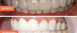
- Instead of a filling, the carious cavity is closed with veneers .
This is a special plate made of polymer or ceramic that effectively masks damage, as it can easily be matched to the color of the enamel. In addition, veneers can correct minor deformations in the shape of teeth. They are selected individually and, if installed correctly, last a long time. - If caries develops deeply and severely destroys the lateral surfaces of the incisors, crowns are installed . They are usually made of ceramics and placed under local anesthesia.
- Severe tooth decay affecting the inner layer of dentin often requires tooth extraction. In this case, implants or prostheses are placed.
We will tell you how circular caries of primary teeth is treated in a new review.
In this article we will talk about how to strengthen molars that have begun to become very loose.
What to do if a piece of front tooth enamel breaks off, read the link https://www.vash-dentist.ru/krasota-i-uxod/narashhivanie/chto-delat-esli-otkololsya-kusochek-zuba.html. What do recovery methods depend on?
Features of restoration of anterior teeth
The features of the carious process on the front teeth, in particular, its localization and degree of neglect, largely determine the approaches to treatment: the stages of cavity treatment, the equipment used and the materials used. If you are already getting ready to go to a dental clinic for treatment, then you will probably be interested to know what can await you there...
So, the most “favorable” option for your front teeth is initial caries or, in other words, caries in the stain stage, that is, when the pathology has barely touched the enamel and has not yet gone deep into the hard tissues. The first stage of the carious process, when a person can independently see the process of tooth destruction, is focal demineralization of the enamel or, more simply, the appearance of a matte or pigmented (yellow, brown) spot on the surface of the front tooth. Practice shows that usually future patients in such cases have thoughts: is it possible to somehow whiten, grind, or otherwise treat this stain on the front teeth at home.
Well, caries in the spot stage can indeed be treated at home, but it is much faster, more effective and safer to do this under the guidance of a dentist.
This is interesting
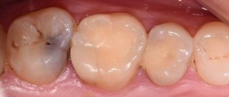
A modern approach to the treatment of caries in the spot stage without a drill is the Icon technology. This microinvasive treatment is indicated for initial caries of the anterior teeth localized on the anterior and contact surfaces of the incisors (that is, between the teeth). This technique is especially relevant after braces are removed, when in almost 60-80% of cases stains are detected on one or all front teeth. The treatment procedure usually takes no more than 20 minutes and does not require pain relief. Consecutive use of the drugs included in the Icon kit allows you to cure a carious spot in one visit.
Photo of the front tooth before and after caries treatment using Icon technology:
Superficial caries. To treat superficial caries on the front teeth, simple grinding with discs and polishers is often used, if the process has not touched the deep layers of enamel. If it is not possible to achieve the desired result, then the classical technology of caries treatment is used.
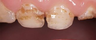
Medium and deep caries. If the carious process goes beyond the boundaries of the enamel and reaches the dentin, then we are talking about medium caries. With deep caries, a cavity is formed bordering the pulp chamber. In all these cases, treatment of the carious cavity is required.
Photo of deep caries on the front tooth:
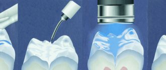
The most popular method in modern dentistry for treating caries on the front teeth is mechanical using burs and hand instruments. Preparation (cavity treatment) using a drill allows you to excise carious tissue as completely as possible to prevent caries recurrence and create an aesthetic filling.
The air-abrasive method using sandblasting of the cavity allows using an aerosol with an abrasive substance to gently treat small carious areas on the front teeth.
The laser method of dental treatment is not yet widely used due to the fact that not all clinics can afford expensive equipment. In addition, a number of researchers doubt the effectiveness of laser treatment in certain clinical cases of caries.
Note: “What should you do if part of your front tooth breaks off due to an impact?”
It is necessary to urgently consult a dentist for an x-ray of the tooth root. The fact is that as a result of an impact, not only the coronal part, but also the root part can be damaged, so it is important to detect a root fracture in time in order to implement an integrated approach to treatment. If the root is in order and the “nerve” is not affected, a one-step restoration of the front tooth is done; if there is damage to the soft tissues inside the tooth, the canal is treated and only then a filling is applied.
The mechanical method of treating caries on the front teeth using a drill provides for maximum excision of not only softened carious tissues, but also pigmented ones. This is due to a principled position regarding future aesthetics. After all, if even a small area of pigmented dentin or a dark spot on the enamel is left, then next to the filling, at a minimum, the appearance of the restoration will be damaged, and, at a maximum, recurrent caries under the filling will soon occur.
In the video below you can follow the main stages in the treatment of deep caries on the front tooth using a drill:
Interesting video: an example of treatment of deep caries on a front tooth
During mechanical treatment of the cavity, an important component of the treatment is the jet supply of water to the tooth, since, first of all, on the front teeth, thin layers of tissue (enamel, dentin) and the proximity of the pulp chamber determine serious risks of its overheating, up to necrosis. In such cases, the tooth begins to hurt under the filling, and in the future it will be necessary to remove the dental “nerve”.
From the practice of a dentist
Often, patients come to see the dentist with complaints of severe pain in the front tooth and think that it is the caries that is “sick,” although in fact the infection has already reached the dental “nerve” and pulpitis has begun. The symptoms that arise in a diseased tooth can sometimes be quite similar to clinical caries: the pain may be insignificant or not at all, external irritants do not provoke a painful reaction in the tooth or it is mild. If the doctor still finds out that it’s pulpitis, then there’s no way to get rid of it with just a filling...
First, anesthesia is mandatory, then the carious cavity is cleared of infected tissue, and the “nerve” is amputated and extirpated (extracted from the root canal). The canal is washed with various antiseptics (hydrogen peroxide, sodium hypochlorite, etc.) and filled to the apex, that is, to the physiological narrowing of the root, with a hardening filling material with or without the use of gutta-percha pins. Work on the complete treatment of pulpitis can be carried out in one visit or in several, depending on the level of technical equipment of the clinic, the characteristics of the tooth itself, the professionalism of the doctor and the approach he practices in such procedures.
After anesthesia, cleansing of the carious cavity, its medicinal treatment with antiseptic solutions and the formation of a cavity for the material used, the color of the future restoration is determined. The materials used for the restoration of anterior teeth with caries are, in most cases, light-curing composites. Each set produced by a particular company has its own color characteristics (sets of syringes with a specific color).
It is also useful to read: The use of caries markers (indicators) in dentistry
Before restoration, the dentist evaluates:
- transparency, tooth tone and color range;
- shades of the tooth by zone: in the cervical part, in the area of the tooth body and at the cutting edge;
- individual characteristics of the tooth (spots of hypoplasia, presence of cracks).
Ideally, after restoration, its surface should not resemble a “patch”, as it did in Soviet times. The finished work must have a set of such individual characteristics that, after polishing the filling, a natural shine characteristic of the enamel appears, each zone of the tooth has its own shade of color, the tooth has the same characteristic lines, microcracks that show through congenital stains. Artistic restoration of a front tooth often takes up to 2 hours or more.
On a note
There are different approaches to assessing the final result: there is an opinion that the ideal restoration after filling should be in harmony with the front tooth that is symmetrical to it. In other words, if there are natural pits, spots, lines, etc. on a symmetrical central incisor, then they are also recreated on a tooth being restored due to caries.
A number of experts are of the opinion that the final restoration of the front tooth should be matched in shape and color to the tooth being restored and to the neighboring one, but nothing more. At the same time, the errors that nature created on a symmetrical incisor and which look natural from the outside are not repeated during restoration with a filling. This is not to say that this restoration technique is considered unpopular.
It is advisable to agree in advance (before treatment) with the doctor which result you prefer and based on the individual characteristics of the tooth.
Cost of treatment
Of course, the treatment of the front teeth must be carried out efficiently so that the filling or crown is invisible. But depending on the economic status of the patient, different treatment methods and materials may be selected.
The cost of treatment also depends on the doctor’s method of work: whether light curing, cooling and saliva ejectors are used, and what kind of anesthesia is used. High-quality fillings are more expensive, but such fillings will last longer.
Typically, treatment of caries between the front teeth costs from 2 to 6 thousand rubles , depending on the materials used and the complexity of the disease.
A light-curing filling of the latest generation at the initial stage of the disease costs 2,000, for medium and deep caries - 2,500, and for advanced periodontitis, treatment will cost 4-5 thousand rubles.
It is also worth considering that this problem usually affects two teeth, so the cost of treatment will be higher .
Crack on front tooth
One of the common complaints among patients is the formation of cracks in the incisors.
Main reasons:
- injuries and shocks;
- poor nutrition;
- non-compliance with hygiene standards and rules;
- increased load on these teeth when chewing.
Depending on the configuration, cracks can be vertical or horizontal. The second type is considered more dangerous, since it leads not only to caries, but even to complete destruction and loss of the tooth.
Shallow microcracks can be sanded down and treated with special fluorine-containing solutions to prevent their further growth. For larger damage, filling is used and veneers are installed.
This problem is typical for teeth located in front. They bear a lot of stress during chewing. When combined with other factors, this becomes the main cause of unsteadiness.
Most often, teeth begin to become loose due to other diseases - from caries to gingivitis. Therefore, the problem must be solved comprehensively, starting with treatment of the underlying disease.
To prevent worsening mobility and subsequent tooth loss, a splinting procedure is used.
Installing a splint helps to secure a mobile tooth and correct the dentition.
Prevention measures
Losing front teeth greatly spoils a person's appearance. And this often happens in cases of advanced caries. Therefore, you need to try to prevent food from accumulating between the front incisors, and they remain strong and healthy.
All measures to prevent tooth decay processes can be divided into two groups: external , aimed at oral hygiene, and internal , helping to strengthen tooth enamel.
What hygiene procedures will help prevent caries:
- After each meal you need to rinse your mouth to remove any remaining food;
- It is advisable to clean the space between the teeth daily with floss;
- use pastes with antibacterial components and minerals that are beneficial for enamel, and they need to be selected individually;
- do not crack nuts, do not bite threads and protect your incisors from injury.
What nutritional rules should you follow to keep your teeth healthy?
- eat less sweets, white bread and confectionery;
- protect teeth from exposure to cold, hot and sour foods;
- include foods rich in calcium and fluoride in your diet;
- eat more vegetables and fruits.
Regular visits to the dentist will help detect caries in the early stages, and timely treated diseases will preserve oral health and the beauty of your smile.
If you find an error, please select a piece of text and press Ctrl+Enter.
Tags diagnostics caries treatment front teeth caries prevention tooth decay
Did you like the article? stay tuned
Previous article
Methods for diagnosing and treating cementum (root) caries
Next article
Is brushing with tooth powder beneficial or harmful?
The role of prevention in preserving anterior teeth
Prevention of caries on the front teeth is currently of key importance for maintaining a beautiful smile area. The complex of such measures can be divided into systemic and local applications.
Systemic preventive measures include:
- limiting carbohydrate intake and prioritizing fruits and vegetables to self-clean the spaces between the front teeth where plaque and food accumulate;
- consumption of foods containing calcium in large quantities (milk and dairy products);
- fluoride prophylaxis under the supervision of a dentist using fluoridated water, salt or milk, as well as taking fluoride tablets.
Local preventive measures:
- cleaning the front and side teeth with fluoride and calcium-containing pastes after each meal;
- the use of mouth rinses with fluoride (see photo below for an example);
- remineralizing therapy and deep fluoridation of the front teeth at a dental appointment and in the future - home use of special mineralizing gels or solutions.