Due to the characteristics of the lesion (absence of defects in the hard tissues of teeth), initial caries must be differentiated from diseases that also cause stains on the enamel. This is a spotted form of hypoplasia and fluorosis. Hypoplasia is characterized by insufficient formation and mineralization of tooth enamel. It occurs as a result of weakening of the histogenesis processes of teeth under the influence of various disorders (general diseases, infections, metabolic disorders, etc.) during tooth formation. Defects with hypoplasia form on the tooth even before it erupts; they cannot occur in a properly formed tooth. Carious spots appear only after teeth erupt. Hypoplasia affects teeth that are initiated and formed at the same time, for example, incisors and first molars. Caries can affect various teeth, regardless of the timing of their formation and mineralization.
An essential differential feature is the localization of the lesions.
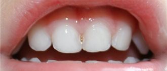
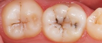
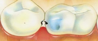
With hypoplasia, spots are often located on the convex vestibular surface of the front teeth and on the tubercles of molars and premolars. Caries, on the contrary, very rarely affects the convex smooth surfaces of teeth and is more often localized on contact surfaces, in the pits and fissures of the lateral teeth. Spots with hypoplasia have pronounced symmetry, affecting the surfaces of the teeth on the right and left. In such cases, even their shape and color may be similar. Caries is also characterized by a certain symmetry of tooth damage, but spots may not appear simultaneously (that is, on one half of the jaw they may appear much earlier). The shape and color of the carious lesion may vary.
Prevention of caries
Regular visits to the dentist's office will help avoid many dental problems. But besides this, we should not forget about preventive measures. First of all, caries prevention involves strengthening tooth enamel with minerals.
To keep your tooth enamel strong and healthy, you need to watch your diet and take vitamins. You need to eat foods that benefit the body in general and teeth in particular. At the same time, you need to avoid harmful foods. In addition, monitor oral hygiene and use special toothpastes with a whole range of minerals and trace elements.
Hypoplasia.
In the vast majority of cases, it occurs on permanent teeth. It is extremely rare that hypoplasia also affects primary teeth, while caries of primary teeth is a fairly common phenomenon.
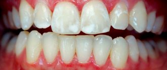
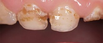
With hypoplasia, the spots are whitish in color, have a shiny surface, are dense and painless upon probing. Acute initial caries is characterized by white, chalky spots with a matte surface. When probing, sensitivity, roughness, and slight pliability of the surface are noted. With chronic initial caries, dense, painless pigmented spots appear on probing, which also do not have the shine inherent in intact enamel.
For diagnosis, in particular differential diagnosis, of initial caries, the method of vital staining of teeth, often with methylene blue, is used. The teeth are covered with cotton swabs, cleaned of plaque and dried. A 2% aqueous solution of methylene blue is applied to the surface of the stained tooth. After a few minutes, the dye is washed off with water. In caries, due to demineralization of the enamel and increased permeability, the stain absorbs the dye, turning blue. Spots with hypoplasia have a denser surface, do not absorb dye and are not stained.
Methods for diagnosing initial caries.
Methods for diagnosing initial caries
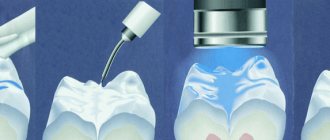
Drying. This method of identifying foci of enamel demineralization in the form of a white spot is the simplest and most effective in a dental office. When dried, water evaporates from the micro-spaces of the enamel and the stain becomes dull and visible. The tooth being examined is cleaned of plaque, isolated from saliva, and the surface is dried with air. Visually determine the size of the foci of demineralization. With careful probing, the enamel surface in the area of the white spot is rough, but dense.
Vital staining is based on increasing the permeability of dental hard tissues affected by caries. The dye penetrates into demineralized hard tissues and colors them; healthy tissues do not change in color. For differential diagnosis of initial caries, a cotton ball moistened with a 2% solution of methylene blue is placed on the tooth surface cleaned of soft plaque for 2-3 minutes. Then the excess dye is washed off with water. To objectively assess color intensity, a standard color scale is used, which provides various shades of blue - from slightly bluish to intense blue.
Colorimetric test. The technique consists of sequentially rinsing the mouth with a 0.1% glucose solution and a 0.15% methylene red solution. In areas of enamel where the pH changes to the acidic side, at values of 4.4-6.0 and below, the color changes from red to yellow. The caries detection rate is 74.8% (Hardwick).
Luminescence study. The luminescence diagnostic method is based on the ability of tissues to change their natural color under the influence of ultraviolet rays. The study is carried out in a darkened room. A beam of ultraviolet rays from a luminescent illuminator (OLD - 41, Pluraflex) is directed onto the dried surface of the teeth from a distance of 20-30 cm.
Healthy hard dental tissues fluoresce in ultraviolet rays with a snow-white, bluish glow. In the presence of a carious spot, a quenching of luminescence is observed against the background of the normal glow of the surrounding healthy enamel.
Didn't find what you were looking for? Use the search:
Best sayings:
Learn to learn without studying!
10961 —
|
8192 - or read all. source
Fluorosis.
A peculiar form of hypoplasia. It occurs due to an excess of fluoride ions in the body, caused by its increased concentration in drinking water. Fluoride ions suppress enameloblasts during the formation and mineralization of teeth. Depending on the concentration of fluoride in drinking water on the surface of the enamel, the disease can manifest itself in different ways: from the appearance of white, brown and even black spots to enamel defects and aplasia. Differential diagnosis of fluorosis and initial caries is carried out according to the same principles as hypoplasia.
Spots on dental enamel due to fluorosis are stationary, have a dense shiny surface, painless and smooth upon probing. When dyeing, they do not absorb the dye. Unlike caries, with fluorosis the spots are much more mineralized and are characterized by high microhardness and resistance to acids (due to the deposition of fluorapatite). Therefore, with fluorosis, caries almost never occurs, while the combination of both hypopoplasia and caries is often observed. Fluorosis does not typically affect certain groups of teeth; depending on the time and duration of residence in an area of endemic fluorosis, several or all teeth may be affected.
In some cases, it is necessary to differentiate between carious spots and pigmentation, plaque, which are localized on the surface of the teeth. Dental plaques on the surface of the teeth can be stained under the influence of food pigments, medicinal products and smoking, etc. In children, dense green plaque (Priestley plaque) is more often detected, firmly attached to the surface of the enamel. It is believed that its cause is the fungus Lichenclentalis, which produces chlorophyll. Plaque is deposited mainly on the vestibular surfaces of the front teeth, that is, on surfaces that are exposed to light. On the lateral teeth its deposits are observed much less frequently.
Differential diagnosis of these formations, as a rule, does not cause difficulties. Often the smooth vestibular or lingual (smoker's plaque) surfaces of the front teeth are affected. Despite their relatively tight attachment to the tooth surface, they are easily removed with an excavator or special brushes or pastes. After removing the pigmented plaque, the intact enamel surface is exposed, which does not have any signs of carious demineralization.
Differential diagnosis of caries with the presence of a cavity in the hard tissues of teeth is carried out with caries of varying depths, hypoplasia, fluorosis, wedge-shaped defects, pathological abrasion of hard tissues of teeth, enamel erosion, necrosis of hard tissues, chronic pulpitis and periodontitis.
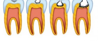
When differentiating carious lesions of different depths, it is difficult to evaluate them by linear or volumetric dimensions, which is associated with variations in the thickness of the hard tissues of different groups of teeth in different areas of the crown. Therefore, it is necessary to evaluate the depth of the carious cavity not by linear dimensions (for example, in millimeters), but by its location in the dentin (mantle or peripulpal) and in relation to the cavity to the pulp. When examining a carious cavity, it is important to determine exactly how thick the layer of dentin separates it from the tooth cavity and pulp. This is very important when differentiating medium and deep caries.
Superficial caries differs from average caries primarily in subjective sensations. With average caries, pain does not occur under the influence of various irritants. In case of superficial caries, the cavity is localized only within the enamel; in case of middle caries, it is also localized in the mantle layer of dentin. With superficial caries, even with acute caries, there are no pronounced overhanging edges of the enamel; with acute moderate caries, they are almost always present.
Fiber optic transillumination (transluminescence)
Transluminescence and luminescent diagnostics, which are closely related to each other, are rather rarely used methods for diagnosing caries in medical practice. They are usually used for other purposes.
- Fiberoptic transillumination is based on illuminating the tooth with bright light. At the same time, the areas affected by caries turn out to be darker and create a clearly visible hemisphere against the background of healthy enamel. This examination is carried out in a dark room and cold light is used.
- Fluorescent diagnostics involves illuminating the tooth with ultraviolet light. Typically, a beam of light is passed through a special filter (Wood filter). In this light, the tongue appears orange, healthy teeth have a snowy white tint, and carious teeth are darker. The boundaries of the affected areas are very clearly visible and therefore easily recognized.
Both of these procedures are completely painless. Devices for diagnosing caries using this method are produced by the domestic industry (OLD-41) and many foreign manufacturers.
It is also useful to read: Chronic caries in adults and children
There is also a method of laser-induced fluorescence. In it, the tooth is illuminated by a laser beam, and based on the spectrum of its own radiation, a conclusion is made about the integrity of the tooth enamel. Devices for such diagnostics are small and are a good help in studying the cariogenic situation in the oral cavity.
Differential diagnosis of medium and deep caries.
It is based on the patient’s complaints, the depth and size of the carious cavity, and the sensitivity of its various areas during probing. Particular care must be taken in assessing the carious cavity located on the contact surfaces of the lower frontal teeth, where, due to the small thickness of the hard tissues, deep caries has small linear dimensions in depth. In acute deep caries, after careful necrectomy of softened dentin, the pink pulp begins to appear at the bottom of the cavity.
Superficial caries may resemble hypoplasia with the presence of erosions, dimples and grooves on the enamel surface. Most of the signs by which they differ are similar to the differential differences characteristic of initial caries: characteristic location, time of occurrence (before or after teething), dynamics of development of defects, simultaneity of damage to different groups of teeth. In addition, hypoplasia is characterized by a peculiar transverse linear displacement of the pits and grooves on the crowns of the teeth. The surface of hypoplastic defects is dense, shiny, painful on probing, softening of hard tissues and overhanging edges of the enamel is not observed. When applying a dye (methylene blue, for example), the hard tissues of teeth demineralized due to caries are stained, but the dye does not penetrate into hypoplastic defects. It should be remembered that in places of defects in hard tissues with hypoplasia, caries can often develop, but in these cases one can observe the appearance of softening of the hard tissues of the teeth in the depths of the defect, pain during exposure and from chemical irritants.
Tooth staining or using caries markers
The principle of using caries markers is that some substances are retained precisely in areas affected by caries. The most common such marker is methylene blue. When it gets on a tooth from a healthy enamel surface, it simply flows off, but on a carious surface, to put it simply, it remains (diffuses into the pores of demineralized enamel). As a result, the doctor can not only identify dental caries, but also reliably identify the shape and border of the damage.
It is also useful to read: About advanced caries: what to do if almost all teeth show signs of destruction
Most modern caries markers (sometimes also called caries detectors or indicators) use magenta, which colors the affected areas pink. Markers are applied to the teeth with special swabs and then washed off. Clinics use both imported products (VOCO, Pulpdent) and domestic products such as Color-test.
Differential diagnosis of caries and fluorosis.
It is carried out using almost the same principles as with hypoplasia. It is often performed with an erosive form of fluorosis. With the chalky-mottled form of fluorosis, teeth of various groups are often affected. Sometimes the entire surface of the enamel is depigmented and has a chalky tint, but, unlike enamel demineralized during caries, it retains its shine. Against this background, individual areas of brown or dark brown enamel pigmentation are often observed. In cases where the enamel loses its shine and becomes dull, small round-shaped enamel defects (chalk-mottled form) may be observed on it. With the erosive form of fluorosis, wider and deeper defects—erosions—are formed in the chalky enamel.
Wedge-shaped defects are often located on the vestibular surface and in the cervical region of teeth that protrude somewhat beyond the dentition (canines, premolars, less commonly incisors and molars). Unlike caries, they have a characteristic wedge shape formed by two planes of the defect. These surfaces are made of sclerotic dentin, smooth when probing, without signs of demineralization characteristic of caries.
With pathological abrasion of teeth, there is no softening of the dental tissues and their pigmentation. The shape of the defects corresponds to the surface of the traumatic factor (mouthpiece, brush, pencil, etc.), often it coincides with a similar surface of the antagonist. The abrasion surfaces during probing are smooth, almost painless (however, increased sensitivity of the hard tissues of the teeth may be noted). Quite often, these defects are localized in areas of the crowns that experience maximum loads during chewing (incisal edges, tubercles), that is, in caries-resistant areas.
DIAGNOdent Pen device
Despite its apparent simplicity, the diagnosis of caries is quite complex, since the symptoms quite often overlap with the symptoms of other dental diseases. In these controversial cases, the dentist comes to the aid of a special device called “DIAGNOdent Pen” . It allows the dentist to accurately identify even hidden forms. The operation of this device is based on laser fluorescence spectroscopy. Even if outwardly the tooth looks quite healthy, the device can show the presence of the initial stage, when it can be very easily and simply cured. The DIAGNOdent Pen device is very convenient and easy to use, allowing the doctor to state with absolute certainty about the presence or absence of caries in the patient.
Erosion of enamel.
It is an oval or rounded enamel defect located in the transverse direction on the most convex part of the vestibular surface of the tooth crown. The cross-sectional defect has a characteristic saucer-like shape, the bottom of the erosion is smooth, shiny, without any signs of demineralization. With further progression of the defect in depth, it penetrates into the dentin, and then its bottom is made of dense, sclerotic dentin. Probing erosion is practically painless, although increased sensitivity of hard tissues may be noted.
Tooth root caries: etiology, classification, diagnosis, prevention, treatment.
Root caries is progressive damage found in any area of the tooth root surface exposed to oral fluid. According to WHO experts, the problem of tooth root caries among the population of older age groups is steadily growing.
Predisposing factors
Billings RJ divided the main risk factors for root caries into 3 groups:
1. Biological factors (composition of dental plaque, level of salivary secretion, pH data of saliva, buffer capacity of saliva, hereditary factors). 2. Behavioral factors (hygienic behavior, general health awareness, eating habits, socio-cultural behavior, psychological awareness). 3. Individual factors (general health, problems of mobility, coordination, understanding, periodontal treatment performed, functional disorders). The more diverse the risk factors, the more pronounced root caries.
Research results
I. Statum, D. Banting, Burt B., Ismail A. showed that with a decrease in the concentration of fluoride in drinking water, the intensity and prevalence of root caries significantly increases. Fejerskov O., Nyvad B. suggested a “local effect” of constant consumption of fluoridated water. Jensen ME., Papas AS., Joshi A.I. et al. report that carbohydrate-containing foods can aggravate the cariogenic situation on the root surface.
The effect of dental plaque microorganisms on hard root tissues is almost similar to that during the development of caries in the area of the tooth crown. Plaque microorganisms are one of the leading factors in the development of periodontal diseases, which in turn contribute to the exposure of root surfaces. Some researchers believe that there are differences between the microorganisms associated with root surface caries and crown caries. Microorganisms of the Actinomyces group attracted particular attention from scientists. In early lesions of the root surface, gram-positive microorganisms predominate, Act. naeslundii. In advanced cases, predominantly gram-positive flora is also present, especially Str. mutans, in addition, there are many Prevotella, Bacteroides.
According to L. Laurisch, soft dentin contains more Str. mutans and lactobacilli than in dense dentin, and skin-like lesions have an intermediate microflora. Str. mutans and lactobacilli are found in greater numbers in the gingival and (or) deep cavities than in the coronal and (or) small ones. Ovrutsky G.D. and co-authors also point to the predominance of Act. viscosus, Act. naeslundii, Act. species (up to 40-50%) in carious cavities of the root surface, without rejecting the significant role of Str. mutans, Str. sangvis.
According to Chepurkova O.A., the species composition of the dental plaque microflora in 96.6% of patients with root caries differs from that in patients without root caries and is represented by an association of various bacteria with Enterococcus faecalis.
Evidence has been obtained of a positive correlation between low salivary flow rates and the incidence of dental caries. The prevalence of root caries was studied in individuals with diseases that cause hyposalivation (Sjögren's syndrome) or require the use of drugs that reduce salivation (antidepressants, antiulcers, cardiological, antihistamines, diuretics, antipsychotics, etc.).
For root caries to occur, a prerequisite is the presence of loss of dentogingival attachment. Consequently, loss of attachment and the predisposing conditions of poor oral hygiene, periodontal disease, advanced age, and anatomical features of the dental system are important risk factors affecting the root environment. However, Muya RJ et al did not find root caries in patients with extensive age-related gingival recession in some populations. The authors rightly believe that gum recession does not lead to root caries without the necessary environmental factors.
According to Chepurkova O.A., the most likely risk factors for root caries, among others, are: a periodontal pocket up to 4 mm deep, high cariogenicity of dental plaque, poor oral hygiene, low resistance of teeth to caries, removable dentures supported by teeth without artificial crowns, frequent intake of carbohydrates (more than 9 times a day), smoking, age over 45 years, male gender, gastrointestinal lesions, endocrine pathology. Narhi TO., Vehkalahti, MM also indicate a more frequent incidence of root caries in men.
The issue of the prevalence and intensity of root caries among persons with systemic pathology (diabetes mellitus, gastrointestinal lesions, Sjögren's syndrome, cardiovascular, psychosomatic, oncological and other diseases) was also considered. The authors do not report the direct influence of general pathology on the occurrence of root caries, but do not deny the indirect role of systemic diseases (through changes in the properties of saliva, the reactivity of the body) in the development of root caries.
Classification of tooth root caries.
Rikota Yu.N.
According to the depth of damage to root tissue:
1. Initial caries - partial destruction while maintaining the cemento-dentin boundary. 2. Superficial caries - a defect with a depth of no more than 0.5 mm 3. Deep caries - a defect with a depth of more than 0.5 mm
By localization:
1. class - caries on the contact surfaces of the root 2. class - caries on the vestibular (oral) surface of the root
Ovrutsky G.D.
According to the depth of damage to root tissue:
1. degree (initial caries) – the surface is soft, there is no tissue defect. 2. degree (superficial caries) – defect depth up to 0.5 mm 3. Degree (cavity caries) – defect depth more than 0.5 mm 4. degree (pulp caries) – defect involving the tooth cavity
Fejerskov O.
According to the depth of damage to root tissue:
1. Without surface destruction 2. Lesions up to 1 mm deep 3. Lesions more than 1 mm deep 4. Lesions with suspected penetration into the pulp
According to the course of root caries:
1. Active lesions 2. Inactive lesions
Laurisch L.
According to the depth of damage to root tissue:
1. No changes in the exposed neck of the tooth 2. Initial change 3. Superficial soft change, depth - 0.5 mm in dentin 4. Cavity more than 0.5 mm deep 5. Cavity with pulp damage (suspected or actual)
By localization:
1. “Gingival” cavities on the tooth root 2. “Coronal” cavities on the tooth root
Leus P.A. , Borisenko L.G.
According to the depth of damage to root tissue:
1. Without cavity formation 2. With cavity formation
According to the course of root caries:
1. Active lesion 2. Suspended caries 3. Secondary caries (active or inactive) 4. Unspecified
Diagnosis of tooth root caries.
To diagnose tooth root caries, a traditional examination scheme for a dental patient is used. In cases of tooth root caries:
- absence of complaints, which is typical for this pathology (often pain occurs only with the development of inflammation of the dental pulp); - complaints about an aesthetic defect (when the cavity is localized on the vestibular surface of the roots of the frontal teeth); - discomfort when eating; -painful sensations from thermal, mechanical, chemical stimuli, disappearing immediately after the removal of the stimulus; - complaints related to the presence of periodontal diseases in the patient, leading to loss of periodontal attachment.
Assessment of the state of hygiene of the oral cavity, dentition, periodontal tissue and mucous membrane is carried out according to generally accepted methods. In addition, when examining patients with tooth root caries, it is recommended to determine indices characterizing gum recession (S.Stahl, A.Morris, 1955), loss of periodontal attachment (Loss of attachment, Glaving, Loe, 1967), dentin sensitivity (KIDCHZ, Dedova L.N., 2004), peripheral blood circulation (IPC, Dedova L.N., 1982), the amount of plaque in the gingival area (PLI, Silness, Loe, 1964). This is necessary to assess the risk of progression of root caries in a given patient.
It is also possible to determine the RCI index (Katz, 1982), which allows you to more accurately assess the degree of damage to exposed root surfaces by tooth root caries. The remineralization index (Fedorov Yu. A., Dmitrieva I. M., 1977, 1994) allows you to evaluate the mineralization of hard tissues before and after conservative treatment of caries. Particular attention should be paid to identifying factors that contribute to the development of gum recession (poor oral hygiene, periodontal disease, dental anomalies, age, iatrogenic trauma).
When examining the affected tissues of the root surface, localization, changes in color, density, tissue relief, depth and area of defects, and the presence of pain when probing the root are determined. A number of requirements have been developed to facilitate the diagnosis of tooth root caries: mandatory removal of supra- and subgingival calculus and soft plaque, elimination of saliva from the examined root surface, use of a sharp probe (allows one to achieve high tactile sensitivity and distinguish the affected surface from a healthy one even without a visible tissue defect) .
When probing rapidly progressing carious lesions on the root surface, a softened or “leathery” consistency is detected. The edges of the carious cavity are sharp, uneven. At any stage of development of tooth root caries, subject to careful removal of microbial plaque, demineralization and even cavitation may stop. The exception is deep cavities, in which it is impossible to sufficiently clean the root surface from plaque. The surface of carious root lesions in remission is usually smooth, shiny, and hard. The edges of the cavity are smooth and dense. According to modern literature, the degree of pigmentation does not always provide reliable information about the activity of caries.
To identify hidden cavities on the contact surface of the root and (or) under the gum, to exclude periapical complications, as well as to assess the condition of periodontal tissues, an X-ray examination method is used. In this case, it is recommended to use a parallel radiography method, a bite-wing radiograph (interproximal method), and an orthopantomogram.
Prevention
Evidence of the decisive role of bacterial flora in the occurrence of root caries and identification of probable pathogens allows us to propose the following prevention program:
1. Professional cavity hygiene every three months, with mandatory polishing of exposed root surfaces with fluoride-containing paste. 2. Covering the exposed root surfaces with varnishes containing chlorhexidine and fluorine (at the 3rd, 6th, 9th months of treatment). 3. Rinse with a 0.1% aqueous solution of chlorhexidine, 10 ml, 2 times a day for 10 days on the 3rd, 6th, 9th months of treatment. 4. Irrigation of periodontal pockets with a 0.1% aqueous solution of chlorhexidine at the 3rd, 6th, 9th month of treatment. 5. Resorption of tablets with fluorides and xylene in the oral cavity twice a day for 12 months (according to Emilson S. G., Ravald N., Birkhed D.) 6. Daily use of toothpaste with fluorides and xylene, (according to Emilson S. G., Ravald N., Birkhed D.)
Treatment and prevention
After making a diagnosis of tooth root caries and identifying possible risk factors, treatment and preventive measures are planned for the patient. Including, first of all, they motivate the patient to maintain oral health, give recommendations on hygienic dental care, and nutrition advice.
Interdental brushes and single-tuft toothbrushes are recommended for such patients as auxiliary hygiene products for cleaning the concave surfaces of the tooth root.
For patients with reduced salivation, pastes, gels, and rinses containing substances found in natural saliva (lysozyme, lactoferrin, salivary proteins) can be recommended.
One of the most important stages in the treatment of a patient with tooth root caries is carrying out professional oral hygiene measures. In addition, it is necessary, if possible, to eliminate factors that lead to plaque retention on the crowns and exposed roots of teeth (overhanging edges of fillings, poor-quality dentures, dentoalveolar anomalies, etc. .).
In case of initial and superficial carious lesions in the period of remission, you can limit yourself to preventive measures, provided that the patient follows medical recommendations:
- use of fluoride-containing varnishes and gels with and without the addition of an antiseptic. Products containing aminofluoride, 0.4% tin fluoride, 0.05-2% sodium fluoride, 4% titanium fluoride in combination with antiseptics - 1-5% chlorhexidine, 1% thymol, triclosan, as well as systems with controlled release of fluorides A clinical and histological study revealed the effectiveness of deep fluoridation in the treatment of superficial tooth root caries using a dentin-sealing liquid containing especially highly dispersed fluoride crystals and copper ions.
- it is advisable to use fluorides in combination with calcium preparations (10% calcium gluconate solution and 0.5-1% sodium fluoride solution in the form of applications, casein phosphopeptide amorphous calcium phosphate paste in combination with sodium fluoride).
— in a practical aspect, it seems quite justified and promising to use protective sealants for exposed dentin, capable of preventing abrasion of tooth necks, reducing the number of cariogenic associations of microorganisms on root surfaces, eliminating or reducing the sensitivity of dentin of exposed roots, and releasing fluoride ions for a long time.
In case of deep caries of the tooth root, it is necessary, and in case of superficial caries of the tooth root, according to indications, preparation and filling of carious cavities should be carried out. In the process of preparing and filling carious lesions of the tooth root, located close to the gingival margin, it is necessary to solve the following problems:
-protection of gums from mechanical and chemical damage; - gum retraction to create access to the carious cavity; - ensuring the dryness of the surgical field (protection from blood, gingival and oral fluid, from exudate from periodontal pockets).
Preparation of carious cavities for tooth root caries has certain features:
-exclusion of the stage of opening the carious cavity. -carrying out necrectomy without prophylactic excision of intact tissues; -formation of an additional platform on the oral surface of the tooth root to improve access to the carious cavity on the contact surface of the tooth root (according to indications); - preparation of the edge of the root cavity with a rectangular ledge to prevent thinning of the edges of the filling (according to indications); -formation of an oval-shaped cavity; -creation of retention grooves in dentin on the occlusal and gingival walls (according to indications); -creating an enamel bevel (2-5mm), if part of the cavity is located in the area of the enamel-cement border.
The issue of choosing filling materials for the treatment of tooth root caries has not been completely resolved. This is due to difficulties in providing access to root carious cavities (especially approximal ones), difficulties in achieving dryness of the surgical field, the peculiarities of fixing fillings to dentin and cement, and the presence of significant compression-tension loads in the gingival area of the tooth.
Currently, glass ionomer cements, amalgams, compomers, and pink-colored composites (according to indications) are considered the most acceptable for filling cavities in the tooth root area. Rikota Yu. N. notes the high efficiency of inlays (porcelain, stainless steel) for replacing defects in the area of the tooth root.
conclusions
1. Patients with loss of periodontal junction and gingival recession should be dynamically monitored using modern methods for diagnosing root caries. 2. Differentiated treatment of root caries depends on the level of development of the carious process. 3. Patients with periodontal diseases need preventive measures aimed at reducing risk factors for tooth root caries. 4. The combination of many predisposing factors influences the risk of root caries.
Chemical (acid) necrosis of enamel.
It also leads to demineralization, pigmentation, the formation of defects in hard tissues and their increased sensitivity to chemical irritants. It differs from caries in its preferred localization on the vestibular surfaces of the frontal teeth and in the planar nature of the spread of the defect. That is, such damage occurs on the surfaces of teeth that are in direct contact with acid-contaminated air and can often be of a professional nature. Therefore, anamnestic data about the nature of the patient’s work may be important.
When differentially diagnosing deep caries with pulpitis, the depth of the carious cavity and the nature of the pain (causal or spontaneous, paroxysmal) are taken into account. It is quite easy to distinguish acute pulpitis, which is characterized by spontaneous, paroxysmal pain, radiating along the branches of the trigeminal nerve. Temperature irritants very easily provoke a painful attack, which is practically not observed with caries. The nature of pain in chronic pulpitis is similar to that in caries, that is, it often occurs after irritation, but, unlike caries, it does not disappear for a long time after the irritant ceases. Anamnesis data about acute pulpitis suffered in the past may be of diagnostic value. When examining and probing a carious cavity, perforation of the tooth cavity is often revealed, through which the condition of the pulp can be assessed. In chronic simple (fibrous) pulpitis, the probe penetrates through the perforation directly into the pulp, which causes pain and bleeding. With gangrenous pulpitis, superficial probing of the tooth cavity is painless, deep probing causes pain; the color of the tooth crown may be changed (the enamel acquires a faded grayish tint).
Chronic caries, especially moderate caries, can occur without significant pain, which necessitates its differential diagnosis with chronic periodontitis. It is characterized by painlessness during probing and preparation of a carious cavity, the presence of an open tooth cavity with necrotic pulp, changes in the color of the crowns, and characteristic radiographic changes in the periodontium. Percussion of such teeth is painless; during electroodontodiagnostics, they respond to a current of more than 100 μA, whereas in case of caries it is 2-6 μA.
Prevention measures
Prevention of caries is aimed at strengthening the mineral structure of teeth. First of all, you need to pay attention to your diet and provide your body with enough vitamins. The minimum content of carbohydrates in food and adherence to the diet have a beneficial effect on the condition of the enamel. It is necessary to eat enough vegetables and fruits, as they are a kind of “natural brush” for cleaning your teeth. Nuts, milk and cottage cheese, onions and sesame seeds help strengthen teeth.
To strengthen the enamel, you can additionally take medications containing fluoride (but only after consultation with your doctor), as well as calcium gluconate, fluoridated salt and fluoridated water.
Regular brushing of teeth will reduce the likelihood of developing the disease. The procedure must be carried out twice a day. The optimal size of the brush should reach the length of three teeth in the human oral cavity. It is not recommended to save on purchasing toothpaste, since high-quality products contain useful microelements that prevent the occurrence of caries.
Healthy sleep, proper rest and lack of stress also minimize the risk of illness.
It is necessary to undergo examination by a dentist twice a year to detect the disease at an early stage and treat it. Only an experienced specialist can make an accurate diagnosis and prescribe treatment.
One of the methods of preventing the disease is timely cleaning of plaque and tartar. The doctor can carry out an effective anti-caries procedure for enamel remineralization using special means, as well as carry out fluoride prophylaxis.