The human oral cavity is his calling card. Teeth should always be in good condition. Also, bad breath is not allowed. But sometimes there are situations when the condition of this cavity leaves much to be desired. This article will tell you why sometimes a person gets a blue tooth. You will learn about the causes of this symptom, and you will also be able to find out the main ways to eliminate it.
Blue tooth: normal or not?
If you once discovered a flaw in your snow-white smile in the mirror, then you should immediately think about the reasons for its appearance. A person can develop a blue tooth for many reasons. Some of them are pathological, while others are absolutely normal. Let's look at the main reasons why a blue tooth suddenly darkened your appearance.
The reason that you have a blue tooth may be due to regular food. Berries such as blackberries or blueberries give a similar color to the enamel. Also, some varieties of grapes and their juice can have a similar effect. It is worth noting that in these cases you will not have just one blue tooth, but the entire enamel will be stained. The supplied fillings will get a more intense color.
A blue tooth in a child or an adult can appear as a result of treatment. Some antibacterial drugs lead to such a reaction. Antimicrobial drugs of the tetracycline series are especially often the cause.
A blue tooth may appear if its roots die off. Often this problem is caused by gum diseases, such as periodontal disease. Root destruction occurs gradually and ends with tooth loss.
Problems in the body
Tooth enamel may turn blue due to certain problems in the digestive system. In this case, you will not be able to solve the problem on your own. Use the services of doctors.
Poor quality filling materials as a cause of discoloration
Everyone knows that white teeth look beautiful and healthy, and even a natural, slightly yellow color does not spoil the appearance.
But everything changes when the enamel darkens, and strongly and noticeably. Of course, you shouldn’t worry if your teeth darken after eating blueberries or bird cherries or eating other foods with food coloring. Sometimes the enamel can not only darken, but also become red, brown and even transparent. This happens when installing fillings containing tin, silver or resorcinol-formaldehyde. Such filling materials are considered obsolete; modern materials do not change color.
https://www.youtube.com/watch?v=ytpolicyandsafetyru
Also, the pigmentation of the crown can be affected by a filling, the materials for which were stored in violation of the conditions. Such substances quickly enter into chemical reactions with dentin, with saliva or food, which leads to a change in the color of the surface.
To treat or not?
What to do if a blue tooth appears? Is there any way to treat the symptom? It all depends on what exactly caused this symptom. To start, try simply using a toothbrush and toothpaste. Thoroughly clean the enamel on all sides and rinse your teeth. After this, evaluate their condition. If the enamel has become lighter, then most likely it is due to nutrition. Review your diet. Eat more dairy products and protein-containing meals.
The filling on the front tooth has darkened, what should I do?
A beautiful smile is the key to success, so you need to monitor the condition of your teeth and oral cavity.
Dental problems and diseases can lead to the appearance of holes in the teeth, and you have to seek help from a dentist to put a filling. Caries can cause holes in teeth. What types does it come in? Are there any special features of dental restoration? What fillings do doctors put on the front teeth? How much will it cost to fill front teeth? Can there be complications after installing a filling? What actions to take if the filling begins to darken? How to care for a filling? There are a lot of questions on this topic, let's answer them.
Types of caries on the front teeth, classes
Based on the depth of tissue destruction, caries is divided into:
- At the initial stage - when darkening appears on the teeth. The cause of this category of disease is the leaching of useful mineral components from the enamel. A person himself will not be able to detect the problem; only a dentist will recognize it.
- At the superficial stage - when caries affects only the enamel. Irreversible changes occur in the structure of the enamel. She will not be able to recover on her own; she will need the help of a specialist. At this stage, the patient may experience discomfort if he eats anything sour, cold, sweet or hot.
- At the middle stage - sharp pain in the teeth occurs, you need to urgently go to the doctor. Dark spots on enamel are getting larger every day. But it may also be that there are no stains, but there are small cracks in the tooth enamel.
- At the deep stage , the pain already affects the nerve endings of the teeth. Black spots appear on the enamel.
If caries affects the front teeth, it is divided into several classes:
- First class - the contact part of the teeth located on the sides is affected. A triangular enlightenment appears on the enamel.
- The second class - it is also called fissure caries, it affects the enamel of the lateral teeth, which are responsible for the chewing process.
- Third class - most often appears among lovers of sweets. Affects the contact enamel of the front teeth.
- Fourth class - the corner of the cutting edge begins to collapse.
- Fifth grade - root caries begins to appear.
DOCTORS RECOMMEND!
In a week, your teeth will be several shades whiter!…Effective teeth whitening at home…. Read more "
Features of restoration of anterior teeth
What treatment the doctor chooses will depend on the stage of development of caries. In the first stage of caries, stains can be removed by using dental gels .
They can also be removed using a drill. At the superficial stage of the disease, the dentist grinds the front teeth using discs and polishes them.
At deeper stages, the dentist treats the carious cavity.
Caries is treated using hand instruments or burs. They help to completely excise tissue affected by the disease. Specialists treat small carious sections on the front teeth using an aerosol containing an abrasive substance.
If the clinic is equipped with modern equipment, the dentist will carry out treatment using
laser preparations .
When carious tissue is destroyed, all pigmented areas are also removed, since due to them, caries can progress again. To ensure that the restoration is painless, the patient is given anesthesia. Restoration of the front teeth can take about one hour.
Silicate fillings
Silicate or silicophosphate cements are the very first type of fillings used by dentists.
Disadvantages of silicate fillings:
- It has a yellow tint.
- Stays on the teeth if the carious cavity is located ideally.
- It cannot be polished.
- Has a sour taste.
This type is still practiced, but only in pediatric dentistry, because they contain the least toxins.
Plastic fillings
Older generation dentists like to use them in their work.
It also has a number of disadvantages:
- They are difficult to place and process correctly.
- It takes a lot of time to install them.
- It settles very strongly when hardening.
- The filling darkens over time.
- They spoil very quickly.
Light seals
They are divided into several categories:
- Consisting of microfiller;
- Consisting of mini-filler;
- They include a nanohybrid composite.
- The composition uses a macrofiller composite.
Light seals have a number of advantages:
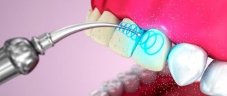
- Harden quickly using a UV lamp.
- Even if the tooth has been severely damaged, they will restore its shape and color perfectly.
- They have high strength.
- You can perfectly match the shade to any enamel.
- The materials used are non-toxic.
- The filling can last more than 5 years.
- It is plastic compared to other types.
- It can be polished well.
- They are safe for health and suitable even for children.
Its disadvantages are minor: you should not use it as a temporary filling, because it costs quite a lot. It should not be placed in hard-to-reach places.
Stages of filling anterior teeth
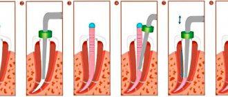
- The dentist examines the teeth for the presence of caries. If it is available, it continues its work.
- Cleans teeth from plaque. It is done using laser machines with ultrasonic attachments if there are massive dental deposits. Specialized brushes and abrasive gels if there is a slight coating.
- The color of the tooth is determined using a specialized scale. This is done to accurately match the color of the filling to the shade of the tooth enamel.
- Anesthesia is administered - the doctor determines the dose based on the stage of caries development and operating time.
- Drills out carious tissue - this is done to remove carious dentin, so that later after installing the filling there are no complications.
- Isolates the tooth from saliva - the service life of the filling will directly depend on this. A so-called latex “scarf” is pulled over the teeth.
- Treats the carious plane with medications.
- Restores the contact point between teeth - for example, the side wall of a tooth is restored if it has been damaged.
- The enamel is etched using acid.
- Dentin and enamel are treated with adhesive so that the filling is better fixed.
- A gasket is placed under the filling. It consists of glass ionomer cement.
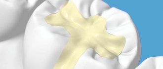
- The filling itself is inserted.
- The filling is ground and polished.
Cost of filling front teeth
Approximately filling one front tooth with a chip costs 2,000 rubles .
The price consists of the following indicators:
- Anesthesia - about 200 rubles;
- Application of a latex “scarf” - from 350 rubles.
- The filling itself costs from 1000 rubles.
- Dentist's work.
Possible complications after installing a filling
- Pain is a normal phenomenon, since the doctor cleaned the canals before installing the filling, which leads to pain after the procedures are completed. The pain may continue for several hours after visiting the dental office.
- If the tooth has not been sufficiently treated before installing the filling, the pain may last for several days. You will have to remove the filling, clean the tooth and the area around it again, and apply a new filling again.
- If the patient was nervous and twitched while the doctor was working, a burn to the pulp could occur when the tooth was prepared.
- The doctor damaged the tooth enamel.
- The acid has reached the dentin.
- An allergy to medications occurs.
What to do if the filling on the front tooth has darkened?
Any filling can darken over time due to food coloring.
It is necessary to consult a doctor so that he can examine her for the presence of:
- Caries;
- Inflammatory processes.
If these phenomena are not observed, the doctor will recommend professional cleaning of the oral cavity to get rid of plaque.
He will recommend a special diet to keep your teeth white for a long time. Replace the filling if it is damaged.
Filling care
- Do not eat solid food after the filling is installed for 1-2 days, depending on what type of filling was placed.
- To reduce tooth sensitivity after visiting the dentist, use a soft toothbrush and special medicated pastes designed for sensitive teeth to care for the oral cavity.
- Do not eat too hot or cold food for several days, this will increase tooth sensitivity.
- Food that is too hot can cause the filling to become deformed.
- Avoid eating sticky foods for a few days because new cavities may develop.
- You should not chew food on the same side of your mouth where the filling was installed, thereby reducing the risk of its deformation.
- When installing a seal, you need to check whether it is comfortable for you, so that there are no problems with it in the future.
- If you have the slightest problem with the filling, you should consult a doctor.
Conclusion
During the discussion of the topic of the article, detailed answers to the questions posed were given.
I would like to give a few tips at the end:
- Before going to any clinic for an appointment, look for reviews about the work of the specialists there, through friends or on the Internet.
- It’s better to put a good filling once and forget about it than to put a new one every 6-12 months.
- Try not to be nervous during the procedure and not to interfere with the dentist’s work, so that unwanted complications do not arise later.
- Listen carefully to the doctor’s recommendations after the procedure and follow them.
- Follow a diet for several days, do not eat solid foods, sour and sweet foods. Food must be cooled before consumption to prevent the filling from becoming deformed.
- Take good care of your oral cavity.
Attention! Teeth whitening strips...A quick way to lighten your teeth by 3-4 shades at home... Read more »
Source: https://glivec.su/2018/06/10/potemnela-plomba-na-perednem-zube-chto-delat/
Contact your dentist
If after all hygiene procedures the blue color of the tooth has not disappeared, then you should visit a doctor as soon as possible. Contact your dentist. The doctor will examine the stained tooth and take an x-ray if necessary. After this we can already talk about treatment. If the root is damaged, the pathological shoot will have to be removed. If everything is not so serious, then the dentist will offer you a suitable correction plan.
If a specialist (dentist or dentist) does not find a pathology in his area, you need to contact a therapist. If you have recently taken certain medications, be sure to tell your doctor. The doctor may ask you to undergo basic tests. They will help you understand the situation and prescribe the appropriate correction. In most cases, the pathological symptom disappears after a few days of treatment.
Blackening of the tooth under a filling: causes and treatment. Reasons for darkening of the filling
If you delay in contacting a doctor, the situation will be aggravated by the appearance of pain. And sometimes darkening indicates destruction of the hard tissues of the tooth. Often the attending physician, not the patient, is to blame for the appearance of darkening. Since before installing a filling, it is necessary to completely clean the carious cavity from dead tissue. A carious cavity or a tooth destroyed in a non-carious way is cleaned using professional equipment. The channels are washed and thoroughly dried for better adhesion of the material.
Before installing a filling, a specialist must check the canals and dentin for the absence of cariogenic microorganisms; for this purpose, a specialized liquid dye is used in dentistry. This procedure allows you to see even the smallest traces of pathogenic microorganisms and re-clean them.
A caries marker shows whether the tooth cavity is well cleaned
If the specialist skipped this stage, then cariogenic microorganisms under the filling will multiply and lead to the appearance of secondary caries.
Bruise on the gum - where it can come from and what to do with it
The appearance of bluish areas on the gums is a symptom that usually occurs against the background of the development of systemic diseases or as a consequence of disruptions in blood circulation in soft tissues. This phenomenon, as a rule, is localized in nature - upon visual inspection it is possible to detect isolated blue spots. To prevent the development of complications, you should see a specialist. From today's article you will learn about what to do if a child or an adult has blue gums, and what this symptom may indicate.
What can cause your gums to turn blue?
So, why can individual areas of the gums change their color, and where do such blue spots appear on the mucous membrane? This could be due to many different factors. So, for example, if we are talking about a child, the reason may be the eruption or change of baby teeth. In adults, a similar symptom is sometimes provoked by certain diseases of periodontal tissue or the oral mucosa as a whole. Also, the area near the tooth may turn blue due to unsuccessful prosthetics or malfunctions in the internal systems of the body. Let's look at possible provoking factors in more detail.
Dark enamel in children
In children, plaque is especially noticeable because the enamel of baby teeth is lighter than that of permanent teeth. Sometimes babies' teeth turn black on the inside, facing the tongue, sometimes dark spots appear on the outside. However, this does not always indicate a carious lesion.
Reasons that can cause enamel discoloration in children:
- long-term use of antibiotics,
- dysbacteriosis, inflammation of the gastrointestinal tract,
- fungal infection of the soft tissues of the oral cavity (plaque looks like mold),
- use of fluoride-containing pastes for hygiene,
- weak immunity.
Treatment for this condition is carried out after diagnosis. Sometimes dental intervention is not even required.
What can you do as an emergency?
It was already mentioned above that the most common cause of symptoms in childhood is the process of teething or changing teeth. To help your baby endure this difficult and painful period more easily, special ointments and gels with an anesthetic effect will help: “Kalgel” or, for example, “Cholisal”. As a rule, such a problem does not require medical intervention - the process of “punching” the tooth out usually takes several days. To speed up this process, you can resort to the help of special baby teethers. The blue spots will gradually resolve on their own.
If the cause of the blue discoloration is a growing tooth, then the symptoms may resolve on their own
As for other causes of hematoma, including in adults, you cannot do without the help of a doctor. During a visual examination and examination of the clinical picture as a whole, the specialist will determine whether the symptom is a consequence of a disease of the oral tissues, a pathology of the body, or an incorrectly fixed crown. Only after this the doctor will be able to begin treatment. If the problem is gingivitis or stomatitis, then the patient will be prescribed a course of appropriate anti-inflammatory and antibacterial drugs - both for oral administration and for external treatment of the affected areas.
If the problem is the result of an incorrectly installed crown or prosthesis, you will have to completely remove the orthopedic device and undergo anti-inflammatory therapy. The prosthetic device is sent for correction or completely replaced with a new design, which is certainly the best option. When a bruise becomes the result of mistakes made by the doctor during filling, a temporary filling is applied and a course of anti-inflammatory drugs is also prescribed.
Taking medications
Darkening of the enamel is a side effect of some medications. Most often these are drugs that are prescribed for iron deficiency anemia (Fenuls, Totema, etc.). They contain gluconates of manganese, copper and iron dihydrate, which cause tissue pigmentation, and since the drugs are used in liquid form, the teeth are the first to take the blow.
We suggest you read: Bitterness in the mouth – causes. What to do when your mouth is very bitter - advice from doctors
Another group of drugs that can deprive crowns of their natural color are tetracycline antibiotics. Most often, this pathology manifests itself in children whose mothers took oxytetracycline, metacycline, minocycline, etc. during pregnancy.
When is it time to go to the doctor?
If a blue spot appears on the mucous membrane, it is better to see a doctor immediately. However, it is not always possible to urgently contact a specialist. Therefore, you need to know exactly when you can’t postpone a visit to the dentist. So, you need to seek help urgently if you have the following symptoms:
- the stain remains on the mucous membrane for more than 3 days,
- hyperthermia with signs of fever is noted,
- the patient complains of severe pain in the gums,
- general condition worsens, appetite is impaired.
A timely visit to the dentist will help avoid problems
If the problem is not resolved in a timely manner, it significantly increases the risk of developing serious complications. Among these are possible suppuration, the appearance of a benign tumor and the formation of a cyst.
Is it possible to determine for yourself why a tooth hurts after a filling?
Secondary caries may not make itself felt for a long time.
- At first, the presence of a problem is only indicated by bad breath, which appears shortly after brushing your teeth.
- Then discomfort or a slight painful reaction to temperature and mechanical stress may appear.
- An attentive patient can detect darkening of the tooth enamel in the area of the filling, the formation of an edge gap, and even mobility of the filling itself.
All these signs indicate ongoing tooth decay and indicate the need to urgently seek professional help.
The recurrent process is more active and appears within a couple of weeks after tooth treatment. The patient complains of aching toothache, which is a consequence of the involvement of the dentinal tubules in the process and the advancement of infection to the pulp chamber. Visually, no changes are observed in the area of contact between the filling material and enamel. In this case, you should immediately consult your doctor to prevent inflammation of the pulp and treat the tooth.
To exclude the possibility of the formation of a new focus of caries and to ensure that the carious cavity has been properly cleaned and treated, dentists use the so-called caries marker. This is a liquid applied using a foam ball and distinguishes between healthy and damaged dentin. It turns the destroyed tissue bright red, showing the dentist exactly which areas need to be removed.
Traditional medicine tips
Many medicinal herbs have proven themselves to be excellent in the fight against inflammation in gum tissue. Infusions and decoctions are made from them, which are then used to rinse the mouth several times a day. Antiseptic plants that additionally have pronounced anti-inflammatory properties include chamomile, sage, and oak bark. Some sources provide recommendations for treating the mucous membrane with aloe juice or honey to relieve pain. In order to restore soft tissues after an incorrectly installed orthopedic structure, stimulation of blood circulation is required. A decoction of nettle leaves, as well as propolis ointment, helps with this.
Chamomile infusion is great for rinsing
It is important to understand that any folk recipes can be used only after consultation with the treating specialist. Otherwise, you can provoke the development of a serious complication and only make everything worse. And certainly traditional medicine cannot become a full-fledged alternative to drug therapy. The use of medicinal plants is justified only as an additional measure.
Causes of dark coloration
At some point in his life, the patient noted that the filling had darkened, and of course he would begin to worry about the reasons for the appearance of the changed color. There may be several etiological factors in the development of color transformation. However, the main factors contributing to this are either the filling material or a violation in the doctor’s tactics during preparation.
Features in the use of materials
A change in tooth color under the material indicates the development of secondary caries. Why does the tooth under the filling turn black when using the material? It's all about what substance is used for restoration work. At the dawn of the development of dentistry, various cements and amalgam were used for filling. The view was not aesthetic.
Over time, chemically cured composites emerged. They are easy to use and come in a set of two pastes and an adhesive. One paste is basic, the other is catalytic. After combining the material, a short period of time is given to stage the material of this group. Based on this, it is extremely difficult to ensure an ideal marginal fit when filling with a chemically cured composite. In addition, over time, polymerization shrinkage of the material occurs, which also contributes to the creation of voids between the tooth and the filling.
Modern light-curing composites are an order of magnitude better than chemical ones. They are easy to use, flexible, and the modeling time is sufficient to complete the restoration. As long as the filling substrate is not exposed to light, it can be instrumented to create anatomy. The reasons that can provoke the development of secondary caries when using light composites are due to a violation in the technology of application or the insensitivity of the components of the material. To install light fillings, it is important to use the technology of layer-by-layer application of the composite, while the thickness of a single application and modeling on the tooth should not exceed a thickness of 2 mm. It is necessary to use a bond (a special glue that holds the filling and tooth in place) and expose it to light. Insufficient modeling of the material along the border with the tooth tissue leads to inadequate marginal fit. Over time, germs from the mouth will begin to penetrate this weakened area of the filling.
Errors at the preparation stages
After the doctor has diagnosed a tooth disease of a complicated or uncomplicated nature, he begins preparation. The reasons that contribute to the appearance of blackness, as a rule, lie in the leaving of non-viable dentin. This is possible in the case of using local anesthesia, when the patient cannot accurately identify pain when exposed to irritants. With this option, necrotic areas of dentin can be left at the enamel-dentin border (medium caries) or at the bottom of the cavity (deep caries).
The doctor uses visual distinctive and instrumental signs during the preparation process. An acute process in a tooth looks like light brown or brown-colored tooth tissue, loose. When removed, a normal shade is observed in the tooth structure in which it is located (enamel - whitish, dentin - yellowish). The chronic process will be represented by altered dark brown tissue, usually dense. However, there are transitional processes in necrosis of dental tissue. That is, the tissue may not yet be indicated by a variable color, but will already be filled with microorganisms. In such situations, it is possible that an infectious part may remain in the tooth.
In order to eliminate the possibility of error at this level, the doctor uses a “Caries Detector”. This drug is able to identify areas of the tooth that cannot be preserved and remove them using a handpiece and bur connected to a dental unit.
The doctor may also make mistakes in creating a cavity for the filling. The cavity must be formed based on the class of damage to the tooth tissue. Minimal preparation possible. However, in both cases, a fold is created at an angle of 45 degrees to the tooth tissue at the level of the enamel. This is necessary to create a complete marginal fit of the material into the tooth tissue. It is very important to consider IROPD (index of destruction of the occlusal surface of the tooth) when choosing a replacement material. A filling can be placed with IROPD up to 55%. If the setting is carried out at a higher index, over time this will contribute to the development of cracks and disturbances in the marginal adhesion of the material to the tooth.
Other mistakes that can create a darkened shade of the tooth under the filling are violations in cooling the tooth and the use of medications. When the pulp horns are close together and preparation is performed for deep caries, overheating can be created without using air-water cooling, burns when using strong solutions, or long-term exposure to etching containing acid.
The cumulative effect of the noted errors will quickly provoke changes in the tooth, manifested by a change in color under the filling.
How to prevent the problem
High-quality care and systematic teeth cleaning are two basic rules, the observance of which will, to a certain extent, protect oneself from inflammatory and carious processes. In addition, do not forget that every time after eating you need to thoroughly rinse your mouth and use dental floss. It is also important to choose the right toothpaste and brush, especially if you have sensitive gums. And of course, don’t forget to visit the dentist regularly.
If a bluish spot appears on your gum, do not put off your visit to the dentist. The longer you ignore the problem, the higher the likelihood of developing truly serious complications.
- Balakhnichev, D.N. The influence of fixed orthopedic structures on the periodontium of abutment teeth, 2011.
How can you restore the whiteness of a dead tooth?
The first method, which requires the least intervention in the tooth structure and is technically the simplest, is bleaching inside the canal. It has a relatively low cost, up to two thousand rubles.
But this method has many disadvantages:
- It is 100% impossible to predict the result; it can be seen at the end of the procedure;
- Several procedures using whitening gel may be necessary;
- the shade of the whitened tooth will still be dull, and it will stand out among healthy ones;
- the active substances of the whitening gel have a bad effect on dentin; after such a procedure it will be more fragile.
Endobleaching: before and after procedures
An alternative method for whitening a blackened tooth is direct restoration or extension using composite materials.
First, the entire blackened surface is removed, then a filling is created, which in size can occupy the entire visible part of the tooth. You can build it up in one trip to the dentist and at a fairly low cost, about 3,000 rubles.
But this method also has its disadvantages:
- the material is short-lived, over time you will have to repeat the procedure;
- it quickly absorbs coloring elements, and accordingly will stand out among the rest;
- repeated restoration reduces integrity.
Many patients are faced with the fact that the extended tooth has darkened, what to do in such a situation? Most likely, it may darken due to the accumulation of dyes; first of all, we exclude products that contain such substances, and then proceed with repeated restoration.
Also, the cause of darkening during extensions may be the formation of caries; do not delay its treatment and immediately consult a doctor to avoid the development of complications.
Another whitening method is the installation of a thin translucent ceramic plate on the outer surface of a blackened tooth - a veneer. First, a layer of enamel is removed to the thickness of the plate.
An experienced dentist can perform this work so that the tooth under the veneer cannot be distinguished from a healthy one . This does not reduce the strength. Among the negative qualities of veneers are the high cost, the possibility of showing through strong darkening, and the fact that it is not recommended to place them on pulpless teeth.
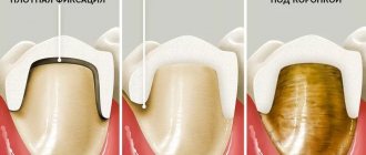
Tooth prepared for crown
If none of the previously proposed whitening options are suitable, then the most radical option is used - installing a crown.
The required tooth is ground and polished, then plaster casts are made. The denture should fit tightly to healthy teeth, but without excessively deepening into the gum.
They are most similar to natural metal-ceramic crowns , or ceramic ones with a porcelain base. Under a crown, the tooth is well protected from destructive factors and can withstand much more load than without a crown. You can also hide aesthetic defects under the crown. The cost of installing crowns is quite high and it takes place over several visits to the doctor.
Modern dentistry has reached a high level of development and can offer many options for eliminating blackening in teeth under a filling. You just need not to delay your visit to the doctor, and he will help establish the cause of the darkening and solve your problems.
Causes of blue gums
There are many different factors that accompany the appearance of blue areas on the gums, and they can be either harmless to health and cause discomfort only from an aesthetic point of view, or indicate the development of more serious diseases. Dentists identify the following main causes of blue gums:
- Stomatitis. It often happens that the gums acquire a bluish tint - just one of the symptoms of developing stomatitis. In this case, blue discoloration occurs around the rash or erosions and is accompanied by a number of other symptoms: pain in the affected area, plaque on the mucous membrane, fever.
- Chronic gingivitis. With inflammation of the gums, bluish areas are rare and mainly appear only in the area of the necks of the teeth. Gingivitis can occur without disturbing the dental-gingival attachment, but bring many unpleasant symptoms, including an unpleasant odor from the oral cavity, abundant deposits of plaque, bleeding and redness.
- Poor quality restoration or prosthetics. In the first case, the problem of the appearance of bluish gums is caused by improper application of the material to the subgingival margin and a violation of trophism. In the case of prosthetics, gum trophism is also disrupted, blood stagnates in the veins, causing swelling. The blood is not oxygenated enough to be scarlet, so areas of the gums turn blue.
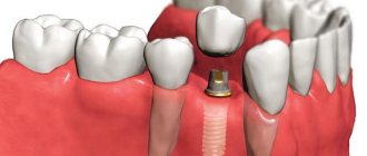
- Silver metal or alloys. Dentures with a metal frame are a fairly common cause of cyanosis of the gums, as there is constant contact of the metal edge of the bridge with soft tissues. What aggravates the situation and leads to disruption of blood flow is the fact that the structure was initially too deeply advanced into the subgingival area. Installed fillings, inlays or intra-root pins made of silver alloys stain not only the gums, but also the tooth tissues bluish.
- The acquisition of blue, blue, violet and other non-standard colors for gums may be a consequence of changes in the mucous membrane. That is, first the mucous membrane changes its color, and then reflects this on the gum.
- Fragile and thin blood vessels or pathology of the coagulation system. These factors for blue gums are hereditary or congenital.
- Mechanical trauma (a fall, a blow, or brushing your teeth with a hard brush) can cause a change in the color of soft formations. In this case, cyanosis and swelling go away on their own over time without the help of specialists.
- Insufficient supply of vitamins and microelements, deficiency of minerals and other beneficial enzymes.
- Drinking colored drinks (wine, tea, carbonated drinks) or food with dyes causes temporary blue gums, which can be eliminated by brushing your teeth.
- Blue gums can be a symptom of various diseases, including rickets and anemia.
Other reasons for darkening of crowns under fillings
Darkening of teeth is a common clinical case in dentistry. Often the problem is associated with the use of low-quality filling materials or their improper installation. But there are reasons for tooth discoloration.
External factors
Smoking damages the human body, provokes the development of many dangerous diseases, and affects the color of tooth enamel. Drinking large amounts of coffee and black tea, and other coloring drinks provokes aggravation of the situation.
Sometimes the reason why a tooth under a filling turns black is a disease called fluorosis. Fluorosis occurs with prolonged consumption of foods and water with elevated levels of fluoride. Dark areas form on the enamel and it gradually deteriorates.
Smoking stains fillings
Pathological processes in the oral cavity
Among the factors influencing the darkening of the hard tissues of the crown under the filling are pathological conditions of the oral cavity:
- The presence of a fungal infection. Oral candidiasis is a dangerous fungal disease that provokes discoloration of dental crowns and the development of pathogens.
- Rupture of the nerve bundle due to trauma to the dental crown. When a vascular bundle ruptures, it fills the delicate dentin tissue with blood, which stains the tooth from the inside.
Necrosis of tooth tissue under a filling due to improper treatment