Histological structure of dental tissues
HISTOLOGICAL STRUCTURE OF DENTAL TISSUE
Hard tooth tissues consist of enamel, dentin and cement, which differ significantly from each other in structure and chemical composition. The pulp is located in the cavity of the tooth. The root of the tooth is surrounded by a connective tissue formation - theriodont, with the help of which the tooth is strengthened in the alveolus (see Pulpitis, Periodontitis).
Enamel (enamelurn ). The main structural formations of enamel are enamel prisms, running in the direction from dentin to the enamel surface. Their thickness varies: on average from 3 to 6 microns; at the enamel-dentin junction they are thinner, while closer to the surface their diameter increases. The shape of enamel prisms varies significantly. In cross-section, they can have oval, hexagonal, polygonal, arcadoid shapes, etc. However, arcadoid prisms are most common. They are formations with rounded heads that turn into a narrower elongated part. Such enamel prisms in cross section resemble the shape of a keyhole. The prisms are arranged in rows, so that their elongated part is inserted between the heads of the underlying prisms. On histological preparations of enamel, where enamel prisms are located longitudinally, one can see an alternation of wider stripes - the heads of the prisms with narrow stripes, which are the tail, elongated part of the prisms. Previously, it was believed that these narrow stripes are an independent formation - an interprismatic substance, and only after studying the enamel under an electron microscope was it proven that this substance is part (extensions) of enamel prisms that are embedded between adjacent prisms. Enamel prisms are folded into bundles (10-20 pieces each), which follow from the enamel-dentin junction in the radial direction. They travel a certain distance in an S-shape and, twisting in a spiral, again take a radial direction and reach the surface of the enamel. Sometimes the prisms bend at the very surface of the enamel, and the shape of the enamel prisms is lost and the surface layer is formed only by radially arranged crystals of hydroxyapatite.
In other cases, as a result of the fact that the enamel prisms from the enamel-dentin junction do not run strictly radially, alternating light and dark stripes (Schröger lines) are visible on polished sections of teeth. These lines arise due to the fact that tortuously directed bundles of enamel prisms are dissected either in the longitudinal or in the transverse direction. Longitudinally dissected bundles of prisms are called vapor zones, and transversely dissected bundles of prisms are called bottom zones. On longitudinal sections of teeth, one can also observe lines that are sometimes brown in color and directed more obliquely from the enamel-dentin junction to the periphery. On a transverse section of the tooth crown, these lines are located in the form of concentric circles in the enamel layer. These are the so-called lines of Retzius, the appearance of which is associated with uneven mineralization of the enamel during tooth development. In this regard, the lines are especially noticeable in teeth with reduced mineralization. Ending on the surface of the tooth, these lines form strips located in the transverse direction and surrounding the crown of the tooth, which are called perekims. In addition to the lines of Schröger and Retzius, enamel plates can be noted in the enamel - less calcified formations passing through the entire thickness of the enamel, and enamel bundles - formations present in the enamel at the enamel-dentin junction.
Enamel prisms consist of hydroxyapatite crystals—[Ca10(PO4)6(OH)2], the length of which varies between 50–200 nm. Crystals have a hexagonal or polygonal shape. The direction of the crystals in the peripheral parts of the prism heads approaches radial, while in the central part of the prism heads they are located longitudinally. In the tail part of the prism, the crystals are directed at approximately 45-50° with respect to the long axis of the prism (Fig. 18-20).
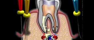
Dentin (dentinum) . In the main substance of dentin there are variously located collagen fibers. Some of them are oriented radially (Korff fibers), while others are tangentially oriented (Ebner fibers). The superficial zone adjacent to the enamel-dentin junction and cement, the width of which is several microns, consists exclusively of radially directed fibers. In the middle zone, radial fibers are collected into bundles, and the bulk of the fibers are located tangentially. These two zones are called mantle dentin. The third, widest zone adjacent to the tooth cavity is called peripulpar dentin and is represented mainly by tangential fibers. Between the collagen fibers there is an adhesive calcified substance.
The degree of dentin mineralization varies. The layer of dentin adjacent to the tooth cavity, as well as the zone of interglobular dentin, remains poorly calcified. This zone is located closer to the enamel-dentin junction (between the mantle and peripulpar dentin) and is characterized by the fact that the calcified areas are located in the form of spherical lumps. In the root area, these lumps are smaller, and the zone is called the granular layer (Toms). On transverse sections of the tooth, contour lines (Owen) are visible, corresponding to dark lines (Retzius) in the enamel. Owen's lines also occur as a result of uneven mineralization of dentin. The entire main substance of dentin is penetrated by dentinal tubes (tubules), the diameter of which ranges from 1 to 5 microns. Many branches extend from the dentinal tubules, and closer to the enamel-dentin junction they branch into thinner branches. Some of them penetrate the enamel-dentin junction and end in the enamel. There is evidence of anastomosis of dentinal tubules with each other using branches. On the side of the tooth cavity, the number of dentinal tubules reaches 75,000 per 1 mm2, and at the enamel-dentin junction there are fewer of them - up to 15,000 per 1 mm2. The inner surface of the dentinal tubules is covered with a membrane (Neumann), which transforms into a similar formation covering the dentin from the side of the tooth cavity (Kölliker-Fleischmann membrane).
The submicroscopic structure of dentin is represented by hydroxyapatite crystals, which are located between collagen fibers. However, in well-mineralized dentin, the collagen fibers are masked by a mass of hydroxyapatite crystals. Collagen fibers are visible only on some preparations in the walls of dentinal tubules or on decalcified preparations. Around the dentinal tubules there are areas of dense, structureless substance—the so-called zones of hypermineralization. These zones are more pronounced in well-mineralized teeth and less pronounced, and sometimes completely absent, in individual dentinal tubules in insufficiently mineralized teeth. Branches extend from the dentinal tubules, which penetrate the main substance of dentin (intertubular zone); closer to the shawl-dentin junction, hydroxylapatite crystals are arranged more densely. This zone is penetrated by branching dentinal tubules (Fig. 21).
Depending on the timing of dentin formation, and sometimes the pathological processes that cause its increased deposition, dentin is divided into primary, secondary (replacement) and tertiary (irregular). Primary is called dentin, formed during the development of the tooth, secondary is dentin, which is deposited throughout a person’s life as a consequence of the physiological activity of the pulp. In its structure, secondary dentin does not differ from primary dentin, and only under a microscope can one see the boundary between them in the form of a prenatal line. In older people, deposits of secondary dentin cause a decrease in the size of the tooth cavity, and sometimes its complete obliteration. Tertiary dentin, which is also called replacement or irregular, is formed in a shorter time as a result of the protective activity of the pulp during pathological processes in the hard tissues of the tooth, and sometimes during general diseases. In tertiary dentin, there may be an irregular arrangement of collagen fibers, and in some cases, dentinal tubules or their complete absence.
Cement (cementum) .
Its structure is similar to coarse fibrous connective tissue. Cementum, like dentin, contains collagen fibers and a mineralized adhesive substance. Some collagen fibers are located in the longitudinal direction, others in the radial direction. These fibers, on the one hand, transform into radial (Korf) dentin fibers, and on the other, into periodontal fibers (Sharpey's). The entire surface of the roots is covered with cement. Directly adjacent to the dentin is the so-called acellular, or primary, cement, in which there are no cells. At the apices of the roots and on the interroot surfaces of multi-rooted teeth, this cement is covered with a layer of cellular, or secondary, cement, which contains many cement cells, cementocytes, with a large number of processes. Unlike primary cement, secondary cement has a pronounced radial direction of collagen fibers.
Eruption of baby teeth
By the age of 2, all baby teeth, as a rule, have erupted . This type of bite is called a milk bite. It should be mentioned that the term “baby teeth” was proposed by Hippocrates, who assumed that they are formed from mother’s milk.
Calcification of the roots of primary teeth ends, as a rule, by the age of 3-4 years. By the age of 6, the process of loss of temporary teeth and eruption of permanent teeth begins. The stage of mixed dentition begins, which ends when the last milk tooth falls out. After this, the teeth are already in a permanent bite.
Eruption of permanent teeth
The eruption of permanent teeth on the upper and lower jaws occurs in different orders. The teeth in the upper jaw erupt in the following order:
- first molars
- central incisors
- lateral incisors
- first premolars
- second premolars
- fangs
- second molars
- third molars
Since there are no premolars in the primary dentition, the primary molars are replaced by permanent premolars. In addition, if a baby tooth falls out before the permanent replacement tooth is ready to erupt in its place, some displacement of the teeth may occur, which in turn can lead to twisting of the teeth in the permanent and permanent dentition.
Permanent dentition begins at the age of 12-13 and continues until the end of a person’s life or until they fall out or are removed due to periodontal or pulp diseases. The most commonly removed teeth are the lower first and third molars.
Teething theories
Despite the development of science, teething is one of the most mysterious phenomena in dentistry. Scientists have long known everything about the formation of teeth, but what force causes the tooth to move to the edge of the gum and erupt remains a mystery.
There are several theories about teething. Some authors believe that the tooth is affected by buoyant forces arising as a consequence of the formation and growth of the alveolar bone; others attribute the emergence of these forces as a consequence of the formation of the root itself. It is now generally accepted that periodontal fibers play a major role in tooth eruption. According to this hypothesis, by their contraction they push the tooth into the oral cavity.
Temporary teeth erupt, as a rule, in the following order:
- central incisors;
- lateral incisors;
- first molars;
- fangs;
- second molars;
Every 6 months, 4 teeth erupt. Moreover, in girls they erupt earlier than in boys. During the temporary dentition, the rudiments of permanent teeth are formed. And the eruption of the latter is “provoked” when the roots of baby teeth begin to dissolve.
Dentine
This concept was already mentioned earlier in the discussion of the stages of tooth development. Dentin is its hard tissue, which is the main one. The coronal part is covered by enamel, and the root part is covered by cement.
Dentin consists mainly of hydroxyapatite (approximately 70%). It also contains organic material (20%) and water (10%).
Dentin is the basis of the tooth and supports the enamel. Its thickness varies from 2 to 6 mm. The hardness is impressive - 58.9 kgf/mm².
Within the framework of the topic on histology about tooth development, it is necessary to note that dentin is divided into types. There are three in total: