1.General information
The oral mucosa is characterized by a number of distinctive features.
It is located in close proximity to vital organs, endocrine glands, lymph nodes; serves as a covering for the “entry gate” for the digestive tract and respiratory tract; it is intensively innervated, supplied with blood, moisturized, and renewed very quickly. Therefore, this mucous layer, extremely sensitive to any physiological, hormonal, biochemical changes in the body, can serve as a reliable early indicator of a pathological process, which sometimes begins in a completely different zone and system. Moreover, it is specific changes in the oral mucosa, discovered, for example, during a dental examination or treatment, that often turn out to be the first and at that time the only sign of a particular disease, sometimes formidable and severe, but progressing asymptomatically until a certain stage.
A must read! Help with treatment and hospitalization!
What is important to know about leukemia
The disease can develop quickly (acute form) or, on the contrary, slowly (chronic form). As healthy cells are destroyed, blood functions change, which manifests itself in characteristic symptoms.
What signs indicate the presence of leukemia?
Constant weakness and fatigue.
Constantly feeling tired and weak is the most common symptom. Typically, this condition is caused by concomitant anemia, which aggravates the person’s physical condition. Initially, the patient may feel slight fatigue, but over time it will increase.
Problems with the respiratory system.
Those with leukemia often suffer from shortness of breath or what is known as a “lump in the chest.” Breathing in such patients is heavy and intermittent.
The appearance of bruises of unknown origin.
If bruises form on the body, but the person has not received any injury, this is one of the signs of leukemia. Such manifestations are often the result of a low platelet count or problems with blood clotting. As a rule, bruises appear on the extremities, but can be on any part of the body.
Bleeding.
Unreasonable bleeding from the lungs, intestines, gums or nose indicates a deficiency of platelets in the body and problems with blood clotting. These signs are characteristic of the acute form of leukemia.
The formation of petechiae - small red spots under the skin.
If barely noticeable, painless red dots form on the body, this indicates a decrease in platelet levels. This manifestation is also one of the signs of leukemia. Typically, spots can be found in the ankle area, since it is in the lower legs that the largest amount of fluid accumulates during the day.
Enlarged and swollen gums.
This symptom is observed less frequently and only in patients with an acute form of leukemia.
Bloating.
An increase in the size of the spleen, as well as loss of appetite, may indicate chronic or acute leukemia. Patients can eat small amounts of food and still feel full, this is due to the pressure of the spleen on the stomach.
Pain or other discomfort in the upper abdomen.
Due to an enlarged spleen, some discomfort and even severe pain may be felt in the abdomen. This is due to the location of this organ - in the upper left part of the abdomen.
Chills or fever.
This symptom is nonspecific and occurs rarely, often in acute leukemia. And the presence of a low-grade fever indicates an infection in the body and a weakened immune system, which may be associated with cancer.
Sweating at night.
If a person sweats heavily during a night's rest, a thorough examination of the body is necessary. Since this sign may indicate the presence of cancer.
Frequent headaches.
Leukemic anemia is often accompanied by throbbing headaches. This condition can be life-threatening due to the high risk of head bleeding, so you should urgently consult a specialist.
Paleness of the skin.
A symptom of acute or chronic cancer can be uncharacteristic paleness of the skin. In this case, the patient may also experience fatigue and shortness of breath.
Bone pain.
This symptom occurs in rare cases, but it is definitely worth paying attention to. Especially if the patient feels general weakness and severe pain in the bones, which is due to the production of cancer cells inside the bone marrow.
Enlarged lymph nodes.
If the lymph nodes in the groin, armpits and neck are enlarged, you should undergo an examination. This symptom often occurs during an infection but disappears with recovery. If the nodes remain enlarged or continue to grow, this may indicate the development of leukemia.
Skin rash.
According to statistics, every 20 leukemia patients experience a skin rash similar to plaque or an allergic reaction of the body. Rashes can be of any size and shape, but in the case of cancer they always grow and spread throughout the body.
Frequent infectious diseases.
If a person cannot overcome an illness despite constant use of antibiotics, then it is necessary to do a complete blood test to determine the level of white blood cells, platelets and hemoglobin. With an increased level of white blood cells, the immune system suffers, which provokes frequent infections. If such a symptom is accompanied by bruising and fatigue, this is a reason to seek advice from an oncologist.
Any of the above symptoms should not be ignored, since leukemia is an insidious disease
.
source
2. Reasons
Of all diseases of the hematopoietic system, the oral mucosa (OM) is most sensitive to two groups of diseases, one of which concerns red blood cells (erythrocytes) and the hemoglobin associated with them, the other - white blood cells, or leukocytes.
The group of anemia includes a number of diseases and pathological conditions in which the number of red blood cells decreases and the hemoglobin content in the blood decreases.
The group of leukemias includes malignant diseases of the hematopoietic system, in which the proliferation (abnormal multiplication and growth) of blood cells or bone marrow, as well as the activity of the hematopoietic parenchyma, precedes the maturation and differentiation of blood cells (i.e., their acquisition of one or another functional type), therefore, a large number of altered, atypical, immature leukocytes - leukoblasts, myeloblasts, etc. - appear in the blood.
Both anemia and leukemia, with all their etiopathogenetic and clinical diversity, which certainly deserves separate consideration, affect the state of the oral mucosa in more than 90% of patients, and often a preliminary diagnosis is made precisely on the basis of specific changes in the oral mucosa.
Visit our Oncology page
Acute leukemia and its manifestation in the oral cavity
Acute leukemia most often affects young people under 30 years of age.
At the very beginning of the disease, the symptoms strongly resemble the clinical picture of influenza, sore throat, dyspepsia, and body temperature also rises. In about a tenth of patients, acute leukemia occurs due to various bleedings - from the nose, after removing a tooth from the socket, gums, and also from ulcerative stomatitis. The occurrence of ulcers and areas of necrosis in the oral cavity in young people after suffering an acute respiratory illness is regarded by dentists as Vincent’s stomatitis. In the initial stage of acute leukemia, pain in the bones may occur. These patients may be registered with doctors for neuritis, polyarthritis, and rheumatism. Often the disease is detected accidentally during a blood test.
The diagnosis of acute leukemia is made on the basis of establishing the fact of blast infiltration of the bone marrow. The clinical picture of symptoms in acute leukemia is conventionally divided into 4 types - hyperplastic, hemorrhagic, anemic and intoxicating.
Symptoms of the hyperplastic type are varied. In about half of the patients, the lymph nodes are enlarged, without pain; there is an enlargement of the liver, spleen, and tonsils in about a quarter of the patients. In about 5% of patients, gum hyperplasia is found; as a rule, this is characteristic of severe leukemia and is usually regarded as a bad sign. Often, gingival hyperplasia manifests itself along with the formation of ulcers and areas of necrosis in the oral cavity. Gingival hyperplasia in acute leukemia must be distinguished from hypertrophic gingivitis, as an independent disease.
It should be taken into account that in acute leukemia, hyperplasia occurs together with general clinical symptoms - a sharp deterioration in the patient’s general well-being, increased body temperature, and regional lymphadenitis.
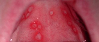
Hemorrhagic syndrome occurs in 50-60% of patients; its etiology is severe thrombocytopenia. Thrombocytopenia and anemia arise as a result of disruption of hematopoietic processes, which in turn develop due to leukemic hyperplasia and bone marrow infiltration. Clinically, hemorrhagic syndrome is manifested by rashes - from pinpoint rashes on the skin and mucous membranes to extensive hemorrhages.
In the oral cavity, it most often manifests itself as very severe bleeding of the gums, even at the slightest touch to them, hemorrhages occur on the mucous membrane of the cheeks in the area of the line of closure of the teeth, and in other parts of the oral cavity. The formation of extensive hemorrhages and hematomas is possible. These symptoms occur along with general weakness, severe fatigue, a sharp decrease in appetite, and fever.
In dentistry, signs of hemorrhagic syndrome of acute leukemia are often taken as a result of hypovitaminosis C, or simply as a result of constant biting of the mucous membrane. The diagnosis can be confirmed by a blood test or bone marrow aspirate.
In acute leukemia, the oral mucosa is most often affected; this fact is explained by its increased sensitivity to trauma as a result of pathological changes, a sharp decrease in the body's defenses, local protective factors of the oral cavity, circulatory disorders, etc.
Patients with acute leukemia are characterized by impaired taste sensitivity, which occurs due to damage to the fungiform papillae of the tongue. Pain may occur in absolutely healthy teeth, as well as in the jaw bones, along with pain in other bones of the body, this is explained by their direct damage due to leukemia.
Since in acute leukemia the body’s defenses drop sharply, candidiasis of the oral mucosa develops, the development of which is also provoked by the patient taking cytostatics, corticosteroids, and some antibiotics.
Based on the characteristic manifestations in the oral cavity, the dentist should suspect leukemia and refer the patient for a blood test to clarify the diagnosis. Treatment of acute leukemia is carried out in special hematology departments.
Local manipulations in the oral cavity for acute leukemia - removal of tartar, treatment and removal of teeth must be coordinated with a hematologist. For ulcerative-necrotic processes in the mouth, antiseptics and painkillers are prescribed - lysozyme, iodinol, rosehip oil, etc. If there are candidal lesions of the mucous membrane, they are treated in the same way as for an independent disease.
source
3. Symptoms and diagnosis
Iron deficiency anemia is characterized by dullness of taste, pallor of the mucous membrane, painful reaction of the mucous membrane to sour or spicy foods, various false sensations (tingling, tingling, etc.), swelling and, less often, bleeding of the tongue, as well as inflammation along the red border of the lips (cheilitis) and multiple caries.
With hypoplastic anemia, hemorrhagic foci, swelling and cyanosis of the interdental papillae, and a tendency to expose the periodontal pockets are observed; in more severe cases, erosions or ulcers and areas of local necrosis appear on the oral mucosa.
B12-folate deficiency Addison-Biermer anemia is characterized by a yellowish-pale tint of the mucous membrane, glossitis (inflammation of the tongue), and in some cases, pinpoint hemorrhages on the mucous membrane.
As for leukemia, their existing classifications (based on etiological, cytogenetic and other criteria) include several dozen clinical forms, the analysis of which is beyond the scope of this material. Even the traditional division in medicine according to the type of course - into acute and chronic leukemia - in this case takes on a slightly different meaning: in fact, these are two different diseases, and they never transform into each other.
If we do not touch upon the main symptoms and limit ourselves only to changes in the oral mucosa (as in the case of anemia, see above), then acute leukemia is manifested by severe bleeding of the mucous membrane on the gums, cheeks (along the line of dental occlusion), on the upper palate, and tongue. Loose hyperplastic growth of the gums in the form of ridges that bleed at the slightest touch or spontaneously is often observed. In addition, pale necrotic areas surrounded by a bluish halo with an off-white or gray foul-smelling coating are often observed. Swallowing is difficult, eating causes pain in areas of necrotic ulceration; trophic disorders are often accompanied by activation of chronic infections (herpes, candidiasis).
Chronic leukemia, which can be latent for a long time, is also characterized by bleeding of the oral mucosa, but it is less pronounced than in acute leukemia, and hemorrhages occur mainly due to trauma or microtraumatization. A diagnostically informative sign is the combination of bleeding, cyanosis, and pallor of the mucous membranes in the absence of signs of inflammation.
In myeloid leukemia, erosive and ulcerative lesions of the oral mucosa often predominate; in chronic lymphocytic leukemia, infiltrating nodes with a loose consistency and bluish tint predominate.
If, during an examination by a dentist, orthodontist, otorhinolaryngologist, therapist or other specialist, there is a suspicion that specific changes in the oral mucosa may be caused by diseases of the hematopoietic system, a consultation with a hematologist is strictly necessary, followed by an in-depth laboratory blood test , the results of which are the most informative and decisive in making a diagnosis. As necessary, other laboratory and instrumental studies are prescribed (imaging diagnostic methods, bone marrow puncture followed by histological analysis, etc.).
About our clinic Chistye Prudy metro station Medintercom page!
Diseases of the gastrointestinal tract
Diseases of the gastrointestinal tract
Due to the fact that various diseases of the gastrointestinal tract do not have specific manifestations in the oral cavity for each of them, the material is presented on the basis of the most common clinical symptoms.
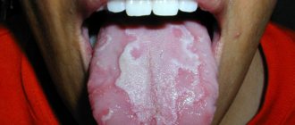
In pathology of the gastrointestinal tract, the condition of the tongue is the most studied. Its appearance, according to many researchers, may have diagnostic value and indicate a hidden pathology of the stomach or intestines. The most common finding is a coated tongue. The severity of plaque depends on various reasons: the severity of the papillae of the tongue, the process of self-cleaning, etc. In the normal state or hypertrophy of the papillae of the tongue, the plaque is dense and significantly pronounced; with atrophy of filiform and mushroom-shaped papillae, on the contrary, plaque is absent or weakly expressed. In the appearance of plaque, an important role is played by disruption of the normal process of keratinization and desquamation of the epithelium due to neurotrophic disorders. The nature of the food consumed (its consistency), the composition of the oral fluid, microbial flora and oral hygiene undoubtedly also influence this process.
Plaque is usually found in gastritis, peptic ulcers of the stomach and duodenum, neoplasms of the stomach, etc. During the period of exacerbation of peptic ulcers, gastritis, enterocolitis, the coating of the tongue is more pronounced.
It is important to note that during the period of remission or during treatment of the underlying disease, the tongue is cleared of plaque to varying degrees and can take on a normal appearance. Plaque on the tongue is usually not accompanied by subjective sensations. However, in the presence of a dense coating, patients experience a feeling of awkwardness and dulling of taste sensitivity. The plaque is based on enlarged, keratinized filiform papillae, food debris, microorganisms, and desquamated epithelial cells. A coated tongue is observed in many infectious and other diseases. In addition, a small plaque, especially in the morning, is possible in healthy people. The plaque does not require local treatment. It is necessary to find out the cause of its occurrence and treat the general disease, sanitize the oral cavity, and give recommendations on proper brushing of teeth.
A sign of gastrointestinal diseases, often found in the oral cavity, is a swollen state of the mucous membrane of the mouth and tongue. Swelling of the tongue and mucous membranes usually does not cause suffering to the patient and is detected by the doctor during examination of the oral cavity. The swelling can reach significant severity, and then patients note awkwardness in the tongue, an increase in size, and sometimes biting of the tongue and mucous membrane of the cheeks. Upon examination, tooth marks are found on the tip, lateral surfaces of the tongue, and the mucous membrane of the cheeks; the tongue is increased in size. The mucous membrane of the cheeks becomes pasty and acquires a whitish color. The edematous state of the mucous membrane in people suffering from gastrointestinal diseases is confirmed by the positive results of the McClure-Aldrich blister test: the resorption time of the blister is shortened (the norm is 45-60 minutes). Often, a blister test turns out to be positive in gastrointestinal diseases without visible disturbances in the relief of the mucous membrane and allows us to judge the presence of hidden edema, which is important in the diagnosis of early changes and prognosis of diseases.
Swelling of the mucous membrane of the mouth and tongue is a pathognomonic sign of chronic intestinal damage (colitis, enterocolitis) and is detected in 80% of cases. This condition is explained by a violation of the absorption capacity of the intestine and its barrier function. In addition, disruption of water metabolism is important.
Changes in the color of the oral mucosa and tongue are also a common manifestation of gastrointestinal pathology in the oral cavity. It is noted that the color of the oral mucosa changes depending on the nature, duration and severity of the underlying disease, and the age of the patient. Catarrhal glossitis, stomatitis in the form of areas of hyperemia of a bright red color or with a predominance of cyanosis are found in peptic ulcers, colitis, enterocolitis and other diseases. Paleness of the oral mucosa is noted in persons suffering from peptic ulcer with complications (bleeding, etc.). Patients are usually concerned about burning or pain in the tongue, mainly when eating hot and spicy food, and changes in the appearance of the tongue and the mucous membrane of other parts of the oral cavity. Typically, catarrhal glossitis and stomatitis do not require special treatment. Anesthetic substances are prescribed: oral baths from a solution of novocaine, trimecaine, a solution of dicaine, anesthesin is used for application. Treatment of the underlying disease is necessary. In addition, given the role of hypovitaminosis, namely B vitamins, in dysbiosis of the oral cavity and intestines, vitamins of this group should be prescribed orally (preferably parenterally) in the form of multivitamin complexes (pangexavit, decamevit). Thiamine, Cyanocobalamin, pyridoxine, etc. are used for injections.
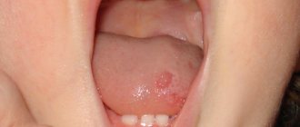
Lesquamation of the epithelium of the tongue in diseases of the gastrointestinal tract is common and is expressed in different ways. In 1932, Glessner described the so-called ulcerative tongue. At the same time, bright spots of different sizes and shapes were found on the back of the tongue, which, against the background of the coated tongue, created the appearance of superficial ulcers. Such changes occurred due to atrophy of the filiform papillae or increased desquamation. Focal desquamation has been described - limited areas along the midline of the tongue in its posterior third in the form of red spots against the background of a coated tongue. Such changes resemble rhombic glossitis, but differ from it and desquamative (geographic) tongue in that they appear during an exacerbation of gastric and duodenal ulcers. Foci of desquamation disappear during the treatment of peptic ulcer disease, are absent during remission, and do not tend to migrate.
In chronic colitis, desquamation of the tongue was found in 33% of patients, in chronic gastritis - in 17.2%. Focal desquamation of the epithelium of the tongue may not be accompanied by unpleasant sensations and patients may not be aware of it. However, more often this condition causes burning and pain in the tongue (not only in areas of epithelial desquamation), which intensifies when eating hot food or smoking.
Paresthesia of the oral mucosa or isolated tongue (glossalgia) often accompanies various diseases of the gastrointestinal tract. Often, burning, tingling, etc. can occur without visible changes in the tongue (see Glossalgia).
Significant violations of taste sensitivity in diseases of the gastrointestinal tract. To determine the taste sensitivity of the tongue, the method of functional mobility of tongue receptors is widely used.
The taste buds of the tongue perform a perceptive function and are the effector—end—link of the gastrolingual reflex. It is known that the number of functioning receptors of the tongue depends on the functional state of the digestive tract. Their maximum activity is observed on an empty stomach. After eating, demobilization of taste buds occurs (the level of mobility of taste buds decreases by 50%). This occurs as a result of centrifugal impulses from the interoreceptors of the stomach to the exteroreceptor apparatus of the tongue and is observed as a normal reaction in practically healthy people.
Changes in taste sensitivity have been found in all diseases of the gastrointestinal tract. In case of peptic ulcer, several types of functional mobility impairment have been identified [Budylina S. M., Artemyev E. N., 1970]: 1) after eating there is no demobilization of taste buds; 2) a perverted reaction is observed - an increase in the level of mobilization of taste buds after eating; 3) a reaction similar to that observed in practically healthy people. These disorders are a consequence of changes in the normal reflex connection between the receptors of the tongue and the stomach as a result of disorders of the secretory and motor functions of the stomach characteristic of peptic ulcer disease. In gastric cancer, a decrease in taste sensitivity to bitter and sweet has been described.
Morphological studies of the oral mucosa in some diseases of the gastrointestinal tract indicate degenerative changes in the nerve fibers of the tongue, which increase with the duration of the disease. In the epithelium, hyperkeratosis with desquamation of the surface layers, swelling of the underlying tissue, and sometimes ulcers were detected. Deeper morphological changes were determined in the tongue compared to other parts of the oral cavity. Their severity is directly dependent on the severity of the underlying disease. The resulting structural disturbances of the nervous apparatus of the oral cavity in gastrointestinal diseases lead to partial or complete denervation of individual areas of the oral mucosa and create the preconditions for trophic changes and ulcerative lesions of the mucous membrane.
Along with the described changes in the mouth, salivation disorders are noted: sometimes increased salivation occurs (with an exacerbation of peptic ulcer with high gastric secretion); Xerostomia is more often observed in peptic ulcers, gastritis and other diseases. Salivation disorders are usually combined with other described changes in the mucous membrane.
Thus, numerous clinical observations and experimental studies indicate frequent damage to the oral cavity due to gastrointestinal pathology. These examples confirm the presence of a neurohumoral connection between them, as well as the importance of local and general in pathology.
In the pathogenesis of changes in the oral mucosa (edema, paresthesia, desquamation of the epithelium, etc.), along with reflex and humoral mechanisms, hypovitaminosis (especially B vitamins) and increased vascular-tissue permeability are important. These provisions are confirmed by biochemical studies, as well as functional tests.
It is important to note that treatment of the underlying disease often leads to the disappearance or sharp decrease in the severity of dental manifestations and normalization of biochemical parameters. Significant changes in the oral cavity with diseases of the liver and gall bladder. With Botkin's disease and chronic hepatitis, various changes in the oral mucosa of an inflammatory-dystrophic nature are noted: hyperemia, dryness, swelling and desquamation of the epithelium of the mucous membrane, especially pronounced in the increasing phase of jaundice (Botkin's disease). In addition, yellowness of the soft palate, floor of the mouth, and the appearance of hemorrhages and telangiectasia are characteristic. Bitterness, perversion of taste (sour or metallic taste in the mouth), as well as hyperesthesia of hard dental tissues are noted.
The connection between liver diseases and manifestations in the oral cavity is confirmed by experimental studies in reproducing the model of hepatitis and hepatocholecystitis. In experimental animals, the mucous membrane became yellow, areas of hyperemia, small erosions and ulcers covered with plaque appeared.