Classification of periodontal diseases
Currently, in our country, the terminology and classification of periodontal diseases approved at the XVI Plenum of the Board of the All-Union Society of Dentists in 1983
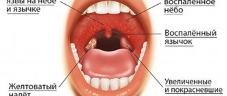
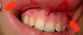
I. Gingivitis is an inflammation of the gums caused by the adverse effects of local and general factors and occurring without compromising the integrity of the dentogingival junction.
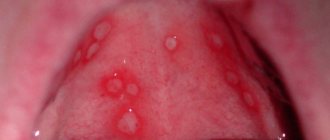
Forms: catarrhal, ulcerative, hypertrophic.
Severity: light, medium, heavy.
Course: acute, chronic, aggravated.
Prevalence: localized, generalized.
II. Periodontitis is an inflammation of periodontal tissues, characterized by progressive destruction of the periodontium and bone of the alveolar process of the jaws.
Severity: light, medium, heavy.
Course: acute, chronic, exacerbation, abscess, remission.
Prevalence: localized, generalized.
III. Periodontal disease is a dystrophic lesion of the periodontium.
Severity: light, medium, heavy.
Course: chronic, remission.
Prevalence: generalized.
IV. Idiopathic diseases with progressive lysis of periodontal tissue ( periodontolysis ) - Papillon-Lefevre syndrome, neutropenia, agammaglobulinemia, uncompensated diabetes mellitus and other diseases.
V. Periodontomas - tumors and tumor-like diseases (epulis, fibromatosis, etc.).
This classification is based on the nosological principle of systematizing diseases, approved by WHO. Currently, the nomenclature and classification of periodontal diseases adopted at the meeting of the Presidium of the Periodontology Section of the Russian Academy of Dentistry in 2001 are being introduced into clinical practice:
1. Gingivitis is inflammation of the gums caused by the adverse effects of local and general factors, which occurs without compromising the integrity of the periodontal attachment and manifestations of destructive processes in other parts of the periodontium.
Forms: catarrhal, ulcerative, hypertrophic.
Course: acute, chronic.
Phases of the process: exacerbation, remission.
Prevalence of the process: localized (focal), generalized.
Severity: - decided not to highlight. Only in relation to hypertrophic gingivitis, the degree of soft tissue growth is additionally indicated: up to 1/3, up to 1/2 and more than 1/2 of the height of the tooth crown. Additionally, the form of hypertrophy is also indicated: edematous or fibrous.
2. Periodontitis is an inflammation of periodontal tissue, characterized by destruction of the periodontal ligamentous apparatus and alveolar bone.
Course: chronic, aggressive.
Phases of the process: exacerbation (abscess formation), remission.
The severity is determined by the clinical and radiological picture. Its main criterion is the degree of destruction of the bone tissue of the alveolar process (in practice, it is determined by the depth of the periodontal pockets (PC) in mm).
Degrees of severity: mild (PC no more than 4 mm), moderate (PC 4-6 mm), severe (PC more than 6 mm).
Prevalence of the process: localized (focal), generalized.
There is an independent subgroup of periodontal diseases - aggressive forms of periodontitis (prepubertal, juvenile, rapidly progressive. The latter develops in individuals aged 17 to 35 years).
3. Periodontal disease is a degenerative process that extends to all periodontal structures.
Its distinctive feature is the absence of inflammation in the gingival margin and periodontal pockets.
Course: chronic.
Severity: light, medium, heavy (depending on the degree of exposure of the roots of the teeth) (up to 4 mm, 4-6 mm, more than 6 mm).
Prevalence is only a generalized process.
4. Syndromes manifesting in periodontal tissues.
This classification group was previously designated as idiopathic periodontal diseases with progressive bone lysis. This group includes periodontal lesions in Itsenko-Cushing, Ehlers-Danlos, Shediac-Higashi, Down syndromes, blood diseases, etc.
5. Periodontomas are tumor-like processes in the periodontium (gingival fibromatosis, periodontal cyst, eosinophilic granuloma, epulis).
Course: chronic.
Prevalence of the process: localized (focal), generalized.
Forms: distinguished only for epulis based on its histological picture.
Did you like the article? Share with friends
0
Similar articles
Next articles
- The structure of the oral mucosa
- Pathological processes in the oral cavity
- Oral injuries
- Oral burn
- Oral leukoplakia
Previous articles
- Periodontal structure
- Filling root canals using the Termafil system
- Lateral (side) condensation method
- Single pin method
- Filling with one paste
Add a comment
The relationship between periodontal diseases and occlusion pathology
E. V. Bykova assistant at the department of orthodontics of North-Western State Medical University named after. I. I. Mechnikova
P. P. Zhdanov Associate Professor of the Department of Orthodontics of Northwestern State Medical University named after. I. I. Mechnikova
The relationship between periodontal diseases and occlusion pathology is currently beyond doubt. The pathology of occlusion is accompanied by incorrect positioning of the teeth, their inclination and rotation, in which the axis of application of occlusal forces does not coincide with the axis of the tooth, and the physiological forces associated with chewing become excessive. Such forces applied to the teeth, with intact periodontal tissues, cause primary occlusal trauma.
With healthy periodontium, occlusal trauma leads to increased tooth mobility without destruction of the periodontal ligament. With damaged periodontal tissue, excessive occlusal load promotes the spread of inflammation into deeper periodontal structures, increasing the severity of periodontal processes (Klinberg I. et al., 2006; Kalamkarov H. A. et al., 1972; Goldman HM, 1956; Jin LJ et al. ., 1992).
At the same time, Geiger AM (2001) refutes the long-existing theory about the pathology of occlusion as an etiological factor in periodontal diseases. However, according to his data, pronounced jaw discrepancies and traumatic occlusion contribute to the aggravation of the pathological process in the periodontium. As dental crowding increases, the risk of developing periodontitis increases in patients over 35 years of age (Pugaca J. et al., 2007).
Physiological occlusion and correctly positioned teeth are anatomically and physiologically necessary to maintain periodontal health and prevent the development of periodontitis (Loginova N.K. et al., 1993; Popov S.A., 1999). Ramfjord SP et al. (1973) argue that with the loss of supporting teeth as a result of the formation of a deep incisal overlap, the resulting overload of this area leads to the development of periodontal diseases.
Thus, the opinions of scientists regarding the relationship between the pathology of occlusion and periodontal diseases are contradictory.
Purpose of the study: to assess the relationship between dental deformations and pathological changes in periodontal tissues.
Material and research methods
126 people (102 women and 24 men) aged 20-54 years were examined. The condition of periodontal tissues was assessed as: healthy periodontium, generalized periodontitis of mild and moderate severity. The comprehensive examination included: clinical, laboratory, morphometric, and radiological research methods.
Research results
The indicators of the recession index and the degree of gum retraction indicate (Table 1) that in patients with dentition deformations, with an increase in inflammatory manifestations in the periodontium, the prevalence and volume of recessions significantly increase. Of the total number of patients examined, 54 (42.9%) were patients with distal occlusion, 44 (34.9%) were patients with neutral occlusion, and 28 (22.2%) were patients with mesial occlusion (Table 2).
Table No. 1. Recession index (RI) and degree of retraction index (DR) of the gums in patients at the diagnostic stage (n=126)
| Sign | Intact periodontium (n=38) | HP of mild severity (n=42) | GP of moderate severity (n=46) |
| IR (%) | 0 | 13,38±5,18 | 59,75±7,05 1 |
| CP (mm) | 0 | 0,13±0,06 | 0,75±0,15 2 |
Note: 1 - t=5.22; p<0.001; 2 - t=3.71; p<0.001 (compared to the group of patients with mild periodontitis).
Table No. 2. Distribution of patients according to occlusion pathology and clinical examination data (n=126)
| Sign | Intact periodontium (n=39) | Mild GP (n=42) | Moderate GP (n=45) | Total (n=126) |
| Neutral occlusion | 15 (39,5 %) | 17 (40,5 %) | 11 (26,1 %) | 43 (34,9 %) |
| Distal occlusion | 17 (43,6 %) | 16 (38,1 %) | 22 (47,8 %) | 55 (42,9 %) |
| Mesial occlusion | 7 (16,9 %) | 9 (21,4 %) | 12 (26,1 %) | 28 (22,2 %) |
| P.I. | 0,26±0,04 | 0,38±0,07 ** | 0,38±0,05 ** | − |
| PBI | 0,49±0,09 | 0,99±0,10 ** | 1,64±0,12 *, ** | − |
| Tooth mobility | 0 | 0,36±0,08 | 0,75±0,12 * | − |
| ZDK depth | 2,14±0,14 | 2,67±0,14 * | 3,51±0,24 ** | − |
Note: 1) there is no relationship between the condition of periodontal tissues and the nature of occlusion (χ2=2.73; p>0.10); * — p<0.01 (compared to mild HP); ** — p<0.001 (compared to intact periodontium).
At the same time, distal occlusion in 47.8% of cases is combined with generalized periodontitis of moderate severity. Mild generalized periodontitis was diagnosed in 42 people (33.3% of cases), and in 17 patients it was combined with neutral occlusion, and in 16 people (38.1% of cases) it was found in combination with distal occlusion. Mesial occlusion in a greater percentage of cases is combined with generalized periodontitis of moderate severity and is observed in 26.1% of cases.
To analyze the relationship of the jaws and options for dentoalveolar compensation of occlusion, the inclination angles of the incisors of the upper and lower jaw, teleradiography of the skull was performed in a lateral projection
The diagnosis of occlusion pathology, made according to a clinical examination, does not fully reflect the discrepancy in jaw size, since dentoalveolar compensation can significantly reduce this discrepancy. It represents a change in the inclination and height of the dentoalveolar complex to achieve optimal or satisfactory occlusion and is implemented in three planes. It manifests itself most significantly in the area of the anterior teeth in the sagittal plane (Slavichek R., 2008; Sadao S., 2008). Dental alveolar compensation leads to changes in the inclination of teeth and the development of periodontal diseases.
For a more accurate analysis of the relationship of the jaws and options for dentoalveolar compensation of occlusion, the inclination angles of the incisors of the upper and lower jaw, we performed teleradiography of the skull in a lateral projection.
The relationship between the apical bases of the upper and lower jaw was determined on a teleradiogram using WITS and A-Po analysis. Of the entire group of patients examined, 64 (50.8%) patients had Engle class I, 36 (28.6%) had Engle class II, 26 (20.6%) had Engle class III. The relationship between Angle class and periodontal pathology is presented in Figure 1.
Rice. 1. Correlation between Angle class and periodontal pathology (n=126)
As can be seen from Figure 1, with intact periodontium, Engle class I occurs in 35.9% of cases, Engle class II - in 33.3%, Engle class III - only in 11.5% of cases. Generalized periodontitis of moderate severity is combined with Engle class III in 42.3% of cases.
The assessment of the inclination of the incisors of the upper and lower jaws was carried out on a teleroentgenogram in the lateral projection according to the angles: internal angle I-NL - the angle of inclination of the upper incisors to the plane of the base of the upper jaw, IMPA - the angle of inclination of the lower incisors to the plane of the base of the lower jaw. The results obtained were compared with the norm presented in the works of Proffitt U.R. (2008), Alexander R.G. (2008). A relationship has been identified between the angle of inclination of the incisors and the condition of periodontal tissues, which is presented in Table No. 3. From Table No. 3 it follows that with normal inclination, intact periodontium occurs in 48.5% of cases, protrusion of incisors in 47.8% of cases is combined with generalized periodontitis mild severity and in 34.3% - with generalized periodontitis of moderate severity.
Table No. 3. Dependence of periodontal pathology on the inclination of the maxillary incisors
| Periodontal condition | Total research | Norm | Protrusion of incisors | Retrusion of incisors |
| Intact periodontium | 38 | 16 (48,5 %) | 12 (17,9 %) | 10 (38,5 %) |
| mild GP | 42 | 9 (27,3 %) | 32 (47,8 %) | 9 (34,6 %) |
| Moderate GP | 46 | 8 (24,3 %) | 23 (34,3 %) | 7 (26,9 %) |
| Total | 126 | 33 (100 %) | 67 (100 %) | 26 (100 %) |
Note: there is a relationship between the condition of periodontal tissues and the inclination of the maxillary incisors (χ2=11.01; p=0.026).
By assessing the position of the incisors of the upper and lower jaw, we established the relationship between the inclination of the teeth and the relationship of the jaws. Thus, in Engle class I, incisor protrusion occurred in 29 (45.3%), normal position - in 21 patients (32.8%). With less frequency, normal inclination of the incisors occurred in patients with distal occlusion (22.2% of cases) and mesial occlusion (15.4% of cases) (Fig. 2).
Rice. 2. Correlation between teeth inclination (normal and protrusion) and Angle class (n=126).
An analysis was carried out of the dependence of the condition of periodontal tissues on the relationship of the jaws in the vertical and transversal planes, the position of individual teeth and groups of teeth. No dependence was found on the degree of periodontitis: on the depth of incisal overlap (?2=7.74; p>0.10), the presence of cross-occlusion (?2=1.15; p>0.10), three and crowding of teeth (?2 =5.02;p>0.10). A relationship has been found between the inclination of teeth and the degree of periodontitis.
Conclusion
As the analysis of clinical data shows, distal occlusion (47.8%) and mesial occlusion (42.9%) in a greater number of cases are combined with generalized periodontitis of moderate severity. Neutral occlusion occurs with almost the same frequency in patients with intact periodontium and generalized periodontitis of mild severity, and only in 26.1% of cases is combined with generalized periodontitis of moderate severity.
Analysis of the recession index shows the predominance of maximum prevalence values for mesial occlusion
When assessing the prevalence of periodontal diseases depending on the pathology of occlusion, it was found that the highest prevalence of generalized periodontitis of moderate severity is observed in patients with distal occlusion and occurs in 47.8% of cases. Generalized periodontitis of mild severity was combined with neutral occlusion in 40.5% of cases and with distal occlusion in 38.1% of cases.
There was no dependence of the degree of periodontitis on the depth of incisal overlap (?2=7.74; p>0.10), the presence of cross-occlusion (?2=1.15; p>0.10), three and crowding of teeth (?2= 5.02; p>0.10). Analysis of the recession index shows the predominance of maximum prevalence values with mesial occlusion. A relationship between the prevalence and degree of gum retraction and the pathology of occlusion was revealed. The recession index values are higher with mesial occlusion. The degree of gum retraction is more pronounced with distal occlusion.
- Kalamkarov Kh. A., Gantsev G. A., Ershov V. N. Relationship between dentofacial deformations and periodontopathies in children // Dentistry. - 1972. - No. 5. - P. 47-50.
- Klinberg I., Jaeger R. Occlusion and clinical practice. - M.: Medpress-inform, 2006. - 200 p.
- Loginova N.K. Functional prerequisites for the development of a mechanical theory of the etiology and pathogenesis of periodontal diseases // New in dentistry. - 1993. - No. 1. - P. 2-7.
- Popov S.A. Diagnosis and treatment of incorrect position of individual teeth in children using modern orthodontic technology: Abstract of thesis. dis. ...cand. honey. Sci. - St. Petersburg, 1999. - 22 p.
- Sato S., Akimoto S., Matsumoto A., Shirasu A., Yoshida Y. Guidelines for the clinical application of the IVD technique. — M.: Azbuka, 2008.—158 p.
A complete list of references is in the editorial office