Examination of the oral mucosa
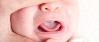
Inspection of the oral mucosa and periodontal tissue begins from the vestibule. Pay attention to the condition of the frenulum of the upper and lower lips, tongue, and the depth of the vestibule of the oral cavity. To determine the depth of the vestibule of the oral cavity, using a graduated trowel or periodontal probe, measure the distance from the gingival margin to the level of the transitional fold. The vestibule of the oral cavity is considered shallow if its depth is less than 5 mm, deep - more than 10 mm. The frenulum of the upper lip is attached 2-3 mm above the base of the interdental papilla between the central incisors of the upper jaw. The frenulum of the lower lip is attached 2-3 mm below the base of the interdental papilla between the central lower incisors. The frenulum of the tongue is attached behind the Wharton's ducts on the floor of the mouth and to the lower surface of the tongue, departing from the tip by 1/3 of the length of its lower surface. When the frenulum of the upper lip is shortened, it is determined to be short and thick, woven into the gum in the interdental space between the central teeth. The attachment of the frenulum of the lower lip is considered abnormal if, when the lip is pulled back, the interdental papilla and the gingival margin at the attachment site turn pale and separate from the teeth.
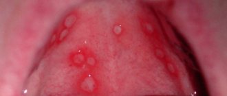
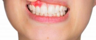
When examining the oral mucosa, it is necessary to pay attention to the presence of bad breath, the nature of salivation (increased, decreased), bleeding of the gingival margin. The purpose of the examination is to determine whether the mucous membrane is healthy or pathologically altered. A healthy mucous membrane of the oral cavity has a pale pink color (more intense in the area of the cheeks, lips, transitional folds and paler on the gums), is well moisturized, there is no swelling or rash elements.
In case of diseases of the oral mucosa, it becomes hyperemic, swollen, bleeding and elements of rashes may appear, which indicates its involvement in the inflammatory process.
A visual examination allows you to roughly assess the condition of the gums. The gingival papillae in the area of single-rooted teeth have a triangular shape, and in the area of molars they are closer to trapezoidal. The gum color is normally pale pink, shiny, and moist. Hyperemia, swelling of the mucous membrane, bleeding indicate its damage.
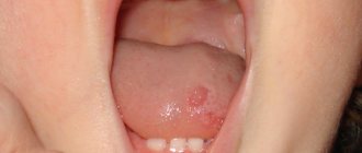
Among the elements of the lesion, there are primary and secondary ones, which arise at the site of the primary ones. The primary elements of the lesion include a spot, nodule, tubercle, node, vesicle, pustule, vesicle, blister, cyst. Secondary elements - erosion, ulcer, crack, crust (found on the red border of the lips), scale, scar, pigmentation.
Atrophy of the gingival margin, hypertrophy of the gingival papillae, cyanosis, hyperemia, bleeding of the papillae, the presence of a periodontal pocket, supra- and subgingival tartar, and tooth mobility indicate a pathological condition of the periodontium. Among periodontal diseases, inflammatory processes are of greatest importance, which are divided into 2 large groups: gingivitis and periodontitis.
Dental examination
When examining the oral cavity, it is necessary to examine all teeth, and not just the one that, in the patient’s opinion, is the cause of pain or discomfort. Violation of this rule may lead to the fact that the cause of the patient’s anxiety will not be detected on the first visit, since the pain may radiate. An examination of all teeth on the first visit is also necessary in order to outline a treatment plan that ends with oral sanitation.
Sanitation of the oral cavity is mandatory when visiting a dentist.
It is important that during the examination all changes in the tooth tissue are detected. For this purpose, a specific inspection system is recommended. For example, the examination should always be done from right to left, starting with the upper teeth (molars), and then from left to right to examine the lower teeth.
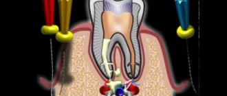
When examining teeth, they use a set of instruments (Fig. 4.12), most often they use a dental mirror and a probe (necessarily sharp). The mirror allows you to examine poorly accessible areas and direct a beam of light to the desired area, and the probe checks all the depressions, pigmented areas, etc.
If the integrity of the enamel is not broken, then the probe glides freely over the surface of the tooth without getting stuck in the recesses and folds of the enamel. If there is a carious cavity in the tooth (invisible to the eye), a sharp probe is retained in it. You should especially carefully examine the contact surfaces of the teeth, since it can be difficult to detect an existing cavity with an intact chewing surface. Such a cavity can be detected by probing. Currently, the technique of transilluminating tooth tissue by supplying light through special light guides is being used. Probing helps determine the presence of softened dentin, the depth of the carious cavity, its connection with the tooth cavity, the location of the canal mouths, and the presence of pulp in them.
Tooth color can be important in making a diagnosis. Typically, teeth are white with many shades (from yellow to bluish). Regardless of the shade, the enamel of healthy teeth is characterized by a special transparency - a vibrant shine of the enamel. In a number of conditions, the enamel loses its characteristic shine and becomes dull. Thus, the beginning of the carious process is a change in the color of the enamel: the appearance first of cloudiness, and then of a white carious spot. The enamel of pulpless teeth loses its usual shine and acquires a grayish tint. A similar color change, and sometimes even more intense, is characteristic of teeth in which pulp necrosis has occurred.
Tooth color can change under the influence of external factors: smoking (dark brown color), metal fillings (dark color), chemical treatment of canals (orange color after using the resorcinol-formalin method).
Pay attention to the shape and size of the teeth . Deviation from the usual form is due to treatment or an abnormality. It is known that some forms of dental anomalies (Hutchinson's, Fournier's teeth) are characteristic of certain diseases.
Did you like the article? Share with friends
0
Similar articles
Next articles
- Percussion, palpation and temperature diagnostics
- Electroodontodiagnosis
- X-ray examination of teeth
- Luminescent diagnosis of caries
- Functional tests
Previous articles
- Examination of the oral cavity itself
- Oral examination
- External examination of a dental patient
- Anesthesia technique
- Composition of anesthetics. Carpule anesthesia kit
Add a comment
Oral examination
When examining the oral cavity, the condition of the teeth (number, missing teeth, dentures, carious teeth), gums (coloring, plaque, ulceration - aphthae, bleeding), tonsils (shape, coloring, presence of plaque), and tongue are assessed.
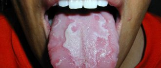
When examining the tongue, pay attention to its color, the presence of plaque, and the severity of the papillary pattern. In a healthy person, the tongue is pink, there are no coatings on it, and it is quite moist.
Dryness of the tongue with the presence of cracks and a dark brown coating is observed with inflammation of the peritoneum (peritonitis), renal failure, severe intoxication, and dehydration.
The coating of the tongue , mainly its root, with a white, sometimes grayish-white, brownish coating is observed in diseases of the gastrointestinal tract, febrile conditions, some infectious diseases, and constipation.
A crimson (the color of a “cardinal’s robe”) tongue is observed in liver diseases.
A “varnished” tongue with a bright red shiny surface, caused by atrophy of the papillae, can be seen in patients with stomach cancer, chronic colitis, pellagra, and with pernicious (B 12-deficiency) Addison-Birmer anemia.
“Geographical” tongue (desquamative glossitis) is characterized by alternating foci of epithelial desquamation with foci of local thickening. It is observed in patients with exudative diathesis and group B vitamin deficiency.
Examination of the patient's body odors
When examining a patient, the smell of acetone, urine, a sweetish characteristic odor of liver, rotten eggs, a putrid fetid odor, a sourish odor emanating from his body may be detected.
The sweetish smell of acetone ( the smell of rotten apples ) is experienced by patients with diabetes mellitus who are in a precomatous and comatose state.
The smell of urine ( urinary odor ) is observed in patients with end-stage renal failure and uremic coma.
The sweetish smell of a peculiar specific shade is inherent in patients with liver diseases who are in a comatose state.
The smell of hydrogen sulfide ( the smell of rotten eggs ) is usually observed in patients with narrowing (stenosis) of the pylorus, when belching.
Bad breath ( foetur ex ore ) occurs in the presence of carious (decaying) teeth, decomposition of plaque on the tongue, purulent diseases of the tonsils, some diseases of the stomach (stomach cancer with decay, phlegmonous gastritis), cancer of the esophagus, diverticula of the esophagus.
A fetid ( sweetish-putrid ) odor is observed in patients with pulmonary gangrene, which makes it possible to make a diagnosis upon entering the ward. The same smell is present in patients suffering from fetid runny nose (ozena).
Patients with diseases accompanied by increased sweating, as well as some patients with tuberculosis, have a sour smell of sweat
Examination of eyes and eyelids
When examining the eyes, the eyelids, orbital fissures, eyeballs, conjunctiva, cornea, and pupils are examined.
Swelling of the eyelids is observed in diseases of the kidneys, heart, myxedema, and coughing attacks. Swelling of the eyelids can appear in women during menstruation, puffiness - as a result of sleepless nights, with trichinosis, and nutritional dystrophy.
Drooping of one eyelid (ptosis) is often observed as a result of hemorrhage in the brain, with cerebral syphilis.
The appearance of dark coloring of the eyelids is characteristic of adrenal insufficiency and increased thyroid function.
Blueness under the eyes (periorbital cyanosis) is a symptom of fatigue, can be due to venous stagnation, decreased venous tone, increased intracranial pressure.
Protruding eyes (exophthalmos) are observed in diseases of the thyroid gland (thyrotoxicosis), some brain tumors, and severe myopia.
Recession of the eyeballs (enophthalmos) is observed with myxedema, significant loss of large amounts of fluid by the patient’s body, inflammation of the peritoneum, and also with agonal conditions.
Unilateral retraction of the eye with simultaneous narrowing of the palpebral fissure of the pupil and drooping of the upper eyelid (Horner's syndrome) can be observed due to compression of the sympathetic nerve, its cervical part, a tumor of the mediastinum, or an aortic aneurysm.
A wide palpebral fissure with rare blinking (Stellwag's symptom) is observed in thyrotoxicosis (Graves' disease).
When examining the pupils, pay attention to their shape, uniformity, reaction to light, and accommodation.
Constriction of the pupil ( miosis ) is observed in renal failure (uremia), tumors and inflammatory processes of the brain, in case of poisoning with morphine drugs and intoxication (nicotine), in patients with glaucoma (suffering from increased intraocular pressure), who regularly instill pilocarpine, with tabes dorsalis (usually uneven ).
Dilation of the pupils ( mydriasis ) occurs in agonal, comatose states (except uremic), cerebral hemorrhage, poisoning with atropine and its derivatives, and less often in very severe pain and helminthic infestations.
Uneven dilation of the pupils ( anisocoria ) - with damage to the nervous system, migraine.
Pupil pulsation - rhythmic constriction and dilation of the pupil, synchronously coinciding with heart contractions, is observed with aortic valve insufficiency.
Pupil reaction to light
is detected as follows: one eye of the patient is covered with a hand. After removing the hand, when light rays enter the eye, the pupil constricts. This indicates that the pupil's reaction to light is preserved.
A change in the reaction of the pupil to light is observed in cases of poisoning with morphine drugs, poisoning with chloroform, atropine, various comatose states, and brain diseases. In these cases, the reaction of the pupils to light disappears.
Yellowish rings around the cornea appear due to lipid metabolism disorders, atherosclerosis, and diabetes mellitus.
The presence of a greenish-brown Kayser-Fleischer ring along the periphery of the cornea is characteristic of Konovalov-Wilson disease - a hereditary disease characterized by a decrease in the synthesis of ceruloplasmin (copper transport protein) in the liver and its deposition in tissues.
Head and neck examination
Examination of the head reveals changes in its movement, shape and size.
Rocking the head back and forth synchronously with the heart rhythm is a symptom of Musset . This symptom is observed with aortic valve insufficiency. Its appearance is due to the hemodynamic characteristics of this defect.
Involuntary movements of the head in the form of “ shaking ” - in Parkinson’s disease and in old age, as well as in chorea.
Abnormally large head sizes are observed with water on the brain (hydrocephalus). Abnormally small head sizes (microcephaly) are observed with congenital developmental disorders, usually combined with mental retardation (oligophrenia).
A square head shape with a flattened upper part and prominent frontal tubercles is observed in patients who suffered from rickets in early childhood or who suffer from congenital syphilis.
The so-called “tower” skull , narrow and high, is usually combined with congenital hemolytic jaundice. Observed in mental retardation.
Examination of the neck area reveals:
a) characteristic deformation in the anterior part of the neck associated with an enlargement of the thyroid gland and cervical lymph nodes;
b) pronounced pulsation of the carotid arteries (“carotid dance”) with aortic valve insufficiency;
c) pulsation and swelling of the jugular veins, the so-called positive venous pulse, detected with tricuspid valve insufficiency.
Severe swelling of the face, neck and upper half of the chest, reminiscent of a “dape” ( Stokes collar ), is observed with effusion of pericardium, as well as tumors of the mediastinum.
Scars after opened lymph nodes ( scrofuloderma ) are detected in patients with the glandular form of tuberculosis
Examination of hands and feet
Changes in the shape and size of the terminal phalanges of the hands with their thickening in the form of “drumsticks” are observed in chronic suppurative lung diseases, prolonged septic endocarditis, congenital heart defects, cirrhosis of the liver, peripheral lung cancer, due to the state of chronic tissue hypoxia. Often, deformation of the terminal phalanges of the fingers is combined with a characteristic deformation of the nails in the form of “hour glasses” (convex upward) .
Thickenings at the ends of the phalanges of the hands in the form of small nodules ( Heberden's nodules ) are more often observed in elderly patients suffering from the so-called metabolic-dystrophic polyarthritis (gout).
A pronounced deformation of the hands with atrophy of the interosseous muscles and the fingers moving outward, reminiscent of “seal flippers ,” is observed in patients suffering from rheumatoid arthritis.
Pointed and shortened terminal phalanges of the hands, pulled inwards (the so-called “clawed paw” ) are observed in patients with systemic scleroderma.
A significant disproportionate increase in size of the hands and feet is typical for patients with acromegaly.
Hyperemia of the palms of the hands, especially in the thenar and hypothenar areas, due to the expansion of small vessels of the skin ( “liver palms” ) is detected in cirrhosis of the liver and active hepatitis.
Examination methods in dentistry
The first stage of a dentist’s work is the collection of factual material, i.e., identifying the symptoms of a disease state. The development of science contributes
the emergence of new methods for diagnosing the condition of hard dental tissues and periodontal tissues. In this regard, it is important to formulate a procedure for examining the patient in which all factors contributing to the development of oral diseases will be taken into account and recorded. The final diagnosis depends on how complete and objective the information received is.
Examination methods are divided into basic (clinical) and additional (instrumental and laboratory).
When examining a patient, there is a certain order. Clinical examination consists of the following stages:
Interview: collection of complaints, collection of life history, collection of medical history.
Objective research:
examination of the skin of the maxillofacial area, examination of the vestibule of the oral cavity,
examination of the oral cavity itself.