CHANGES IN THE ORAL MUCOSA WITH DERMATOSES. Lichen planus
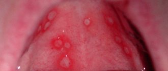
• Lichen planus (lichen ruber planus) is a chronic inflammatory disease of the skin and mucous membranes, manifested by the formation of keratinized papules. This disease is more common in women aged 40-60 years. With lichen planus, along with skin lesions, changes in the oral mucosa and the red border of the lips are often observed. Isolated damage to the oral mucosa is quite common. According to E.I. Abramova, simultaneous lesions of the skin and oral mucosa were noted in 25% of patients, and isolated localization on the oral mucosa without skin lesions was found in 75% of patients. Lichen planus is also localized on other mucous membranes: genital organs, anus, conjunctiva, esophagus, stomach, urethra. Etiology. It is not completely clear. There are non-virogenic, viral, toxic-allergic theories of the occurrence of this disease. Lichen planus, as a rule, occurs against the background of chronic diseases of the gastrointestinal tract, hypertension, diabetes mellitus, neuroses, imbalance of the immune system, etc. In most patients, a significant increase in the permeability of the vascular walls and a decrease in immunological reactivity were revealed. Trauma (sharp edges of teeth, poor-quality dentures, galvanism phenomena, allergic reaction to the plastic of dentures, etc.) has a certain significance in the development of lichen planus of the oral mucosa. Clinical picture. The main morphological element of the lesion is a keratinized papule of round or polygonal shape up to 2 mm in diameter. On the skin, papules are usually flat, with a waxy sheen, and have a pinkish or bluish-red color. On the oral mucosa, due to keratinization of the epithelium and constant maceration, they acquire a whitish-gray color, standing out against the background of normal or hyperemic mucous membrane. A characteristic feature of lichen planus is the tendency of papules to merge in the form of a pattern resembling a lace mesh, snowflakes, tree-like branches, sometimes rings, stripes. Papules slightly rise above the level of the mucous membrane, giving it roughness. On the dorsum and lateral surface of the tongue, papules, merging, often form hyperkeratic plaques of various sizes, reminiscent of leukoplakia; the papillae in this area are smoothed. In smokers, the papules are more pronounced and larger; leukoplakia spots are often layered on them. On the red border of the lips, papules can merge, forming a whitish stripe, sometimes taking a star-shaped shape. Lichen planus on the vermilion border and mucous membrane of the lips often leads to glandular cheilitis. The most typical localization of lichen planus is on the mucous membrane of the cheeks at the junction of the large molars with the capture of transitional folds, on the lateral surfaces of the tongue and the back with a transition to the lower surface in the area of the large molars. Less commonly affected are the lips, gums, palate, and floor of the mouth. Due to the variety of clinical manifestations of lichen planus in the oral cavity, the following forms are distinguished: typical (simple), exudative-hyperemic, erosive-ulcerative, bullous, hyperkeratotic. Typical form. It occurs more often than others. Whitish-pearly papules are located separately or in the form of patterns, lace, fern leaves, rings, stripes on the apparently unchanged oral mucosa (Fig. 11.39). With such a typical picture of lichen planus, subjective sensations are minimally expressed and can be manifested by a feeling of burning, tightness, roughness, and dryness of the oral mucosa. Quite often, the disease is asymptomatic and can be detected accidentally during a dental examination.
Rice. 11.39. Typical form of lichen planus. Multiple papules forming a pattern on the buccal mucosa.
Exudative-hyperemic form. Less common. Papules are located on the hyperemic, edematous mucous membrane (Fig. 11.40). This form is accompanied by more pronounced pain, burning, pain, which intensifies when eating spicy food or talking. Against the background of an inflamed, hyperemic mucous membrane, the pattern of papules may lose the clarity of their outlines and even partially disappear. But in the process of reverse development, when the swelling and hyperemia of the mucous membrane decrease, the pattern of papules reappears.
Rice. 11.40. Exudative-hyperemic form of lichen planus. Against the background of severe hyperemia of the gum mucosa, multiple papules.
Erosive-ulcerative form. The heaviest of all forms. It can occur as a complication of the typical and exudative-hyperemic forms as a result of erosion of the area of the affected mucous membrane by various traumatic factors (sharp edges of teeth, dentures, galvanism, etc.). In this form, on the hyperemic and edematous mucous membrane of the mouth there are erosions, sometimes ulcers, around which, against the background of pronounced inflammation, papules typical of lichen planus are located in a pattern (Fig. 11.41). Erosions or ulcers of irregular shape are covered with fibrinous plaque, after removal of which bleeding easily occurs. They can be single, small, slightly painful, but they can also be multiple with pronounced pain. Such erosions and ulcers last a long time, sometimes for months, even years, without epithelialization. Often, under the influence of treatment, they partially or completely epithelialize, but soon recur again in the same or another area of the mucous membrane, sometimes even immediately after cessation of treatment. Sometimes, at the site of long-term erosions and ulcers, areas of mucosal atrophy appear. In some cases, long-term erosions and ulcers can become malignant.
Rice. 11.41. Erosive-ulcerative form of lichen planus. Erosion on the hyperemic mucous membrane of the cheek along the line of closure of the teeth, along the periphery of which papules are visible, forming a patterned pattern.
Bullous form. Rarely seen. It is characterized, along with the typical rashes of whitish-pearly papules, by the appearance of blisters with a diameter of 1 to 10 mm. The blisters may contain serous or hemorrhagic contents and open quite quickly. Their lifespan ranges from several hours to 2 days. The erosions that form at the site of the blisters quickly epithelialize, which distinguishes the bullous form of lichen planus from the erosive-ulcerative form. The duration of the bullous form can vary, sometimes blisters can appear for many months. Blisters may appear on the oral mucosa simultaneously with papules or join them later. Sometimes they precede the appearance of papules, which creates difficulties in diagnosing lichen planus. Hyperkeratotic form. It is observed very rarely. It is characterized by the presence of hyperkeratotic plaques of various shapes and outlines, rising above the level of the mucous membrane with sharp boundaries (Fig. 11.42). Around the foci of hyperkeratosis there are papular rashes, typical of lichen planus. Most often, this form is localized on the mucous membrane of the cheeks and the back of the tongue. The described forms of lichen planus can transform one into another. So, as a result of complications, the typical form can turn into exudative-hyperemic and erosive-ulcerative. The transformation process is determined by the influence of general (somatic diseases) and local factors. The presence of sharp edges of teeth and dentures, dissimilar metals, dentoalveolar anomalies and deformations, untreated periodontitis, and tartar contribute to the transition of lichen planus from a typical form to a more severe one.
Rice. 11.42. Hyperkeratotic form of lichen planus. An extensive hyperkeratotic plaque on the dorsum of the tongue, around which there are multiple papules.
Lichen planus is a chronic disease characterized by a persistent long-term course. It can last for decades with alternating exacerbations and remissions, the duration of which is influenced by severe general diseases and the presence of local traumatic factors in the oral cavity. Lichen planus on the oral mucosa can become malignant. More often this occurs in elderly people who have suffered for a long time from erosive-ulcerative or hyperkeratotic forms. Signs of malignancy of lichen planus are the formation of a compaction at the base of the lesion, increased keratinization processes, and the appearance of vegetation on the surface of long-term non-healing and untreatable erosions and ulcers. Pathological anatomy. Pathohistological changes in the epithelium are characterized by the presence of hyper- and para-keratosis, as well as acanthosis. In some cases, the formation of a granular layer is detected. In the stroma, edema and a diffuse inflammatory infiltrate (mainly from lymphocytes and plasma cells) are detected, the cells of which penetrate through the basement membrane into the epithelium (exocytosis), as a result of which it is not clearly distinguishable. The infiltrate almost never penetrates into the deeper layers and the lamina propria of the mucous membrane; it comes close to the epithelium, as if supporting it from below. Depending on the type of lesion, some differences are determined. With erosive-ulcerative lesions, in addition to the phenomena of hyper- and parakeratosis, ulceration and destruction of the epithelium, hemorrhages in the mucous layer itself are noted. The infiltrate is determined directly under the basal membrane. Bullous lesions develop beneath the epithelium as a result of extensive swelling in the connective tissue. The blisters are located subepithelially, beneath them there is a massive round cell infiltration into the lamina propria of the mucous membrane. Changes in the epithelium characteristic of lichen planus in erosive-ulcerative and bullous forms are determined in the areas bordering the defect. Diagnostics. In typical cases, especially in the presence of lesions on the skin, the diagnosis of lichen planus is not difficult. It can be more difficult to make a correct diagnosis with isolated lesions of the oral mucosa. Differential diagnosis. Differential diagnosis of lichen planus should be carried out with: leukoplakia; ? candidiasis; And lupus erythematosus; ? papular syphilis; And allergic stomatitis; A chronic injury; And Bowen's disease. In erosive leukoplakia, in contrast to lichen planus, the keratinization area has the appearance of a solid grayish-white plaque slightly rising above the level of the mucous membrane, around which there is no inflammation and no papules. Unlike lichen planus, leukoplakia is localized mainly in the anterior parts of the oral cavity, most often in the mucous membrane of the corners of the mouth and cheeks. Differential diagnosis with candidiasis is based on differences in clinical manifestations. With candidiasis there is no clear pattern of papules; when scraped, the white plaque is removed partially or completely, but the papules of lichen planus are not removed. The final diagnosis is made on the basis of bacterioscopic examination data. Lesions on the oral mucosa in lupus erythematosus are localized mainly on the lips, palate, and less often on the cheeks. They are hyperemic, infiltrated, hyperkeratosis is present only within the focus of inflammation, in the center of which there is atrophy, which does not happen with lichen planus. Syphilitic papules are significantly larger in size and have a round or oval shape. Their surface is covered with a grayish-white coating, which is easily removed by scraping. Treponema pallidums are found in smears from the surface of exposed erosion. Lichen planus is differentiated from allergic stomatitis on the basis of medical history, the disappearance of the lesion after eliminating the action of the allergen, and the results of allergy tests. It is necessary to distinguish lesions of the oral mucosa in lichen planus from similar changes under the influence of local traumatic factors, in which the affected area corresponds to the action of the irritant, after which the source of inflammation disappears. The bullous and erosive-ulcerative form of lichen planus must also be differentiated from other diseases in which there are blisters and erosions. These are exudative erythema multiforme, acantholytic and non-acantholytic pemphigus, herpes simplex, erosive-ulcerative form of chronic lupus erythematosus. The hyperkeratotic form must be distinguished from verrucous leukoplakia and cancer. Treatment. Patients with lichen planus should undergo a thorough examination to identify somatic diseases. Particular attention is paid to the state of the gastrointestinal tract, nervous system, blood pressure and blood glucose levels are determined. If a pathology is detected, the patient is subject to treatment by appropriate specialists. All patients with lichen planus undergo thorough sanitation of the oral cavity, which the dentist begins with the elimination of traumatic factors. Sharp edges of teeth and dentures must be ground down. If there are dissimilar metals in the oral cavity, it is necessary to eliminate them or at least one of the dissimilar metals. Removable plate dentures should be made of colorless plastic with an internal elastic layer. It is advisable to replace amalgam fillings with cement ones, especially if their placement coincides with the period of appearance of lichen planus. It is recommended to stop smoking and eat irritating foods, and maintain good oral hygiene. Drug therapy for lichen planus depends on its form. For all forms of the disease, it is advisable to prescribe sedatives (valerian, bromine preparations, tranquilizers). For typical and exudative-hyperemic forms, vitamin A is prescribed orally (3.44% solution of retinol acetate in oil or 5.5% solution of retinol palmitate in oil) 10 drops 3 times a day with meals, in courses of 1 .5-2 months with 2-month breaks. In addition, long-term intake of B vitamins, especially nicotinic acid, is prescribed. At the same time, an oil solution of vitamin A is used for applications to the oral mucosa in the affected area. For exudative-hyperemic and erosive-ulcerative forms, the use of 1% nicotinic acid is effective, which is injected under the lesions in 1 ml along with a 1% solution of novocaine or trimecaine every other day, for a course of 15-20 injections. If the injections are poorly tolerated by the patient, nicotinic acid is prescribed orally at 0.05-0.1 g 3 times a day after meals, and a novocaine (trimecaine) blockade is given under the lesions 2-3 times a week, for a course of 10- 12 injections. A good effect is obtained by prescribing tigazon (Tigason) 10 mg 3 times a day, taken with meals and washed down with milk. After 2-4 weeks of treatment, if well tolerated, the dose can be increased: 25 mg 3 times a day. The entire course of treatment lasts 6-8 weeks. In case of relapses, repeat courses of treatment are carried out. For the treatment of the erosive and ulcerative form of lichen planus, honsurid is used, which is used in the form of applications 2-3 times a day or injections of 1 ml under the lesion every other day (0.1 g of honsurid is diluted in 10 ml of a 0.5% solution novocaine or isotonic sodium chloride solution). Khonsurid stimulates the process of epithelization of erosions and ulcers and has an anti-inflammatory effect. In cases of severe inflammation of the oral mucosa, erosions or ulcers, anti-inflammatory rinses (chamomile solution, 0.5% chloramine solution, 0.02% furatsilin solution, etc.) are applied topically, followed by applications of vitamin A oil solutions to the lesions. , E, ointments "Solcoseryl", "Ak-tovegin", corticosteroid-containing, 1% dibunol ointment. To increase the resistance of the oral mucosa and stimulate its regeneration, applications or oral baths with a 0.25% lysozyme solution are used. Treatment of erosive-ulcerative and bullous forms at the Department of Skin Diseases of the Moscow Medical Institute is carried out using a combined method: prednisolone 20-25 mg per day every other day (triamcinolone 16-20 mg, dexamethazone 3-3.5 mg), hingamine (delagil) 0.25 g 1-2 times a day for 4-6 weeks and nicotinic acid (0.05 g 3 times a day after meals) or xanthinol nicotinate (com-plamin, teonicol) 1 tablet 3 times a day or intramuscularly for 1.5 months. The dose of prednisolone is reduced by 5 mg every 7-10 days. Such treatment, in the absence of contraindications, accelerates the epithelization of erosions and ulcers and reduces the inflammatory process. You can treat with only one of these drugs (prednisolone or delagil), but it will be less effective. In the presence of limited erosive and ulcerative lesions, it is effective to inject them with a suspension of hydrocortisone (or a solution of prednisolone) and a 5-10% solution of hingamine. Injections are carried out once every 3 days, 1 - 1.5 ml for each erosion, for a course of 8-12 injections. Similar courses can be repeated after 3-4 months. When treating with corticosteroid drugs, their possible side effects should be taken into account, and therefore during treatment it is necessary to conduct a blood test (general clinical and glucose), measure blood pressure, prescribe potassium, calcium, multivitamins, and recommend a diet with a reduced amount of table salt. If there are contraindications to the use of corticosteroids, then for the erosive-ulcerative form of lichen planus, it is recommended to carry out hypo-sensitizing therapy with repeated courses of hist-globin (several courses with a two-month break; 2 ml under the skin 2 times a week, for a course of 8 -10 injections). It is necessary to remember about the possibility of malignancy of long-term and untreatable erosions, ulcers, and foci of hyperkeratosis. In these cases, their excision followed by histological examination is necessary. Cryodestruction gives good results. Lichen planus is a disease that does not always respond well to treatment. Relapses often occur again after stopping treatment. Crucial importance in their prevention belongs to the exclusion of local traumatic factors and the successful treatment of somatic diseases. In order to increase the body's defenses, repeated courses of vitamin therapy and auto-hemotherapy are carried out; pyrogenal, prodigiosan, thymogen and other drugs are used in a hospital setting.