Signs and symptoms of scarlet fever in children and adults
Incubation period
The incubation period for scarlet fever is most often 1 - 3 days, but sometimes there are fluctuations within 1 - 12 days.
Signs and symptoms of scarlet fever in the initial period of the disease
The disease begins acutely, suddenly, with a sharp increase in body temperature. There is pain when swallowing, headache and single vomiting. And after a few hours, a rash typical of the disease appears on the face, torso and limbs.
Fever with scarlet fever
Body temperature rises to 38 - 39oC for several hours. The more severe scarlet fever occurs, the longer the febrile period. With a mild course, it lasts no more than 2 - 3 days. In severe cases - 9 days or more.
Symptoms of intoxication with scarlet fever
Symptoms of intoxication appear suddenly and include chills, weakness, headache and loss of appetite. Sometimes nausea and vomiting occur. The child becomes lethargic and drowsy. In severe cases, seizures, delirium and meningeal symptoms appear. With a mild form of scarlet fever in children, body temperature rises moderately, and signs of intoxication are mild.
Signs and symptoms of oropharyngeal lesions in scarlet fever
Angina
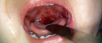
Simultaneously with the appearance of symptoms of toxicosis, damage to the oropharynx develops. On the first day of the disease, hyperemia of the pharynx appears. Necrotizing tonsillitis develops on days 2–4. There is swelling of the follicles (places where lymphocytes accumulate) in the area of the soft palate. They look like bright red tubercles up to 1.5 mm in diameter. Very quickly, the redness of the tubercles merges into continuous hyperemia. Hyperemia of the pharynx has clear boundaries and is called “flaming pharynx.” The mucous membrane of the hard palate is not affected by the inflammatory process.
Sore throat with scarlet fever is a typical symptom of the disease. Toxigenic streptococci on the surface of the tonsils multiply quickly, damaging the tissue. In response to this, inflammation occurs, the signs of which are swelling and redness caused by dilation of the capillaries, and pain caused by compression of the nerve endings by the swollen tissues.
In dilated capillaries, blood flow sharply slows down, which causes the formation of microthrombi. Areas with impaired blood supply become necrotic. The palatine tonsils are covered with grayish-dirty films, which are easily removed with a spatula. There is an unpleasant odor from the mouth.
With necrotizing tonsillitis, the inflammatory process can spread deep into the tissues, affecting the floor of the mouth, arches and soft palate. Angina can acquire a gangrenous-hemorrhagic form.
Catarrhal symptoms disappear after 5 days. Necrosis disappears slowly, within 8 - 10 days.
Rice. 2. The photo shows a sore throat due to scarlet fever. When the disease occurs, the pharynx acquires a bright red color (“flaming pharynx”). Necrotizing tonsillitis develops on days 2–4. The palatine tonsils are covered with grayish-dirty films.
"Raspberry Tongue"
On the first day of the disease, the patient's tongue becomes dry. A grayish or brown coating quickly appears on it. On days 2-3, the tongue begins to clear. Cleansing begins from the tip of the tongue and sides. The cleaned surface acquires a bright red color (“crimson tongue”). Swollen papillae protrude on the surface of the tongue. The symptom of a “scarlet fever” tongue clearly manifests itself on the 3rd – 5th day of the disease. Further, the intensity of its color weakens and disappears on the 7th - 10th day of the disease. Enlarged papillae persist for up to 2–3 weeks of illness.
Rice. 3. In the photo, the symptoms of scarlet fever in children are “crimson tongue” and enlarged papillae.
Scarlet fever rash
- Scarlet fever rash is the result of streptotoxicosis. When exposed to streptococcal toxins, small blood vessels of the skin dilate, the upper layers of the dermis become inflamed, followed by the development of necrotic processes. The rash characteristic of scarlet fever appears 6 to 12 hours after the onset of the disease.
- The rash is pinpoint, located on a hyperemic background, which creates a picture of erythema. The rash covers the entire body except for the area of the nasolabial triangle, and thickens in the area of natural folds of the skin. When you press the area of hyperemia with your palm, the rash temporarily disappears. Initially, the rash appears on the skin of the face, neck and upper half of the torso. Next, the rash quickly spreads to the flexor surfaces of the limbs, the lateral surfaces of the chest and abdomen, and the inner surface of the thighs. Stripes of a dark red color are often observed in the folds of the skin, which occurs due to the impregnation of areas of the skin with blood.
- The rash on the face is concentrated in the cheek area. Their flaming color contrasts sharply with the nasolabial triangle, which is pale in color (Filatov's triangle).
- The more severe scarlet fever occurs, the more intense the rash. In severe toxic forms of the disease, the rash often becomes hemorrhagic. Increased permeability of skin capillaries is manifested by petechial hemorrhages in the area of articular bends and places of friction and compression of the skin. The symptom of a tourniquet becomes positive when petechiae appear after a short compression of the skin in the shoulder area with a tourniquet. The rash with scarlet fever is often accompanied by scratching.
- The rash usually lasts 3 to 7 days. Then it disappears and peeling of the skin begins. Peeling of the skin begins in the area of the earlobes and neck. Peeling in the form of thin scales is observed on the face. Pityriasis-like peeling is observed on the skin of the trunk, neck and ears. Lamellar peeling - on the soles and palms. The rash does not leave pigmentation behind.
- The rash with scarlet fever may be atypical. Sometimes small blisters filled with cloudy liquid appear on the skin. In severe forms of scarlet fever, the rash can be maculopapular in nature and take on a cyanotic hue.
- The disease causes white dermographism, the formation of which is associated with spasm of the subcutaneous vessels. After passing a blunt object over the skin, a mark appears in the form of a white swelling.
Rice. 4. The photo shows a scarlet fever rash. The more severe the disease, the more intense the rash.
Lymph nodes in scarlet fever
Lymph nodes always enlarge during illness. They have a dense consistency and are painful on palpation. First of all, the paratonsillar and anterior cervical lymph nodes enlarge. The inflammation is necrotic in nature. Molten purulent masses in the lymph nodes can capture the gland capsule and fiber, with the subsequent development of adenophlegmon of the neck, sinusitis, inflammation of the middle ear and cellular structures of the mastoid process.
Rice. 5. The rash usually lasts 3 - 7 days. Then it disappears and peeling of the skin begins.
Changes in the cardiovascular system
In the first stages of scarlet fever, the tone of the sympathetic nervous system predominates, which is manifested by tachycardia and increased blood pressure. After 4 - 5 days, the tone of the parasympathetic nervous system begins to predominate, which is manifested by bradycardia and a decrease in blood pressure. During this period, heart sounds become muffled and the boundaries of the heart expand. These changes occur as a result of exposure to streptococcal toxins, last 2-4 weeks and disappear without a trace. The pathology was first described by the Russian doctor N.F. Filatov and was called “scarlet heart.”
Rice. 6. One of the symptoms of scarlet fever is peeling of the skin. Lamellar peeling is observed on the soles and hands.
Signs and symptoms of extrabuccal scarlet fever
When toxicogenic streptococci penetrate the human body through a wound or burn surface, through damage to the genital organs, an extrabuccal form of scarlet fever develops. After a short incubation period, a bright red rash appears around the entrance gate. Regional lymph nodes are slightly enlarged. The disease is mild.
Rice. 7. The rash on the face is concentrated in the cheek area. Their flaming color contrasts sharply with the nasolabial triangle, which is pale in color (Filatov's triangle).
Plaque research
intensity, color and thickness
If the plaque is sticky and difficult to remove from the surface, then there is a reason to take care of examining the patient for gastroenteritis; the presence of a putrid odor promises confirmation of the diagnosis. A thin whitish layer, easily removed, with a metallic taste in the mouth, indicates damage to the stomach and intestines.
A greasy deposit like sludge indicates stagnation of food in the intestines, accumulation of excess mucus, and poor digestion of food. A whitish layer at the base of the tongue indicates enterocolitis, the same at the edges and on the front, not reaching the tip, shows pulmonary problems. If the latter type of deposits turns into a foam-like layer, it means that the patient has chronic bronchitis.
A yellowish coating indicates problems with the gallbladder and ducts, and if it is located in the lower region of the organ, then jaundice begins. If the entire tongue is covered with yellowness, the liver is not working properly, there is excess bile, cholecystitis and problems with the digestive system develop.
The brown color of the layer indicates serious problems with the lungs and stomach; the same sign evenly in the middle part of the tongue, symmetrically with respect to the middle groove, indicates bilateral pneumonia. A black-brown coating, difficult to remove, resembling a chessboard, occurs with pellagra, namely, a deficiency of B vitamins and nicotinic acid. At a late stage of the disease, the organ turns red, and the surface resembles a varnished coating.
Diagnosis of diseases of internal organs by language has been worked out over decades of medical practice. But it is the doctor, seeing the first signs of the disease, who will prescribe an examination and determine the true cause of the appearance of changes on the surface of the tongue. The patient can only raise the alarm on his own; all other actions remain the prerogative of the specialist.
Tongue diseases
Classification of scarlet fever
Scarlet fever can occur with typical symptoms characteristic of this disease and have an atypical course.
Typical forms of scarlet fever
Typical forms of scarlet fever can be mild, moderate or severe.
A mild form of the disease occurs with normal or subfebrile body temperature and mild symptoms of intoxication. Catarrhal sore throat and rash bother the patient for no more than 4 - 5 days. Currently, up to 80% of all cases of scarlet fever are mild.
The moderate form of the disease occurs with febrile body temperature and moderate symptoms of intoxication. The child is worried about weakness, headache, short-term vomiting and tachycardia up to 140 beats per minute. Sore throat can be follicular or purulent. The rash is pronounced. The disease lasts 6-8 days.
The severe (toxic) form of the disease occurs with high (up to 41°C) temperature, repeated vomiting, tachycardia up to 160 beats per minute, and low blood pressure. There is a disturbance in the patient's mental status, meningeal symptoms and loss of consciousness appear, and infectious-toxic shock may develop.
The severe septic form of the disease occurs with a predominance of the septic component. Necrotizing tonsillitis develops. The inflammatory process spreads deep into the tissues, involving the floor of the mouth, arches and soft palate. Angina can acquire a gangrenous-hemorrhagic form.
Inflammation of the lymph nodes is necrotic in nature. Molten purulent masses in the lymph nodes capture the gland capsule and fiber. Adenophlegmon of the neck, sinusitis develops, the middle ear and the cellular structures of the mastoid process become inflamed.
Rice. 8. The photo shows scarlet fever in children. Rash and peeling of the skin are symptoms of the disease.
Atypical forms of scarlet fever
Atypical forms include scarlet fever, which occurs in mild, rudimental, hypertoxic and hemorrhagic forms.
In the subclinical (erased) form, the disease is asymptomatic or with slightly pronounced individual symptoms characteristic of this disease.
In the rudimentary form of the disease, the symptoms inherent in this disease are present, but weakly expressed. The disease goes away in 1 - 2 days.
Hypertoxic and hemorrhagic forms of the disease are severe, often under the guise of meningoencephalitis or foodborne toxic infection. The patient's death occurs 1 to 2 days from the onset of the disease. Hypertoxic and hemorrhagic forms of scarlet fever are extremely rare.
Extrabuccal forms of the disease are characterized by a short incubation period; a bright red rash appears around the entrance gate. Regional lymph nodes are slightly enlarged.
Rice. 9. The photo shows scarlet fever in children. A skin rash and “crimson tongue” are symptoms of the disease.
A lacquered tongue is observed when
Changes in the condition of the mucous membrane of the tongue are an indicator of pathogenic processes in the body
A special symptom of the mucous membrane of the tongue is the so-called “lacquered tongue”. It occurs when the taste buds atrophy.
The tongue becomes smooth, which causes the shine characteristic of other mucous surfaces of the mouth to appear. A lacquered tongue occurs with loss of the ability to absorb vitamin B2, chronic colitis and stomach cancer.
Due to the first reason, there is a deficiency of vitamins PP and B2 - pellagra, the second symptom of which is a brown, hard-to-remove plaque covered with cracks in a checkerboard pattern. Advanced pellagra causes the return of the symptom of a varnished tongue and its severe redness. The bright red lacquered tongue is called "cardinal tongue".
A varnished tongue is a sign of anemia
Another cause of a lacquered tongue is anemia. This disease in most cases occurs due to a lack of vitamins B9 and B12, as well as iron, in the body. With anemia, the tongue, in addition to redness, acquires a glossy shine. The appearance of shine is explained by the fact that during anemia the papillae of the tongue atrophy and their smoothing is observed.
The main symptoms of anemia are:
- drying and peeling of the skin;
- deterioration of hair and nails;
- redness of the tongue;
- in severe cases of the disease, cracks may appear on the palms and soles;
- weakened immunity, which can, in parallel with anemia, lead to colds, flu, bronchitis and other similar ailments;
- lethargy; mood swings;
- dizziness, sometimes even fainting;
- vomit; constipation or diarrhea.
Complications of scarlet fever
- In more than 80% of cases, scarlet fever has a mild course and ends in complete recovery.
- With necrotizing tonsillitis, the inflammatory process can spread deep into the tissues, affecting the floor of the mouth, arches, soft palate, esophagus and stomach.
- Inflammation of the lymph nodes in scarlet fever is necrotic in nature. Molten purulent masses in the lymph nodes can capture the gland capsule and fiber, with the subsequent development of adenophlegmon of the neck, sinusitis, inflammation of the middle ear and cellular structures of the mastoid process.
- The necrotic process that has developed in the wall of blood vessels is the cause of fatal bleeding.
- Streptococci, entering the bloodstream, settle in many internal organs, where purulent-necrotic inflammatory foci (sepsis) are formed.
- The peculiarity of streptococcal infection to cause an autoimmune response leads to the development of serious complications of internal organs: rheumatic damage to the heart muscle,
- joint damage (arthritis),
- kidney damage (glomerulus and pyelonephritis).
Rice. 10. The photo shows a complicated course of scarlet fever. Damage to the heart and joints due to rheumatism.
Lacquered tongue
When examining the tongue, the doctor determines its color, shape, swelling, thickness, moisture, teeth marks, the severity of the vessels under the tongue, the color and nature of plaque. A healthy person's tongue is pink, clean and shiny; plaque indicates the presence of some disease. An equally important indicator is the violation of the relief of the tongue, the appearance of various grooves. This often indicates a violation of vitamin metabolism.
The organ appears varnished as a result of atrophy of its papillae. With some pathologies, the number of papillae decreases, sometimes they disappear altogether. As a result, it looks smooth, shiny, as if varnished. This condition is typical for malignant tumors of the stomach, vitamin B12 deficiency, and chronic colitis.
The smooth surface of the tongue, the so-called varnished tongue, often appears with iron deficiency and B12 deficiency anemia
A type of “varnished” tongue includes the so-called “chessboard” tongue, which is covered with a difficult-to-remove coating of black-brown color, with cracks resembling a chessboard.
This happens with pellagra (deficiency of nicotinic acid and vitamin B). In the late stage of pellagra, the tongue acquires a red tint with a varnished surface - “cardinal tongue”.
A disease that causes a varnished tongue
A smooth surface of the tongue, the so-called “polished” or “varnished” tongue, often appears with iron deficiency and B12 deficiency (pernicious) anemia, as well as with a deficiency of vitamins B2 and PP. The factors that lead to such changes in the mucous membrane of the tongue also affect the mucous membrane of the stomach and intestines: the same changes occur there - atrophy of the gastric mucosa.
Vitamin B12 is necessary for normal hematopoiesis, the formation of epithelial cells, the functioning of the nervous system (participates in the formation of myelin), growth and regeneration processes.
The lacquered tongue is observed:
- megaloblastic anemia (pernicious anemia, pernicious anemia, Addison-Biermer anemia);
- lesions of the digestive tract (the tongue becomes bright red, smooth, highly sensitive to chemical irritants, atrophy of the gastric mucosa and achylia are noted);
- disorders of the nervous system (paresthesia, pain, gait disturbances).
The crimson lacquered tongue is characteristic
The reasons for changes in the color and tongue to raspberry color in adults may be gastritis, pneumonia, poisoning, lack of vitamin B12
The reasons for the change in the color of the tongue to crimson in adults may be gastritis; pneumonia; poisoning; lack of vitamin B12. A raspberry tongue may be associated with chronic liver disease.
Problems with the gastrointestinal tract, in particular gastritis, will be indicated by a bright crimson tongue coated. At the same time, the intensity of plaque accumulation will be uneven: there will be less of it along the edges and at the tip, and more in the center. At the same time, the appearance of a burning sensation on the tongue indicates a decrease in stomach acidity.
With a lack of vitamin B12, a severe form of anemia may occur. It develops against the background of poor absorption of vitamin B12 in the body or its insufficient intake from food. Nerve cells and bone marrow especially suffer from vitamin B12 deficiency. One of the signs of anemia is a raspberry lacquered tongue.
Red lacquered tongue
“Red lacquered” tongue, or atrophic glossitis (the surface of the tongue is bright red, shiny, smooth due to atrophy (death) of the taste buds) - stomach cancer (a malignant tumor arising from the epithelium (surface cells of the stomach)), chronic colitis (inflammation colon), malabsorption of nutrients in the intestine, vitamin B12 deficiency, xerostomia (dry mouth), candidiasis.
- https://si-yanie.com/smartblog/308_diagnostika-po-yazyku.html
- https://www.remedium.ru/doctor/detail.php?ID=2993
- https://buzovanovosti.ru/aksessuary/raspberry-tongue-in-adult-causes-diagnosis-of-diseases-by-language.html
Post Views: 448
Immunity in scarlet fever
- Immunity in scarlet fever is developed to the toxin of the causative agent of the disease - toxicogenic strains of b-hemolytic streptococci of group A (Streptococcus pyogenes) and after the illness remains for life.
- If, during infection with toxicogenic streptococci, antitoxic immunity is present in the body, then superficial and invasive infections (sore throats and pharyngitis, bronchitis, pustular skin diseases) and soft tissue infections (abscesses, phlegmons, boils, erysipelas) develop. If there is no antitoxic immunity during infection with toxicogenic streptococci, then scarlet fever develops.
- The antitoxin is transmitted to newborns through the placenta from a mother who previously had scarlet fever and protects him from the disease during the first 6 months of life. Antitoxin in adults accumulates as a result of “household” immunization.
- If antibiotics are prescribed in a timely manner during an illness, antitoxic immunity does not have time to develop and then the person is at risk of recurrent illness.
Rice. 11. The photo shows scarlet fever in adults. Lamellar peeling is observed on the skin of the palms and feet.
Diet
With any changes in the language, it is first necessary to normalize the diet. Excluded from the menu:
- Fast food;
- Fatty meat (pork, lamb, duck);
- Salty and spicy foods;
- Smoked meats and marinades;
- Carbonated drinks;
- Alcohol;
- Bakery and sweets;
- Mushrooms.
Pathological diseases of the oral cavity are very diverse and do not always have characteristic symptoms.
Some diseases develop gradually, unnoticed by the patient, and only a doctor can detect them during the next examination.
This happens with, or more precisely, its variety - Gunter's glossitis.
Glossitis is an inflammatory disease that develops on the mucous membrane of the tongue.
In this case, pathological changes in the mucous membrane occur, it takes on an unusual appearance, and sometimes this process is accompanied by unpleasant sensations.
Discomfort in the oral cavity is the first sign of the development of glossitis
As a rule, glossitis is not an independent disease, but one of the signs of serious disorders in the body. If we talk about Gunter's glossitis, it is one of the symptoms of anemia that occurs due to a lack of vitamin B12 and folic acid in the patient's body.
Changes in the mucous membrane of the tongue during the development of Gunter's glossitis acquire the following features:
- the surface of the tongue becomes very smooth, as if polished;
- the color of the mucous membrane becomes very bright, crimson;
- There may be a slight burning sensation in the mouth.
The papillae of the tongue, mainly in the root zone, become atrophic and smoothed. The mucous membrane of the tongue becomes covered with reddish areas of inflammation, located mainly along the edges and at the tip; aphthous rashes and cracks may appear. Similar changes can occur in the mucous membrane of the gums, cheeks, pharynx, soft palate, and even the esophagus.
In parallel, atrophy of the gastric mucosa is observed, which is most pronounced in the fundus area; the folds of this mucosa become smooth or not defined at all, the walls of the stomach become thinner, and polypous growths may appear on them.
Development occurs gradually; first, symptoms similar to the inflammatory process appear, the tongue becomes scalded or inflamed. Then atrophic processes occur on it, leading to changes in the mucosa, which can subsequently “spread” to the gums, cheeks and other mucous membranes of the mouth and esophagus.
Features of scarlet fever in children of the first year of life
Cases of scarlet fever in newborns are extremely rare.
- If, when infected with toxicogenic streptococci, a child has transplacental immunity received from a mother who previously had scarlet fever, then the disease will be mild, in the form of a rudimentary form. The symptoms of scarlet fever are mild. Body temperature rises slightly and for a short period. The rash is weak, unnoticeable, and disappears quickly. There is no peeling. Diagnosis of scarlet fever in such cases is very difficult.
- In children who lack immunity, scarlet fever is severe. As a rule, necrotizing tonsillitis and numerous purulent-necrotic complications develop.
Diagnostics
The child’s tongue requires constant examination, since it clearly indicates the baby’s condition. A change in the color of the tongue is a clear sign of the presence of a pathological process in the body.
If the red bumps on the child’s tongue are caused by bacteria or fungi, then the doctor makes a smear on the microflora from the surface of the tongue. This will allow you to identify the infectious agent and prescribe treatment appropriate to the strain.
Even before visiting a doctor, it is recommended to examine the child in detail, studying information about the nature of the redness, location, intensity and accompanying symptoms.
Scarlet fever in adults
- Scarlet fever in adults occurs mainly in mild forms with rare complications, which is due to the timely administration of antibacterial drugs.
- General toxic symptoms of scarlet fever in adults are mild. Catarrhal phenomena in the pharynx resemble a cold. The rash is subtle and disappears quickly.
- Diagnosis of the disease is based on data from an epidemiological investigation, clinical manifestations and peripheral blood picture (increased number of leukocytes and eosinophils, neutrophil shift, decreased hemoglobin and accelerated ESR).
- Very rarely in adults (not in children) a toxic-septic form of scarlet fever occurs. It develops with delayed diagnosis of the disease and untimely prescription of antibiotic therapy. Patients are admitted to the hospital with pronounced changes in the cardiovascular system - low blood pressure, thready pulse and cold lower extremities. The patient has hemorrhages on the skin. Heart sounds become muffled. Infectious and allergic complications quickly develop.
Rice. 12. The photo shows scarlet fever in adults.
Rice. 13. The photo shows scarlet fever in adults. The rash usually lasts 3 to 7 days. Then it disappears and peeling of the skin begins. Lamellar peeling on the soles and palms persists for up to 2–3 weeks of illness.
Causes of a red tongue
Possible reasons for the tongue to change color to red:
- tongue injuries: physical, thermal, chemical;
- tartar;
- caries;
- bad habits;
- uncontrolled use of antibiotics;
- bacteriological and viral damage to the oral mucosa.
Diseases that cause the tongue to become red
- blood diseases ();
- vitamin B-12 and folic acid deficiency;
- endocrine diseases;
- allergic diseases (stomatitis, aphthous rashes);
- Kawasaki disease - the disease occurs in children under 5 years of age;
- diseases of the gastrointestinal tract (gastritis and duodenum);
- venereal diseases (, );
- geographic tongue - benign migratory glossitis;
- infectious diseases (,
- worms.
The causes of a red tongue are varied; you should be thoroughly examined and consult a gastroenterologist, endocrinologist and therapist.
Diagnosis of scarlet fever
Laboratory diagnosis of scarlet fever in the early stages of the disease is extremely difficult, therefore, when making a diagnosis, it is first necessary to take into account the clinical picture and carry out a thorough differential diagnosis.
Usually, making a diagnosis of scarlet fever is not difficult. Difficulties arise when diagnosing atypical forms of the disease and when a patient is admitted to the hospital in serious condition, when the rash has already disappeared or turned pale.
Diagnostic signs of the clinical picture of scarlet fever
Typical clinical signs of scarlet fever
Acute onset of the disease, fever, intoxication, sore throat and skin rash are the leading symptoms that provide the basis for the clinical diagnosis of scarlet fever. Information about contact with a sick child also has important diagnostic value.
Rice. 14. In the photo, a child has scarlet fever. Hyperemia of the pharynx has clear boundaries and is called “flaming pharynx.” The mucous membrane of the hard palate is not affected by the inflammatory process.
Rice. 15. A crimson-colored tongue with hypertrophied papillae is one of the main diagnostic signs of scarlet fever.
Rice. 16. Rash on the face due to scarlet fever. The flaming color of the cheeks contrasts sharply with the nasolabial triangle, which is pale in color (Filatov's triangle).
Rice. 17. The photo shows a scarlet fever rash. The rash is one of the main diagnostic signs of the disease. It is small-pointed, located on a hyperemic background, which creates a picture of erythema, covers the entire body except for the area of the nasolabial triangle, and thickens in the area of natural folds of the skin.
Rice. 18. Scarlet fever rash usually lasts 3 - 7 days. Then it disappears and peeling of the skin begins.
Diagnostic signs of scarlet fever in the later stages of the disease
- When diagnosing scarlet fever in the later stages of the disease, when the rash has faded or disappeared, it is necessary to pay attention to the skin folds of the body, where the rash is more intense and lasts much longer.
- The more severe the disease, the more intense the rash. In severe toxic forms of scarlet fever, the rash often becomes hemorrhagic.
- The intensity of the color of the tongue weakens and disappears on the 7th - 10th day of the disease. Enlarged papillae persist for up to 2–3 weeks of illness.
- Lamellar peeling on the soles and palms persists for up to 2–3 weeks of illness.
- Changes in the heart that occur as a result of exposure to streptococcal toxins persist for up to 2–4 weeks. The pathology was first described by the Russian doctor N.F. Filatov and was called “scarlet heart.”
Diagnostic signs of erased forms of scarlet fever
Diagnosis of erased forms of scarlet fever is based on data from an epidemiological investigation. It is necessary to pay attention to the condition of the pharynx. Pronounced delineation of pronounced hyperemia of the pharynx from the hard palate, which has a pale color, is an important diagnostic sign of scarlet fever.
Laboratory diagnosis of scarlet fever
Laboratory diagnosis of scarlet fever includes the following studies:
- Isolation of β-hemolytic streptococci when inoculating biological material on nutrient media.
- Serological reactions carried out to determine the titer of antibodies to pathogen antigens.
- Detection of changes in the patient’s peripheral blood (increased number of leukocytes and eosinophils, neutrophil shift, decreased hemoglobin and accelerated ESR).
Rice. 19. The photo shows streptococci (view under a microscope). Often arranged in chains, less often in pairs. Gram stained blue.
Rice. 20. The photo shows colonies of streptococci. They are small in size, flat, translucent, smooth, shiny. When growing on blood agar, a hemolysis zone is formed around the colonies (photo on the right).
Differential diagnosis of scarlet fever
Differential diagnosis of scarlet fever is carried out with diseases that occur with a rash on the skin. These include rubella, pseudotuberculosis, allergic dermatitis, measles, menigococcemia, rashes due to enterovirus infection and other diseases.
Rice. 21. The photo shows a rash with menigococcemia (left) and rubella (right).
Rice. 22. The photo shows a measles rash (left) and drug-induced dermatitis (right).
Rice. 23. In the photo there is a rash due to prickly heat (on the left) and yersiniosis (on the right).
Rice. 24. The photo shows a rash on the face due to diathesis (left) and scarlet fever (right).
What diseases does a red spot on the tongue indicate?
A spot on the tip of the tongue is a neoplasm (usually benign), which was formed as a result of certain (viral, bacterial, traumatic) processes on the surface of the mucous membrane of the masticatory organ.
If you have ruled out all everyday causes of redness in the internal organ of your child’s mouth, then the cause is more serious. The red tip of the tongue and the stain formed on it are an indicator of a number of ailments, among which heart disease is considered a dangerous ailment.
Let's place the suspected diseases in order from the most common in childhood to the more rare.
The disease and its symptoms
Stomatitis
This disease involves damage to the mucous membrane, after which red spots in the form of painful ulcers appear on the tip of the tongue. The disease is accompanied by inflammatory processes resulting from decreased immunity, infectious burns, vitamin deficiency, and allergic manifestations.
Aphthous stomatitis on the tongue is very painful
Herpes
A disease that occurs frequently. This viral disease mainly affects children aged 5-7 years. It actively manifests itself when the body is infected with a cold or feels unwell. Red spots that appear as a result of herpes on the tip of the tongue later take the form of dropsy, which burst, causing pain.
Herpes on the tongue manifests itself as redness and rashes
Scarlet fever
An infectious disease that is characteristic of childhood is accompanied by symptoms of redness of the tongue and the formation of spots on it. Infection usually comes from a patient with tonsillitis. Scarlet fever is accompanied by a fever in the child, a rash on the body, and a sore throat.
The tongue with scarlet fever is bright crimson in color
Dysbacteriosis
The appearance of spots may have been caused by prolonged use of medicinal antibiotics. Since the child’s body is sensitive to any medication, it is they that cause red spots of various shapes and sizes.
Geographic language
The name comes from the spotty appearance of the tongue, which it acquires after inflammatory diseases of the gastrointestinal tract. In infants, spots on the tongue cause teething or the appearance of worms.
Geographical tongue appears as white streaks
Hypovitaminosis
This disease is a consequence of the child’s body taking insufficient amounts of vitamins. Spots on the tip of the tongue in most cases are a symptom of a lack of vitamin B12, which is found in plant and animal products.
Allergy
Spots as a consequence of an allergic reaction occur in children who suffer from asthma. There are other irritation factors: food, household chemicals, etc.
Glossitis - inflammation of the tongue
Kawasaki syndrome
What do red spots look like:
- The red tip of a child’s tongue comes in different sizes, shapes, and color saturation:
- Single dots of medium size (typical of infectious diseases).
- Red spots with white contour lines (in case of intestinal diseases).
- Spots framed by bubbles (allergic reactions).
- Watery, bright red formations (a symptom of herpes).
- Spots that are covered with a layer of blue plaque (typical of children who are infected with sexually transmitted diseases).
Treatment of scarlet fever in children and adults
The basis of treatment for scarlet fever is antibiotic therapy.
Place of treatment
Scarlet fever, which occurs in mild or moderate form, can be treated at home. The patient should be given a separate room, separate dishes and household items. The room must be routinely disinfected. Final disinfection of the outbreak is not carried out.
Patients with severe forms of the disease, as well as children who cannot be isolated and provided with proper care, are subject to hospitalization. In a hospital, patients with scarlet fever should be in a separate ward, without the possibility of contact with patients from other wards. Contact between recovering and newly admitted patients is unacceptable.
Regime and diet
For 5 to 10 days, while the acute period lasts, the patient must remain in bed. The diet for scarlet fever should be gentle or semi-gentle. During the acute period, food should be semi-liquid, easily digestible, rich in vitamins and appropriate to the patient’s age. During the acute period, table No. 2 is assigned, during the recovery period - table No. 15. Drinking plenty of fluids is recommended for detoxification purposes.
Antibiotics for scarlet fever
Antibiotics for the treatment of scarlet fever are prescribed to all patients, regardless of the severity of the disease. Thanks to the timely prescription of antibiotics, the patient’s body is quickly sanitized from toxicogenic streptococcus, the risk of complications is reduced, the recovery period is shortened and a quick recovery occurs.
Penicillin is the drug of choice for the treatment of scarlet fever in children and adults. The duration of treatment with penicillin is 5 - 10 days.
For home treatment, Phenoxypenicillin, Amoxicillin and Amoxicillin + clavulanic acid tablets are used.
When treating scarlet fever in a hospital setting, antibiotics are administered intramuscularly. For severe forms of scarlet fever, 3rd generation cephalosporins are used.
Replacing penicillin with erythromycin, tetracycline or 1st generation cephalosporins is done if the main drug is intolerant.
Antibiotic therapy is the mainstay of treatment for scarlet fever
Pathogenetic treatment of scarlet fever
Complex pathogenetic therapy is aimed at combating intoxication and compensating for hemodynamic disorders.
- To reduce allergization of the body, antihistamines are used.
- Vitamin therapy (ascorbic acid and B vitamins) is indicated.
- The pharynx is irrigated with disinfectant solutions.
- In case of severe intoxication, detoxification therapy is carried out (glucose, hemodez, rheopolyglucin, polyionic solutions, albumin).
Treatment of red tongue
If plaque is formed as a result of glossitis and stomatitis, then the patient is treated by a dentist. Local treatment is indicated in the form of rinsing the mouth with antiseptic solutions. It is recommended to apply applications of ointments that have disinfecting and wound-healing properties.
In other cases, consultation with a cardiologist, therapist, oncologist and gastroenterologist is required. Self-medication is excluded, even if the coating on the tongue is insignificant. The therapeutic regimen is determined by the doctor. After all, sometimes serious cancer is hidden under a small area of red plaque on the tongue. Treatment even involves surgery, so it is so important not to waste time.
Spicy, salty and fried foods are excluded from the diet. This heals the gastrointestinal tract and helps the red coating on the tongue disappear as quickly as possible. This also applies to bad habits - drinking alcohol and smoking.
If the cause of the red plaque is an allergy, then an appointment with an allergist is necessary to determine the irritating factor. The doctor will conduct the necessary tests and prescribe antihistamines.
Of course, it is not the tongue that is being treated, but the disease that led to the change in color of the tongue. When the symptoms of the disease are eliminated, the tongue will again look healthy.
WHAT TO DO WHEN A CHILD HAS A CARRIBON TONGUE?
A crimson tongue can be a symptom of a disease, but the procedure largely depends on its cause. If you have alarming symptoms, you should consult a doctor.
Treatment of scarlet fever will be based on the use of an appropriate antibiotic
For Kawasaki disease, it is important to start treatment as early as possible. It involves the use of infusions of immunoglobulins, acetylsalicylic acid, and sometimes steroids
Vitamin deficiencies may require their use, changes in diet or treatment of diseases that result (for example, nonspecific intestinal inflammation). Large amounts of vitamin B12 are found in animal products such as meat, milk, fish, and eggs. Vegetables and fruits rich in folic acid include spinach, broccoli, legumes, kiwi, and oranges. The source of iron for the body can be, for example, liver, red meat, legumes, nuts, cocoa. If the cause of glossitis is a fungal infection, it requires treatment with appropriate antifungal drugs.
Causes of pimples on the tongue
The main causes of rashes:
- Traumatic injuries, burns. It is not uncommon for the tip or side surface of the tongue to be damaged by cutlery, excessively hot food, or one’s own teeth.
- Weakened immunity. When pimples appear on the tongue closer to the throat, at its base or near the frenulum, this may indicate reduced immunity. A variety of pimples and bumps are localized here: condylomas and warts, candidal bumps.
- Development of benign and malignant tumors.
- Poor nutrition, bad habits. If you violate the rules of nutrition, excessive consumption of spicy, hot, rough foods, an irritating and traumatic effect on the mucous membrane occurs, which provokes the appearance of pimples. Excessive smoking and alcoholic libations also cause diseases of the mucous membranes in adults.
- The causes of lumps at the end and root of the tongue may be allergic. Allergic disease manifests itself not only on the skin, but also on mucous membranes.
Large bumps located close to the pharynx make it difficult to swallow food and can lead to breathing blockage.
- If pimples appear on the tongue closer to the larynx, they could be caused by various diseases of a chronic, viral and infectious nature.
- Neglect of hygiene rules. Eating unwashed foods and irregular oral hygiene not only in children, but also in adults, causes irritation of the mucous membrane and, as a result, the appearance of inflamed pimples.
In most cases, bumps at the base of the tongue, pimples on the side, growths on the tip of the tongue are manifestations of the following diseases:
Diseases of the mucous membrane of an inflammatory nature (glossitis) are accompanied by the formation of painful red rashes on the mucous membrane, in severe form - abscesses, phlegmons.
- Metabolic disorders, vitamin deficiency. Such conditions are characterized by redness and enlargement of the taste buds, which often become inflamed and acquire a red-white color.
- Herpes, characterized by the appearance of painful red pimples and blisters.
If red bumps or nodules appear on the base of the tongue, this may be a symptom of pyogenic granuloma. The formations consist of blood vessels and are localized in the wounded area. Pathology occurs due to injuries and damage to the mucous membrane. When touching the formations, a person feels a sharp pain.
- Digestive disorders (malabsorption). Pimples on the root, all over the surface of the tongue, are one of the symptoms of this pathology.
- Candidiasis (thrush). A disease that often affects newborns. The child's tongue becomes covered with red bumps and a cheesy coating forms. The baby behaves restlessly, cries, and has difficulty swallowing; these symptoms are caused by itchy red-white pimples and dry mouth.
- HPV – human papillomavirus. Formations are localized in any area of the tongue: at the tip, root, side. The bumps on the tongue closer to the larynx cause particular discomfort, as they interfere with swallowing and create the sensation of a foreign object in the mouth.
- Allergic diseases. The rashes do not hurt, but they interfere with swallowing and speaking.
- Diseases common to children: chickenpox, measles.