Why is it worth coming to the Dental Practice clinic?
We monitor our equipment, its novelty and quality.
We monitor the experience and qualifications of the clinic’s staff. Come to the Dental Practice clinic. We monitor the equipment and its novelty - only people in the clinic are older than 2012.
Often, during endodontic dental treatment, dentists are faced with obstruction of the root canals. But in most cases, this problem can be solved thanks to the high professionalism of specialists and the excellent equipment of the DentaBravo clinic.
Why does it hurt if a nerve is removed?
Dentists usually try to avoid removing the nerve. A dead tooth, devoid of pulp, ceases to receive the necessary nutrition and decays faster. The pulp consists of conducting nerves and a bundle of blood and lymphatic vessels. After extirpation of the nerve, the canals must be cleaned and filled with filling material. Many patients are interested in the question of why a dead tooth may hurt when pressed after treatment for pulpitis is completed (see also: what to do if it hurts when you press on a tooth after filling it?)?
Moderate pain is expected after nerve removal, but only if it is not unduly inconvenient and gradually decreases in intensity. Prolonged pain, intensifying and throbbing, is not considered normal and indicates a complication. This pain can occur for the following reasons:
- A foreign body in the dental canal causes inflammation and pain.
- Perforation of the walls of the tooth root. The filling material extends beyond the canal and injures the periodontium.
- Poor filling can mean the presence of voids inside the tooth in which bacteria multiply.
- Poor quality treatment of the canal and cavity of the tooth.
Thus, the causes of acute pain after the nerve has been removed are most often medical errors and carelessness of the dentist.
Why does tooth root canal obstruction occur?
There are several causes of root canal obstruction:
- Anatomical structure of the root. The canals may be flattened, curved, with small branches or transverse bridges.
- Age-related changes. Over the years, deposits of dentin or dentin-like tissue (denticles) accumulate on the walls of the canals. They narrow the lumen and often contribute to root canal obstruction.
- Treatment using phosphate cement. This is a dense material that cannot be removed by ultrasound, solvents, or mechanical methods.
Inflammatory phenomena in the pulp. Chronic, subacute and mechanical overload of the tooth provoke overgrowth of the canals.
Dentist advice: how to determine how well the canals are filled?
If you have extensive carious lesions on your tooth or inflammatory processes (pulpitis, periodontitis), then, unfortunately, removal of the nerve from the diseased tooth cannot be avoided.
And at the same time, filling the canals. Root canal filling is a complex and responsible procedure. And not only because the doctor must carry out all manipulations with high precision and caution.
Even if the operation went without surprises, after a while you may encounter very unpleasant complications.
And in 60%-70% of cases, the cause of these complications is poor-quality canal filling!
The consequences of such treatment may be:
- severe toothache due to “overfilling” - when excess filling material extends beyond the upper border of the canal;
- gumboil, the development of acute abscesses, the appearance of cysts on the tooth due to “underfilling” of the canals - then an infection develops in the voids of the canal;
- complete tooth removal , which is caused by inaccurate treatment of the canals before filling and perforation of the root wall.
First of all, let's figure it out -
How to determine tooth canal obstruction
Diagnosis of root canal obstruction is very important, because quality treatment requires careful preparation. And in order to freely manipulate during cleaning and expansion, the doctor must determine the working length.
The most common research method is. A photograph of the root of a tooth with an instrument inserted into it makes it possible to see:
- tooth length;
- direction of movement of the endodontic instrument;
- obstruction of the root canals of the tooth;
- canal curvature;
- presence of perforation;
- periodontal condition, etc.
If there are symptoms of root canal obstruction, they need to be opened to the apical narrowing so that endodontic treatment can be carried out.
What difficulties may arise at each stage of canal treatment?
This way you will understand what the dentist does and in what order, and for what reasons complications arise.
After all, the tools and materials that the doctor uses directly affect the success of the entire operation. A minor mistake and the whole procedure will have to start all over again.
In modern dentistry, all manipulations are performed under anesthesia.
1. First of all, the doctor removes the tooth tissue affected by caries.
To open access to the root canals and pulp, diseased and (partially) healthy tissue must be removed.
To do this, the doctor drills the tooth in the same way as when filling.
2. After this, you need to remove the tooth pulp.
This is a bundle of nerves that fills the crown of the tooth and the canals themselves.
It is important to remove the pulp completely, because it is incomplete removal of the nerve that often leads to prolonged pain.
3. Determine and record the length of the root canals.
This is an important stage of treatment, because the correct filling depends on how accurately the doctor determines the working length of the canal.
Incorrectly determined canal length is the main cause of all the complications that you already know.
An unfilled or insufficiently densely filled canal, in the remaining space of which bacteria can multiply.
In this case, you need to reseal the canals as quickly as possible. Otherwise, you risk losing your tooth!
Filling material (gutta-percha or paste) that extends beyond the root apex can lead to severe pain for a month (!) after surgery.
In some cases, the pain will go away on its own, while in others, resection of the root apex and removal of excess material will be required.
In their work, specialists at our clinic use high-precision methods for measuring the length of canals:
- X-ray - based on X-ray images;
- electrometric - using special apex locator devices;
- a combination of both methods.
Today, using an apex locator together with x-rays is the most accurate way to determine the length of the canals.
In this case, much fewer x-rays are needed for diagnosis, which means that your treatment will be more relaxed and comfortable.
Too much canal treatment can lead to post-operative pain. Therefore, having accurately determined the length and shape of the canal, DentaBravo specialists fix it with a limiter, after which all manipulations are carried out within this length.
4. Now let's move on to processing the channel.
As a rule, the channels are too narrow, and their walls have unevenness. Therefore, before filling, the doctor expands and aligns them in order to evenly fill them with filling material.
Our clinic specialists use two types of mechanical canal treatment:
- using hand instruments that the doctor himself rotates in the root canal;
- machine processing using endodontic tips and a special device.
In our work we use both techniques, but we recommend machine processing. In our clinic, the latest tools are used for canal treatment - nickel-titanium files (tapers):
- more flexible and therefore suitable for processing the most complex curved channels;
- more durable , and therefore safer - because they will not break off inside the tooth and the fragments will not damage the tissue;
- more effective due to their special shape, so they clean the channels better.
In addition, in our clinic this is where doctors are extremely careful to prevent such complications.
Careless movement of the instrument can lead to perforation of the canal wall and then the filling material will get inside the bone tissue.
If measures are not taken immediately, further treatment of this tooth will be ineffective!
After preparing the canals, they are treated with antiseptics and filling begins.
5. At this stage, the canals are filled with filling mass.
There are several methods for filling canals, but we will look at the most common of them - the lateral condensation method.
The root canal is filled with a special paste (sealer). Then gutta-percha pins are inserted - first the main one, and then additional ones. As a result, from 8 to 12 pins are tightly placed in one channel.
6. Final control of the filling is carried out and excess gutta-percha is removed.
At the DentaBravo clinic, they first use an X-ray to check that the material densely fills the entire canal. And only after this the remains of gutta-percha protruding above the mouth of the canal are cut off.
7. A temporary filling is installed.
Immediately after filling the canals, the crown of the tooth cannot be restored, so it is carried out at the next visit to the dentist.
And finally, the most important thing...
How is tooth canal obstruction treated?
Usually, during treatment, the doctor works with a pulp extractor. If there are no problems with the channels, then difficulties usually do not arise. But if the canal is curved, narrowed, closed, or adjacent to an adjacent root, a special approach is needed.
Treatment of a blocked tooth root canal requires the use of special chemicals and a stronger extractor to avoid metal fatigue and instrument fracture. For example, nickel-titanium files (titanium needles) are used - rotating extractors, which make it possible to pass root canals without preparation and reduce the risk of pinching or breaking the instrument.
Also, in case of obstruction of the tooth canal, thin drill burs are used - instruments with spiral blades designed to eliminate old filling material (pastes, cements, gutta-percha).
Unsealing is also done using solvents that work well with soft and flexible materials. Difficulties arise with channels filled with resorcinol-formalin paste. When it hardens, it becomes very hard, and to eliminate the obstruction of the tooth canal it is necessary to use strong substances, drilling with a bur and nozzles with ultrasound. Unsealing a resorcinated canal is a long and labor-intensive procedure associated with the risk of tooth perforation. And if the patient has a phosphate-cement filling, the root is generally amputated or its apex is cut off.
Treatment methods for dental canal obstruction are selected individually. For consultation and diagnostics, visit the DentaBravo clinic!
head of the expert magazine about dentistry Startsmile.ru,
What are the problems in dental treatment?
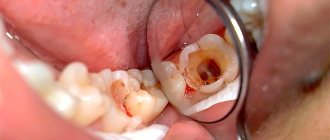
Any mistake during canal treatment is a direct path to future inflammation and loss of the chewing element. Insertion into the cavity with pulp inside the tooth is the same surgical procedure as the others. After all, penetrating a part of the human body with medical dental instruments is the same stress as during surgery. After dental treatment, inflammation often occurs.
Therefore, cleaning the canals and filling the cavity of the chewing element with sealant should be carried out under antiseptic conditions, creating sterile conditions inside.
Photo of the tooth canals, what problems there are in dental treatment
What are the difficulties?
In the case of teeth, it is sometimes easy to get inside the canals, since they are anatomically straight, and sometimes, on the contrary, it is difficult or practically impossible. This is where the problems of root canal treatment .
What problems:
- Endodontic treatment with errors leads to painful sensations when pressing on the tooth and the risk of loss of the chewing element in the future.
- And the proliferation of pathogenic microorganisms causes infection and the risk of complications after filling.
In the first case, when the canal cavity is processed and obturation is carried out, there can be many errors.
Incorrect endodontic treatment, resulting in pain at the top of the tooth
The obstruction of the canals due to the turns at the exit to the apex of the root, that is, in the far part, not only prevents the cavity from being thoroughly cleaned of remnants of blood, vessels and nerve, but sometimes the nerve itself is not completely removed. As a result, after a few months or six months it becomes noticeable that the tooth has darkened. Although this is considered a medical error according to the treatment protocol, it does not depend on the skill of the dentist - endodontist. Obstruction of the root cavities does not make it possible to reach the turn of the canal and clean it. Sometimes because it is not possible to go through the turns of the channels, and sometimes the corners. Uneven canals in the teeth are difficult to navigate during tooth treatment, which causes the instrument used to clean the internal space to break off.
Unsuccessful endodontic treatment
“We did this back in 1990!”
Dentistry, like medicine in general, does not stand still. But individual professionals are still treading water in the same spot where they were presented with a diploma. If, of course, you want to save as much as possible on your health, you can put, for example, a chemical filling instead of a light-curing one completely free of charge. However, good doctors do not cling to the sacred traditions of Soviet dentistry, but willingly use new, advanced techniques in practice - from laser to 3D diagnostics.
“So, I don’t see any problems. Are you sure it hurts?
Visual inspection - that's all? When a piece of a tooth breaks off, the naked eye may be enough. However, if you come with a complaint of hellish pain in half of the jaw, then refusal to conduct an X-ray diagnosis is an argument in favor of immediately leaving the doctor alone with the dental chair. A good dentist not only studies the image before treatment, but also, if necessary, checks the result after, for example, when filling canals. Without proper diagnostics, you can easily miss a serious problem.
Popular
Missed canal in tooth - treatment with a microscope will help
Problem: a 34-year-old patient came to the Family Dentistry with a complaint of pain when chewing. After examining the oral cavity and performing X-ray diagnostics (dental X-ray, CT scan of the teeth), a computer tomograph revealed the causative tooth 2.6, in which the fourth, additional canal was missed, which caused pain when chewing and an inflammatory process at the root of the tooth (the red arrow shows the missed canal, blue – inflammation at the root of the tooth).
Solution: the tooth canals were treated under a microscope, all canals were treated and sealed, the inflammatory process stopped, there was no pain in the tooth, the tooth was preserved (the red arrow shows the sealed canal that was missed earlier, there is no inflammation at the root).
If a tooth hurts after canal filling, most likely the canal treatment was carried out incorrectly - either the canals were not sealed tightly and they became infected, or the canals were not completely cleared of infection, or a canal or its branch was missed. Other causes of tooth pain after root canal filling are much less common. An endodontist can make an accurate diagnosis using modern X-ray equipment, especially a computed tomograph, as well as experience.
This patient complained of tooth pain when chewing. In a calm state, the tooth did not hurt. A computed tomography scan showed inflammation at the root of a tooth on the left upper jaw. The cause of inflammation could be a missed canal in the tooth. Additional canals in the upper sixth and seventh teeth are found in 97% of cases. Experienced endodontists are well aware of this, who quite often encounter missed tooth canals and the resulting inflammation at the roots. Patients turn to a specialist who works with a microscope (dentist-endodontist), when in other clinics they are offered to remove a painful tooth for unknown reasons, since they cannot find out the cause and eliminate the pain in the tooth after filling the canals.
On a CT scan of the teeth, the red arrow shows the missed canal, and the blue arrow shows inflammation at the root of the tooth:
The patient was asked to treat the canals of the teeth under a microscope and have the tooth replaced with a metal-ceramic crown. An alternative to root canal treatment may be tooth extraction and installation of a dental implant, but in this case, root canal treatment is chosen.
Periodontitis (inflammation at the roots of teeth) is successfully treated at Dial-Dent, since a dental microscope and modern endodontic instruments help to cope with the most problematic teeth with narrow, tortuous canals, which many consider “impassable.”
Root canal treatment is performed under local anesthesia. Modern anesthesia drugs make treatment painless. Treatment of tooth canals is carried out using a rubber dam - a protective scarf that isolates the tooth from saliva and the patient’s oral cavity from drugs. The use of rubber dam improves the quality of dental canal treatment and preserves the patient’s health.
First, the endodontist examines the tooth under a microscope and identifies all existing canals, and then measures the length of the canals. The x-ray shows the problematic tooth before treatment; here the missed canal is barely noticeable:
When measuring the length of the canals, the canal missed during the previous treatment is clearly visible:
Unsealing of the canals was carried out using an operating microscope and ultrasound. To eliminate infection in the tooth canals, the canals are treated with 3% sodium hypochloride solution (NAOCL), 17% EDTA solution, 2% chlorhexidine solution. After treatment, the canals were temporarily sealed with calcium hydroxide for 14 days. During this time, the infection in the canals is finally eliminated, the inflammatory process stops, and the pain goes away.
After 14 days, the patient came for a follow-up appointment. The tooth does not hurt, there are no unpleasant symptoms. Akopov R.A. performed root canal filling using AH+ material and gutta-percha using a hybrid method. This root canal filling hermetically seals all areas of the tooth canals.
The x-ray shows permanent filling of the root canals of the tooth:
A follow-up examination and dental x-ray are scheduled after 6 months.
On a computed tomography scan after 6 months, the red arrow indicates a sealed canal (which was previously missed) and the absence of an inflammatory process at the roots of the tooth:
The doctor calculates the cost of treatment at a free consultation, after conducting an examination and taking the necessary photographs. The cost of computed tomography is 2900 rubles.
See other examples of root canal treatment under a microscope here.
“Everything is very bad: two caries, pulpitis, removal”
The opposite situation is when the doctor suddenly finds so much caries and other diseases in your mouth that it is unclear how you survived to your age. Most often, such unexpected discoveries are also accompanied by a refusal to show, at least on an x-ray, where all this horror is hidden.
The goal is to defraud you of as much money as possible for treatment of healthy teeth. This is how you can distinguish a good doctor from a bad one: an honest specialist will always show the real condition of your teeth and gums using an intraoral video camera. When you see for yourself in 56-fold magnification how far the caries has reached in the seven below, the doctor will not need to convince you of anything.
Why does a tooth hurt when pressed after nerve removal?
Many people are afraid to visit dental centers and therefore, when any dental problems arise, they limit themselves to taking painkillers. However, it should be understood that toothache indicates an infectious lesion of the dental pulp , in which the nerve endings responsible for these symptoms are located. Deep caries, pulpitis and periodontitis cannot be cured at home. And the longer a visit to the doctor is postponed, the more painful and longer the recovery period will be after depulpation - root canal treatment.
After removing the nerve, the tooth should hurt, but not severely and not for long. This pain is called post-filling pain and is associated with the body’s reaction to surgery. The depulpation procedure is painful because during it soft and hard dental tissues are injured and sensitive nerve endings are removed. For the same reason, almost all patients experience pain upon completion of canal cleaning. However, in order to accurately determine why a pulpless tooth hurts, you need to seek help from a dental center.
Root canal treatment
Upon completion of the depulpation procedure, you can:
- sensitivity of the enamel to hot, cold, sweets will appear;
- jaw hurts when closing;
- throbbing toothache becomes more frequent in the evening;
- general health worsens (headaches, weakness, fever).
If the acute pain in the tooth that arose after it was depulped did not go away within 7 days, most likely, re-inflammation has begun. In this case, you cannot do without the help of a specialist.
The main causes of toothache associated with root canal cleaning
Why a tooth may hurt after removing the nerve and cleaning the canals:
Dental canals on x-rayThe doctor performed the procedure poorly - particles of the dental nerve remained in the canals, which led to severe pain and increased body temperature.
- Poor quality antiseptic treatment of dental canals or infection after the procedure due to poor oral care.
- The appearance of voids under the filling is accompanied by pain when pressed and the formation of abscesses and fistulas.
- The doctor did not take an x-ray and did not notice one of the canals (their number can be up to 6), as a result of which the infected tissue remaining in them began to rot, and a toothache occurred.
Causes of toothache associated with root canal filling
Why can a tooth hurt after removing the nerve and filling the canals:
- Dentin burn. Occurs when a dentist drills into a tooth root for a long period of time and does not use cooling. In this case, the tooth hurts severely.
- Depressurization of the seal after installation. Most often occurs due to mechanical trauma, in which the material is torn from the dental tissues.
- Allergic reaction. Allergies occur to medications and materials used in the treatment of pulpitis.
- Bite modification. If the filling is too high, it will prevent the jaws from closing, which will cause pain when biting on the tooth.
- Shrinkage of the filling. Any filling material hardens when exposed to light and can shrink. In this case, the filling begins to put pressure on the tooth walls. As a result, cracks appear in the dentin through which air penetrates. If you put pressure on such a tooth, it will begin to ache.
Why does a tooth hurt after root canal treatment - medical errors
The doctor may be to blame for the inflammatory process that occurs after root canal treatment. It is necessary to contact the dentist again so that he can eliminate the errors and heal the tooth correctly. Most likely, a decision will be made to install a temporary filling.
Medical errors that lead to toothache after depulpation include:
- Incorrect therapeutic treatment of pulpitis - the doctor poorly cleaned the dental canals, as a result of which the nerve was not completely removed, which began to decompose naturally.
- Root puncture - the doctor introduced a filling material beyond the root, which provoked the development of an inflammatory process.
- Poor application of filling material – the canals are not filled tightly enough with the material, which is why infection has penetrated into them.
- Violation of the canal filling technique - the tooth may hurt when touched after treatment of pulpitis if the doctor placed the filling beyond the apex of the root.
- Inattentiveness of the doctor - while cleaning the canals, part of the instrument broke off and remained in the gum, which led to discomfort and the development of inflammation.
Fragment of an instrument in a dental canal
Filling beyond the apex of the tooth root
A tooth may hurt after the pulp has been removed from it, due to trigeminal neuralgia. The disease progresses in different ways: pain can torment a person constantly or in attacks. Only a neurologist can identify and cure it.
Why could a long-healed tooth without a nerve with a filling get sick?
A long-healed tooth without a nerve with a filling may hurt for the following reasons:
- the doctor was unable to thoroughly clean the root system, which led to the development of low-grade chronic inflammation;
- the dentist who filled the tooth introduced the filling material beyond the apex of its root;
- during depulpation, the dental canals were poorly washed with an antiseptic;
- the canals are not completely filled with filling material - in this case, acute pain occurs when biting hard foods.
“No need to tell me, let me see for myself”
So, you did something, but after treatment it got worse, or the filling scratches your cheek, braces don’t allow you to close your mouth, and whitening makes you want to bang your head against the wall. A good dentist will always say the magic phrase at the end of the appointment: “If anything bothers you, contact us immediately!” If you experience discomfort, the doctor will try to find the cause, and not throw you out the door so that he can quickly take care of a new patient.
The same rule applies to your individual characteristics: increased sensitivity of teeth and gums, drug intolerance, and even panic fear of a drill. A professional will never brush aside information that a patient tells him.
Causes of pain
During treatment, bone, hard and soft tissues are subjected to severe stress. After therapy, the tooth may hurt, it can be painful to press on it, hot or cold food, mechanical effects when biting cause a reaction of acute, sometimes aching pain.
Painful sensations after completion of treatment are allowed in the following cases:
- if it is moderate dentalgia after the action of an anesthetic;
- the pain is slight, aching or throbbing, and intensifies when you press on the tooth;
- the pain gradually weakens and lasts several days.
How long can a tooth hurt after filling? During therapeutic treatment of caries, pain may bother the patient for no more than a week. According to some doctors, the period of possible pain after tooth filling does not exceed 3-5 days. If the canals were cleaned, painful sensations may occur within two weeks - this is due to the increased sensitivity of the gum and periodontal tissues as a result of injury. Often, the causes of toothache under a filling are medical errors or non-compliance with treatment technologies, which may result in additional complications and repeated dental treatment.
Recurrence of caries
If the doctor cleans the carious cavity poorly, after a few months the tooth may become ill again due to the development of secondary caries. Sometimes, in addition to removing the affected dentin, additional treatment is required to eliminate inflammatory processes (medical treatment of the inflamed tooth with the installation of a temporary filling for a short period). Lack of qualifications of the dentist, fussiness and haste can lead to ignoring this procedure, so after a while signs of inflammation appear again: the tooth aches and hurts, causing a lot of anxiety. Secondary treatment may result in removal of the pulp.
Deep caries resembles the chronic form of pulpitis in appearance, so it is important to be able to distinguish one from the other and, accordingly, choose the right treatment. The dentist should carefully study the x-ray and not rely solely on visual examination - inattention or overconfidence may lead to a distorted explanation of the symptoms of chronic pulpitis. Often, repeated treatment of the affected tooth ends with extirpation of the nerve, cleaning of the canals, and only then can a permanent filling be installed.
Sometimes it is impossible to distinguish inflammation in an image, even with a thorough approach to studying the picture and the dentist’s extensive experience. However, when a tooth hurts after pulpitis treatment, most often the reason lies in a specialist’s mistake.
Medical error
The next medical mistake is a careless attitude to x-ray examination after tooth filling.
“There is no point in treating, only removal”
The goal of a good dentist is to preserve as many of the patient’s original teeth as possible. Tooth extraction is almost always a last resort. The doctor must argue why this tooth needs to be removed, which brings us back to the question of the intraoral camera, X-rays and detailed answers to all the patient’s questions. Acute excruciating pain may turn out to be pulpitis, which is now successfully treated by keeping the tooth in its proper place. When the dentist just sends something for removal, it is better to look for another doctor.
“We cannot do without these procedures. Yes, yes, they are not cheap, but there is no other choice."
There is another category of doctors who avoid intraoral video cameras like hell. Those who certainly want to treat you for a long time, a lot and with the help of the most expensive techniques, devices and materials.
Yes, modern technologies are a good thing: such treatment is faster and more comfortable, and gives reliable, long-term results. But only you have the right to decide whether you want to use these super-sophisticated technologies or whether it’s better to do everything the old fashioned way. Blackmailing a patient with his life and health is unacceptable! It is also important to remember that the doctor should always offer alternative treatment: either traditional methods or modern ones. And the choice is yours.
“Sorry, pregnant women are not allowed anesthesia”
But no. Long gone are those wild times when toothache during pregnancy had to be either endured or treated without pain relief. Of course, there are procedures that expectant mothers cannot undergo: for example, installing an implant, removing complex figure eights, or performing a telex-ray. But, despite the special caution that should be observed when treating teeth in pregnant women, doctors, armed with modern techniques and methods, will always find a way to help. A doctor who, upon learning about your “interesting situation,” changes his face and refuses to look for ways to solve the problem is not your option.


