Anesthesia according to Go-Gates
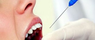
During anesthesia according to Gou-Gates, the anatomy of the peripheral branches of the third branch of the trigeminal nerve is compared with the anterior edge of the branch and the condylar process of the mandible (Fig. 5.23, a). The patient is lying on his back, his face is turned towards the doctor, the intertragal notch of the auricle is facing upward. The doctor is located to the right and in front of the patient. The patient is asked to open his mouth as wide as possible so that the condylar process moves slightly anteriorly and approaches the inferior alveolar nerve. To create a guide for the direction of needle advancement, the patient is asked to place a finger in the external auditory canal. The doctor palpates the anterior edge of the lower jaw branch on the side of the anesthesia with the first finger of his left hand. The syringe barrel is placed in the opposite corner of the mouth. The needle is inserted into the pterygomaxillary (pterygotemporal) recess immediately medial to the temporal muscle tendon. It is advisable to first palpate the medial border of the tendon of this muscle from the side of the oral cavity. On the injection side, the needle is combined with a plane passing from the lower edge of the intertragal notch through the corner of the mouth parallel to the auricle (Fig. 5.23, b).
This is not always easy to do. This difficulty can be overcome using the technique proposed by S.A. Rabinovich. The doctor places the index (II) finger of the left hand in the external auditory canal of the patient or in front of the lower border of the tragus of the auricle at the intertragal notch. With the maximum opening of the mouth, the neck of the condylar process is determined under the finger. The doctor advances the needle toward a point in front of the end of the index finger. This corresponds to the direction towards the tragus of the auricle. The needle is directed to the posterior edge of the tragus of the auricle and immersed into the tissue to a depth of 25 mm until it comes into contact with the lateral neck of the mandible. Pull the needle toward you 1 mm and perform an aspiration test. After this, 2 ml of anesthetic is slowly injected. The patient is left for 20-30 seconds with his mouth open. An anesthetic depot is created at the lateral neck of the mandible. In this case, it is possible to block the inferior alveolar, lingual and buccal nerves. Complications are rare. Anesthesia zone: anesthesia according to Gou-Gates: the same tissues as for mandibular anesthesia, the mucous membrane and skin of the neck, the mucous membrane covering the alveolar process of the lower jaw from the middle of the second molar to the middle of the second premolar, and also “turn off” the buccal nerve. In some cases, infiltration anesthesia is additionally required along the vault of the oral vestibule to “switch off” the peripheral branches of the buccal nerve.
Did you like the article? Share with friends
0
Similar articles
Next articles
- Anesthesia according to Egorov
- Mandibular anesthesia according to Lagardie
- Mental anesthesia
- Case history: exacerbation of chronic granulomatous periodontitis
- Trunk anesthesia
Previous articles
- Thorusal anesthesia
- Mandibular anesthesia
- Incisal anesthesia
- Palatal anesthesia
- Infraorbital anesthesia
Add a comment
Results and discussion
The results of the first clinical signs of pain relief in the form of paresthesia in the area of the anterior third of the tongue and the corresponding half of the lower lip are presented in the table.
Clinical manifestations of anesthesia with methods of blocking the mandibular nerve according to P.M. Egorov and J. Gow-Gates, modified by S.A. Rabinovich and O.N. Moskovets The effectiveness of conduction anesthesia in the lower jaw according to P.M. Egorov was 92%, according to the modified method of J. Gough-Gates - 98%. In conditions of tissue inflammation when performing anesthesia according to P.M. Egorov noted effective pain relief in 89% of cases. When performing anesthesia according to the J. Gow-Gates method as modified, effective anesthesia occurred in 96%. At the same time, the number of complications, hematomas, positive aspiration tests, and post-injection pain was 1.9%. Slightly more (2.2%) local complications were observed during anesthesia according to P.M. Egorov.
When using the modified method according to J. Gou-Gates, when removing impacted and dystopic teeth, additional anesthesia was performed 2.6 times less frequently than using the P.M. method. Egorova. In addition, during anesthesia according to P.M. Egorov, insufficient anesthesia of the mucous membrane on the lingual side was identified in 3 (3.2%) patients, and on the buccal side - in 14 (15.2%) patients. With mandibular anesthesia according to the modified J. Gow-Gates method, insufficient pain relief on the lingual side was noted in 2 (2.1%) patients, and in the area of innervation of the buccal nerve - in 11 (11.9%) patients. Our data confirm that buccal nerve blocking is achieved in significantly fewer cases. A combination of any method of mandibular anesthesia with buccal nerve block is necessary.
Dynamics of somatosensory evoked potentials: after anesthesia according to the method of P.M. Egorov observed a rapid decrease in the amplitude of fluctuations of somatosensory evoked potentials. The amplitude of the sensation threshold reached its minimum values already at the 5th minute. When blocking the mandibular nerve according to the modified J. Gow-Gates method, the greatest decrease in the amplitude of fluctuations of somatosensory evoked potentials at all sensitivity thresholds was noted at the 10th minute, when determining the pain threshold and level of endurance - at the 10-15th minute. The dynamics of the amplitude of N2P2 oscillations of somatosensory evoked potentials at the sensation threshold, pain threshold and pain tolerance level after blockade of the mandibular nerve are presented in the figure.
Dynamics of the amplitude of N2P2 fluctuations (negative and positive waves) of somatosensory evoked potentials at the sensation threshold (TS), pain threshold (PT) and pain endurance level (PAL) after blockade of the mandibular nerve according to P.M. Egorov and according to J. Gou-Gates (modified) using a 4% solution of articaine with adrenaline at a concentration of 1:100,000.
Thus, clinical and physiological studies have shown that the effectiveness of conduction anesthesia in the lower jaw depends on the method of injection anesthesia, the composition of the local anesthetic drug, the type of dental intervention and the presence or absence of an inflammatory process in the anesthetized tissues.
Indications for mandibular anesthesia
Mandibular anesthesia of the lower jaw is appropriate for the following procedures:
- In the treatment of dental caries;
- For endodontic dental treatment;
- When removing teeth on the lower jaw;
- With periostotomy of the lower jaw, however, in this case, mandibular anesthesia alone will not be enough. Two methods should be used together - mandibular and torus;
- When removing a cyst;
- When removing tumors;
- When performing dissection and excision of the hood of wisdom teeth;
- When performing sequestrectomy;
- With a fracture of the lower jaw;
- When removing impacted, dystopic and semi-impacted teeth.
Anesthesia should not be used in the following cases:
- Allergy to used analgesics;
- Hemorrhagic syndrome;
- Infection of tissue in the area of suspected blockage;
- Neurological diseases;
- Septicopyemia;
- Emotional intolerance in the patient.
Technique of the palpation method
With this method, the doctor first palpates the future injection site to determine the position of the mandibular foramen. In this area, the nerve exits the lower jaw.
The approximate time for the onset of the effect of the injection is 10-15 minutes. The duration of pain relief depends on the amount of substance administered and can be 2-3 hours.
Technique:
- The patient should open his mouth as far as possible.
- After this, the doctor places the index finger behind the molars. At the same time, the inner surface of the lower jaw branch is palpated.
- The syringe is located on the second premolar on the other side.
- The needle enters the tissue 1 cm behind the index finger and 1 cm above the teeth.
- First, the injection is made 1-1.5 cm deep into the tissue. As a result, the tongue will be the first to be numbed. The volume of released anesthetic is approximately 0.2-0.3 ml.
- Next, the needle is inserted to the bone.
- If the doctor feels it, then he moves the syringe to the incisors and deepens another 2-2.5 cm.
- An aspiration test is required to prevent injury to the blood vessel. If the test is negative, then the main volume of anesthetic is administered.
- At the end, the needle is carefully removed from the soft tissue.
If everything went well, the patient will develop a feeling of numbness, tingling and coldness on the corresponding half of the lip.
The anesthesia zone corresponds to that described above.
What complications may arise?
If the needle enters more medially than necessary, then there is a risk of rupture of the fibers of the pterygoid muscle.
If a blood vessel is damaged by a needle, there is a possibility of bleeding with its subsequent organization into a hematoma. In the future, an infection may join it. The result will be an inflammatory process that cannot be treated on an outpatient basis.
If a vessel is damaged, then in addition to bleeding there is also the possibility of the anesthetic entering the bloodstream. This is fraught with the development of ischemic zones on the lips and chin. There is also a risk of systemic influence of adrenaline, which is part of the anesthetic. Vascular spasm occurs and blood pressure rises.
Damage to the mandibular nerve itself is possible. This will be manifested by a feeling of numbness, which will persist 8-12 hours after the anesthesia.
One of the very rare complications is disruption of the normal functioning of the facial muscles. This is possible if the branches of the facial nerve are damaged, if the technique of the procedure was grossly violated.
A large number of complications should not be confusing. If you strictly follow the technique, then the likelihood of their occurrence is quite low.
Pros and cons of the tactile method
The following advantages of this method are highlighted:
- the risk of complications is lower, since anatomical landmarks are determined by palpation;
- pain relief occurs even in the most painful situations;
- long duration of anesthesia;
- Half of the jaw is completely switched off, which allows the doctor to use several anatomical zones in his work.
Disadvantages of the palpation method:
- high morbidity in case of equipment violation;
- discomfort for the patient, since half of the jaw and tongue do not move;
- biting soft tissues until the anesthetic wears off;
- Even if performed correctly, the procedure can be very painful.
Possible complications
The consequences that are possible after such a procedure most often manifest themselves in the form of pain in the injection area. It manifests itself against the background of incorrect insertion of a syringe needle into the mucous membrane.
If high-quality treatment with an antiseptic drug is not carried out, then the tissues may become inflamed, swelling and redness may appear.
If these signs do not disappear in the first few days after the procedure, but only increase in intensity, then the help of a doctor is required.
Otherwise, inflammatory processes will lead to detachment of the mucosa and periosteum, which will cause the death of soft tissues.
The developing purulent process will cause infection of the jaw bone and the appearance of osteomyelitis.


