Local anesthesia is an integral part of many dental procedures and is performed before treatment to reduce or completely eliminate pain.
In most cases, it is achieved by exposing the area of intended intervention to drugs and blocking impulses from nerve endings.
Local complications during local anesthesia in dentistry mainly arise as a result of the inexperience of the specialist, and sometimes as a consequence of the negligence of the patient himself.
Indications for pain relief
The anesthesia procedure in dental practice is carried out by a surgeon or therapist (with the appropriate level of qualifications, and usually in a small clinic), the indications for which are:
- tooth extraction;
- treatment of deep carious cavities;
- tooth depulpation procedure;
- preparation for prosthetics;
- curettage of periodontal pockets;
- other surgical and orthodontic interventions.
Types of local anesthesia in dentistry:
- Infiltration is an injection anesthesia based on the impregnation of soft tissues with an anesthetic and the development of a blockade of small nerves and plexuses.
- Application , achieved by blocking nerve endings by superficial application of an anesthetic substance, electrophoresis, physical influences (low temperature, electromagnetic and laser radiation).
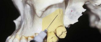
- Conduction – blockade of nerve branches and trunks.
The need for one or another type of local anesthesia is determined by the dentist.
Indications for anesthesia
To avoid local complications during local anesthesia in dentistry, you should know a number of indications for this procedure.
There is a list of certain situations when anesthesia is mandatory:
- Treatment of advanced stages of caries.
- Removal of one or more teeth, including fragments and roots.
- Manipulations in cases where teeth have changed their location or direction of growth.
- Inflammation of the bone frame or soft tissues of a purulent nature.
- Contracture of the temporomandibular joint.
- Carrying out plastic surgeries - piercing (for example, tongue), botulinumplasty, etc.
- Lesions of the peripheral nervous system of an inflammatory or degenerative nature (neuritis).
- Palliative treatment in cases of damage to oral tissues by malignant tumors.
Early complications
Pain and burning sensation at the injection site
They can occur when choosing the wrong needle injection site, rapid administration of anesthetic or its excessive volume.
To increase the comfort of the procedure for the patient, the entire volume of the drug is administered slowly over a minute.
Some medications, in addition to the analgesic component, contain vasoconstrictor substances, which causes vascular spasm in the injection area and discomfort. A preliminary injection of a “pure anesthetic” will help avoid discomfort.
Needle breakage
The most common cause of this complication is sudden movement of the patient at the time of the injection, but needle breakage can also occur due to incorrect injection technique or a manufacturing defect in the syringe.
Short, thin or previously bent needles break more often.
To prevent breakage, it is necessary to check the integrity of its components before using the syringe, warn the patient about the injection and do not insert the needle into the soft tissue the entire length.
If a fracture occurs, the foreign body is removed if possible.
If the fragment remains in the soft tissues, the procedure is delayed using x-ray control.
Local allergic reactions
Observed in case of individual intolerance to the drug or allergy to lidocaine, which is part of most anesthetics. They can occur both during injection administration of the solution and during application of the drug in the form of an ointment or gel.
If itching, burning, redness or swelling of the mucous membranes occurs, it is necessary to stop using the anesthetic and use antihistamines.
Phenomena of residual anesthesia
The condition is also called paresthesia and occurs due to damage to the nerve trunk by a needle, a drug administered under pressure, or a high concentration of the active substance of the anesthetic.
In this case, all types of sensitivity of the innervated zone decrease or completely disappear, which gradually recovers on its own within one to two weeks and does not require additional treatment procedures.
The time for complete recovery depends on the extent of nerve damage.
Complications of induction of anesthesia
Complications of induction of anesthesia include vomiting, regurgitation, aspiration, allergic reactions, laryngo- and bronchospasm, complications during tracheal intubation, respiratory and circulatory disorders.
Vomiting at the beginning of induction anesthesia may be associated with mental agitation before surgery, the influence of analgesics and anesthetics, but most often it occurs in patients with a “full stomach” during emergency operations. Since many diseases are associated with slower gastric emptying (intestinal obstruction, acute pancreatitis, peritonitis), fasting on the eve of anesthesia does not guarantee its emptying. This must be done immediately before anesthesia using a thick gastric tube 5-7 mm in diameter, followed by rinsing with isotonic sodium chloride solution and sodium bicarbonate solution. There are supporters of leaving the tube in the stomach throughout the entire operation; There are also opponents of this method, who claim the greatest possibility of developing regurgitation in this case.
Droperidol and atropine have antiemetic properties, the administration of which is indicated before anesthesia and when vomiting develops at its onset. To prevent this complication, it is advisable to perform intravenous induction anesthesia.
When vomiting, it is necessary to suck out the contents from the mouth, lowering the head end of the operating table, and apply the Sellick maneuver: press on the cricoid cartilage, squeezing the esophagus, which prevents the contents from entering the pharynx and trachea. Then thoroughly dry the oropharynx. After intubation, it is necessary to rinse the trachea with a warm 5% sodium bicarbonate solution.
During the period of induction anesthesia, but not at the beginning, but at the end, when the patient is sleeping, protective reflexes are suppressed by anesthesia and breathing is compensated through a mask with partial penetration of the inhaled mixture into the stomach, regurgitation may occur.
Regurgitation is the passive entry of stomach contents into the mouth and pharynx without gagging. This complication is often asymptomatic, but more often than vomiting it ends in aspiration of gastric contents. All preventive measures (gastric lavage, Sellick maneuver before tracheal intubation, mechanical blockade with an esophageal probe with a cuff) do not guarantee complete success.
Aspiration is one of the most life-threatening complications for a patient. The clinical picture of aspiration was described by S. Mendelson, which is why the syndrome is named after him: difficulty breathing, cyanosis, tachycardia, loss of consciousness, pulmonary edema and collapse. Prevention of aspiration is: premedication (atropine and droperidol), gentle induction of anesthesia, endotracheal method of pain relief.
The tactics in case of aspiration during mask anesthesia are as follows:
1) lower the head end of the table;
2) apply the Sellick technique;
3) suck out the contents of the oropharynx;
4) perform tracheal intubation followed by toilet of the tracheobronchial tree;
5) introduce bronchodilators, hormones, antibiotics;
6) carry out symptomatic treatment of cardiopulmonary failure;
7) perform bronchoscopy and toilet through a bronchoscope.
Often the outcome of aspiration is bronchopneumonia, abscess, and gangrene of the lungs.
During the period of induction anesthesia there may be hemodynamic disturbances - tachycardia, arrhythmia, hypotension - as a result of the action of anesthetics, analgesics on the myocardium and peripheral vascular tone, respiratory depression. These changes also develop during hypoxia. Therefore, symptomatic therapy and compensation for the patient’s breathing with a high oxygen content are necessary.
Complications during tracheal intubation more often develop in patients with a short thick neck, facial injuries, abnormal development of the upper and lower jaw, when the time of the manipulation itself increases significantly. Hypoxia and cardiac dysfunction can be prevented by compensating for breathing through a mask, deepening anesthesia and relaxation. The development of laryngo- and bronchospasm, which manifests itself as difficulty in inhalation and exhalation, cyanosis, sweating, and poor hearing of respiratory sounds occur after rough intubation and hypoxia.
Treatment consists of compensating the patient’s breathing with oxygen under pressure, administering aminophylline (2.4% solution - 5-10 ml drip), prednisolone (100-200 mg), atropine (0.1%-0.5-1 ml), novodrinum (0.2-0.4 mg). In case of cardiac arrest - external massage, in case of ventricular fibrillation - defibrillation. The nurse anesthetist must ensure that the suction and defibrillator are ready when performing any operation.
After intubation, if the endotracheal tube is poorly secured, it may come out of the trachea, become bent, and its lumen may be blocked by sputum, blood clots, or an inflated cuff. If there are signs of obstruction in the patency of the endotracheal tube, it is necessary to check its position in the oropharynx, loosen the cuff, insert a catheter into the tube, suck out the contents, and tighten it. If there is no effect, it must be removed and intubation repeated.
Allergic reactions during induction of anesthesia manifest themselves as hyperemia, urticaria, changes in hemodynamics, and laryngospasm. Allergic reactions and complications can be caused by barbiturates, sombrevin, non-depolarizing relaxants, donor blood, and dextrans.
Treatment: administration of antihistamines, calcium gluconate, prednisolone, bronchodilators.
Delayed complications
Hematoma at the injection site
Formed due to damage to a blood vessel by the sharp end of a needle.
More often, hematomas occur during conduction or infiltration anesthesia of the areas of the lower jaw due to rich vascularization.
Risk factors for developing the condition are blood coagulation disorders and arterial hypertension.
When the first signs of a hematoma occur, the dentist must take measures to stop the bleeding: mechanical pressure on the area of the damaged vessel, application of cold, local administration of vasoconstrictors.
After the hematoma stops growing, the patient can be sent home, postponing dental intervention for several days.
To prevent complications, it is necessary to follow the technique of the procedure, as well as choose the right injection site.
Infection and inflammation of soft tissues
It manifests itself in the form of inflammatory infiltrates, abscesses and phlegmons and is the result of violations of the rules of asepsis and antisepsis during local anesthesia.
The entry of bacterial flora into soft tissues may be associated with contamination of the needle, medicinal solution, or trauma during the intervention of an existing source of infection in the oral cavity.
A preventive measure in this case is the use of disposable syringes and solutions that have retained their sterility and physical properties.
Local soft tissue necrosis
A rare late complication that develops a few days after dental surgery.
Usually associated with the rapid administration of a drug containing a large amount of a vasoconstrictor component, which causes a sharp spasm of blood vessels and disruption of the nutrition of surrounding tissues, the development of ischemia and their subsequent death.
Predisposing factors for necrosis are systemic diseases of the blood vessels and severe concomitant pathology of endocrine and cardiovascular pathology.
Trismus of masticatory muscles
Prolonged spasm of the masticatory muscles can occur due to a high concentration of the drug, damage to muscles or nerve fibers during injection, infection of the wound, or compression of the tissue by the resulting hematoma.
Trismus manifests itself as severe difficulty opening the mouth, which impairs food intake and speech function.
If the muscle fibers themselves are damaged, aseptic inflammation may develop, accompanied by an increase in body temperature, severe pain, and scarring with the further formation of contracture.
Temporary paresis of the facial nerve
It is observed when the technique of anesthesia in the area of the posterior part of the lower jaw branch is violated during dental interventions associated with the extraction of wisdom teeth.
If the anesthetic is administered excessively or the injection site is incorrect, paresis of the facial nerve develops, accompanied by sagging of the upper lip, the inability to close the eyelids on the affected side, and weakness of the facial muscles.
Typically, paresis goes away on its own within a few days and does not require additional treatment.
Sometimes the condition with local complications after unsuccessful anesthesia requires complex medication and physical therapy, and the prognosis depends on the timeliness of diagnosis and the moment of initiation of treatment.
Computer
Errors and complications of local anesthesia in dentistry are largely due to human factors. If you carry out the anesthesia process using a computer using a special electronic system, which includes a system unit and a handpiece, troubles can be avoided. In this case, due to the special design of the needle, the puncture is done as painlessly as possible. This also applies to perforation of the cortical bone.
The dosage of the administered drug is completely controlled by the electronic “brain”, which eliminates the human factor.
Prevention of the development of local complications
Preventive measures have a binary direction and concern both the specialist and the patient:
- Compliance with injection techniques by a dentist.
- Correct selection of drugs for anesthesia, taking into account allergy history.
- Monitoring the expiration date of the medicine used and the integrity of its packaging.
- Use of disposable instruments.
- Compliance with the rules of asepsis and antiseptics.
- Warning the patient about the injection and administration of the anesthetic solution and his adequate behavior during the manipulation.
- The patient’s compliance with medical recommendations for caring for the intervention area.
Local complications during pain relief in dentistry can be a consequence of both a medical error and non-compliance with the recommendations of a specialist on the part of the patient. If any pathological symptoms are detected, it is necessary to contact the doctor who performed the pain relief procedure to determine further treatment tactics.
General information about local anesthesia
Previously, performing dental procedures without pain was only a dream of mankind for many centuries. When the anesthetic properties of cocaine and other drugs were discovered, it became possible to develop different methods of anesthesia. The composition of the funds is different. The doctor must select them for each patient individually, so the risk of adverse reactions is minimal. However, no one is immune from mistakes.
Nowadays, painkillers used in dentistry are already in their fifth generation. However, patient demands for treatment conditions continue to grow steadily. Many people are interested in what local complications may arise during local anesthesia in dentistry.
It should be clarified that anesthesia has an important difference from anesthesia. When it is performed, tissue is affected in a certain place of the human body, which loses sensitivity, but the patient himself remains conscious.
In any modern dental clinic, such a procedure is treated very responsibly. There are even special standards according to which these manipulations must be carried out efficiently, painlessly and as comfortably as possible for patients.
Excessive longitudinal expansion of the canal in the middle third on the internal curvature of the root (“stripping”).
The causes of this complication are usually underestimating the curvature of the canal and working in a curved canal with insufficiently curved instruments.
Prevention . To avoid excessive expansion of the canal in the area of “minor curvature”, you should first bend the files in accordance with the curvature of the canal, and during processing use the “anti-perforation technique”, when the file is pressed against the “greater curvature” of the canal. This complication can also be avoided by using safe drills (“Safety Hedstroem”) and flexible files.
Excessive expansion of narrow, curved channels should be avoided: it is recommended to expand them no more than 2-4 numbers from the original width.
Tool breakage in the canal.
Breakage of an instrument in the canal is one of the most unpleasant complications for the doctor and the patient. Leaving a fragment of an instrument in the canal sharply worsens the prognosis of endodontic treatment, and sometimes causes tooth extraction.
most common causes of
— application of excessive force when working with the tool;
— non-compliance with the recommended angles of rotation of the instrument in the canal;
— work with deformed, untwisted tools;
- improper opening of the tooth cavity.
Prevention consists of following the following rules:
firstly, careful, careful work in compliance with the rules and sequence of use of tools;
secondly, compliance with the maximum angles of rotation of the instruments in the canal: K-reamers - 180°, K-files - 90°; for narrow, curved canals, it is recommended to reduce the rotation angle to 20-30°. H-files cannot be rotated in the canal;
thirdly, the mandatory use of gels to expand root canals; fourthly, timely rejection of unusable instruments.
Let us recall once again the criteria for rejecting endodontic instruments :
— plastic deformation of the tool;
— pre-curved tools;
— deployed tools;
— damage to the cutting edge of the tool;
- a dull blade of the working part, as evidenced by the shine of the cutting edge.
It should be remembered that pulp extractors and instruments smaller than ISO size 10 are disposable and should be discarded after a single use.
Did you like the article? Share with friends
0
Similar articles
Next articles
- Root canal filling
- Filling with one paste
- Single pin method
- Lateral (side) condensation method
- Filling root canals using the Termafil system
Previous articles
- Coronal-apical methods
- Apical-coronal methods
- Methodology for instrumental treatment of root canals
- Retrograde pulpitis
- Chronic hypertrophic pulpitis
Nerve damage during anesthesia
Nerve injuries are a significant source of both patient morbidity and professional liability.
The true incidence is unknown due to the large number of cases not reported in official reports. Much of the data presented below is taken from the ASA Closed Claims Program database (1975-95). Nerve injuries are the second most common cause of claim across the entire database (16% of the total). Of the specific nerve injuries: ulnar nerve (28%), brachial plexus nerves (20%), lumbosacral root (16%) and spinal cord (13%). Less commonly damaged nerves are the sciatic, median, radial and femoral.
Recently, there have been fewer claims for ulnar nerve damage, with spinal cord injuries leading the way.
In many cases of perioperative nerve injury, the mechanism of injury is not identified (only 9% of ulnar nerve injuries had an identifiable mechanism). However, the mechanism of spinal cord injury was established in 48% of cases, and regional anesthesia was performed in 68% of cases of spinal cord injury.
Etiology
- Direct trauma from needles, sutures, instruments.
- Injection of a neurotoxic substance.
- Mechanical factors such as traction and compression.
- Ischemia appears to contribute to all of these causes.
Classification
The extent of the damage will determine the type of intervention required and the likelihood of recovery.
- Neuropraxia is damage to the myelin, the axon remains intact. Recovery will take several weeks to several months. Good prognosis.
- Axonotmesis is the destruction of the axon. The prognosis and likelihood of recovery are questionable.
- Neurotmesis - the nerve is completely destroyed. Surgery may be required. Bad prognosis.
Predisposing factors
Patients with pre-existing generalized peripheral neuropathy are at increased risk for injury/ischemia. Prior to surgery, any pre-existing deficiency should be documented in detail in the medical history.
The operation itself and the concomitant forced positioning can cause specific nerve damage.
Anesthetic factors include direct needle injury to the nerve during regional anesthesia. A good knowledge of anatomy and careful execution of the procedure can reduce the likelihood of this.
The optimal needle design is not yet clear, but it is clear that short bevel needles cause less damage to the nerve bundle and are currently popular.
The resulting paresthesia should alert the doctor, and severe pain when administering the drug is a signal to immediately stop the injection (apparently, the needle got into the nerve or its sheath). These phenomena are a strong argument in favor of performing the blockade on a conscious patient.
The time required to apply tourniquets should be minimized and only pneumatic models should be used. During spinal or epidural anesthesia, solutions of local anesthetics with preservatives should not be used.
Systemic factors include: hypothermia, hypotension, hypoxia and electrolyte disturbances such as uremia, diabetes mellitus, vitamin B12 and folic acid deficiency.
Symptoms
- Symptoms may appear within a day, but sometimes occur after 2-3 weeks.
- The extent and duration of symptoms vary depending on the severity of the lesion, ranging from numbness and mild paresthesia for a few weeks to persistent painful paresthesia, sensory loss, paralysis for years, and eventually reflex sympathetic dystrophy.
Ulnar nerve neuropathy
- More common in men (3:1).
- More often with excess body weight and prolonged hospital stay. It is rare in young patients.
- Prospective studies have shown an incidence in the acute period from 1 in 200 to 1 in 350 patients, but by 3 months. clinically significant lesions are much less common.
- 85% of cases were associated with general anesthesia, 15% - regional (6% - spinal), which indicates that the etiology is not obvious.
- Damage occurs at the level of the superior condylar groove of the ulna. In men, the subcutaneous fatty tissue in this area is thinner and the tunnel is narrower, which explains the difference by gender.
- Recent studies have suggested that nerve compression is less severe in the supinated position. Abduction of the opposite arm is preferable for the ulnar nerve, but there is a greater risk of brachial plexus traction in this position.
- Additional padding was accurately identified in 27% of closed cases!
- In 62% of cases, onset was “delayed” (ie, more than 1 day after surgery).
- In the other, uninjured arm, nerve conduction disturbances are often detected, which indicates subclinical neuropathy, which can manifest itself in the postoperative period.
Brachial plexus injury
- Causative factors include hypertraction (abduction of the arm while turning the head to the opposite side), compression (upward movement of the clavicle and retraction of the sternum), and regional anesthesia (only 16% of closed claims for brachial plexus injury).
- Injection pain or paresthesia is reported in 50% of claims involving regional techniques.
- Among closed claims for brachial plexus injuries, 13% involve damage to the long thoracic nerve (which provides abduction of the scapula). The mechanism is unknown.
- Damage to the upper bundles is more common.
Damage to the lumbosacral root (radiculopathy)
- More than 90% of cases are associated with regional anesthesia (55% - spinal, 37% - epidural).
- Paresthesia or pain when inserting a needle or injecting a drug indicates possible injury. Postoperative radicular pain can be persistent in up to 0.2% of cases, but almost always disappears within a few weeks/months.
- Multiple failed attempts increase the likelihood of damage.
- The most common complaint is persistent paresthesia, with or without motor impairment.
Spinal cord injury
- According to closed lawsuits. 58% of cases are associated with regional anesthesia.
- A specific pathogenesis has been identified in approximately 50% of cases, which is much higher than for other causes of nerve damage.
- The most common mechanisms are epidural hematoma, chemical injury, anterior spinal artery syndrome, and meningitis, respectively.
- Lumbar epidural blocks were four times more common than subarachnoid blocks and eight times more common than thoracic epidural blocks (this is partly related to the number of blocks performed).
- Damage is more common with blocks performed for chronic pain and in the presence of systemic anticoagulation (fractionated heparin, introduced in the USA in 1993).
- There was a significant delay in diagnosing spinal cord or nerve compression. Persistent weakness or numbness was considered to be due to the epidural infusion. All suspicious lesions should be investigated if there is a relevant history and an MRI should be performed if possible, especially in the presence of anticoagulation.
- It is difficult to estimate the true incidence of serious neurological injury after spinal/epidural blockade. Scott suggests that in obstetric practice the incidence of permanent disability is 1 in 100,000. Epidural hematoma in the presence of LUMV may occur at an incidence of 1 in 1000 to 1 in 10,000, but other investigators estimate the risk to be 1 in 150,000 to 1 in 220 000 (after epidural and spinal blockade, respectively). Epidural abscess is less common, but it should be suspected in patients with impaired immunity and anticoagulation, when the epidural catheter is in place for a long time.
- With anterior spinal artery syndrome, there is usually spastic paralysis of the lower extremities below the level of the lesion, flaccid paralysis at the level of the lesion, variable sensory loss and dysfunction of the sphincters. This pathology may be associated with general or regional anesthesia and usually develops with prolonged hypotension. Other causes not related to anesthesia include aortic cross-clamping, thrombosis, embolus, dissecting abdominal aortic aneurysm, periarteritis nodosa, SLE and spinal surgery.
- A rare cause of paraplegia is arachnoiditis, for which no effective treatment has been developed. It is usually characterized by gradually increasing weakness and loss of sensation, occurring several days or months after spinal anesthesia, sometimes leading to complete paraplegia or death. Causes - meningitis, bleeding, spinal cord surgery, may also be associated with drugs administered into the spinal or epidural space, such as preservatives, erroneous administration of the drug, etc.
- Transient neurological symptoms (TNS) are back pain or dysesthesia radiating bilaterally to the legs or buttocks after full recovery from spinal anesthesia and starting within 24 hours. There are usually no objective neurological signs. The pain is moderate and can be relieved with NSAIDs. Symptoms usually disappear after a few days. The clinical significance is unclear and data are conflicting, although PNS appears to develop more frequently following the use of spinal 5% hyperbaric lidocaine (lignocaine)/epinephrine. Some studies have shown that PNS is common after the use of pure spinal lidocaine (lignocaine).
- Cauda equina syndrome (CAS) is characterized by low back pain, sciatic anesthesia, sphincter dysfunction, and motor or sensory symptoms below the knees. There are a number of reports of the development of SCS after the use of spinal microcatheters (28 G) and 5% hyperbaric lidocaine (lignocaine). This was considered to be a consequence of hyperbaric local anesthetic leakage and resulted in the US Food and Drug Administration banning the use of spinal catheters thinner than 24 gauge in 1992.
Prevention
- It is necessary to be aware of complications and the most common causes of injury (eg, careful positioning of the patient).
- A thorough history taking, objective examination and documentation in the medical history of all neurological disorders present before the operation.
- Accurately maintain anesthesia records of all features of regional technique (including needle type, paresthesia, drug used, etc.).
- Performing a blockade on a conscious patient or with preliminary administration of a sedative (not practiced in children).
- Do not perform a regional block if the patient refuses.
- Always stop injection if pain or paresthesia occurs.
Diagnosis and treatment
- Neurophysiological tests such as EMG and nerve conduction studies, coupled with MRI, often help pinpoint the location of the injury, which can indicate the cause and possible liability.
- Treatment and prognosis depend on the severity of the injury and are best left to neurologists and neurosurgeons.
- Spinal cord compression or cauda equina syndrome requires emergency neurosurgical intervention and decompression.
- Medicines used for neuropathic pain are tricyclic antidepressants (amitriptyline at a dose of up to 150 mg / day, starting with 25 mg at night) or antiepileptic drugs (carbamazepine 100 mg 1-2 times a day up to 200 mg 4 times a day, or gabapentin, best tolerated, 300 mg once daily up to 600 mg three times daily).
Source: https://www.ambu03.ru/anesteziya/prakticheskie-voprosy/povrezhdeniya-nervov-pri-anestezii/
