10995
In dentistry, occlusion is the mutual position of the upper and lower dentition when the jaws are tightly closed. Almost all malocclusions entail serious, unpleasant and even dangerous consequences , which can only be avoided by undergoing orthodontic treatment on time. Moreover, problems can arise both in childhood and much later.
Types of physiological occlusion
A bite is considered physiological if:
- The dentofacial system is fully functional,
- The work of the temporomandibular joint is optimized,
- The muscular-ligamentous apparatus of the jaw does not experience overload,
- There are no breathing, swallowing, or speech disorders.
Types of physiological occlusion, from top to bottom: orthognathic bite, straight bite, biprognathic bite, opisthognathic bite
- Direct bite - the upper and lower dentitions close together with cutting edges; according to some dentists, a direct bite is intermediate between physiological and pathological. The contacting edges subsequently push against each other, which leads to disruption of the maxillofacial joint and a decrease in the longitudinal size of the lower third of the face, but such a direct bite also has its advantages: the worn cutting surface is highly resistant to caries.
Direct physiological bite
- Biprognathic bite - the front teeth of the upper and lower dentition are tilted forward, the contact between them is not broken, but when the lips are closed, some movement of the jaws is visually visible.
Biprognathic physiological occlusion
- Opisthognathic bite - the anterior teeth of the upper and lower dentition deviate somewhat posteriorly, towards the tongue, while the contacts between the antagonist teeth are not disturbed.
Opisthognathic physiological occlusion
- Orthognathic occlusion
Most often - in 75-80% of the population - orthognathic occlusion occurs and is considered the standard.
Schematic representation of the position of the anterior teeth in an orthognathic bite
Signs of orthognathic bite:
- The upper dental arch in the anterior part of the oral cavity overlaps ⅓ of the height of the lower one, due to the fact that the upper dental arch is wider than the lower one;
- In the lateral sections, each upper tooth of the upper jaw, excluding wisdom teeth, comes into contact with 2 antagonist teeth (located opposite in the lower row) - the lower and posterior teeth of the same name;
- The lower central incisor and upper wisdom tooth each have only one antagonist tooth;
- The upper dentition has the shape of a semi-ellipse, the lower one is represented by a parabola;
- The external (buccal) cusps of the upper molars and premolars are located outside the same buccal cusps of the lower row;
- The muscles responsible for the movement of the lower jaw contract evenly; at rest this contraction is minimal;
- At rest, the jaws are in a position of central occlusion; after performing various movements, they return to their original position.
Orthognathic occlusion
Types and characteristics of pathologies
Sagittal
Sagittal pathologies of the dentofacial apparatus include disturbances of occlusion that occur in the anteroposterior direction, that is, one of the jaws is pushed forward and noticeably predominates over the other.
The group of sagittal defects includes two types of bites: distal and mesial. Treatment can be carried out using different orthodontic structures, as well as surgical and combined methods. The selection of therapeutic agents is based on the clinical picture. In this case, therapy is aimed at normalizing the area of occlusion and stimulating or suppressing the growth of one of the displaced jaws.
Vertical
A characteristic sign of vertical pathology: too high or low position of a group of teeth relative to occlusion, while the features of the manifestation of pathology depend on its severity and localization.
Such anomalies include open and deep bite types. With a vertical anomaly, the loss of one of the teeth often leads to dentoalveolar elongation - excessive growth of the antagonist tooth until it comes into contact with the oral mucosa.
In children, treatment of vertical anomalies usually comes down to eliminating the causes of the pathology. For this, adults have to resort to the use of non-removable orthodontic structures.
Transversal
Transversal defects include the crossbite type. This type of pathology is characterized by one of the following symptoms :
- Lateral displacement of one of the dentitions;
- The presence of disproportions: one of the dentitions may be significantly wider or narrower than the other;
- Pathological narrowing of the jaw.
Pathologies of this type are corrected with the help of expanding structures, having previously isolated the dental lines. Severe, pronounced defects require surgical intervention.
Attention: it is important to begin correcting transverse anomalies as early as possible when the first symptoms appear.
What different types of malocclusion look like:
Types of malocclusion
Types of pathological bite: 1 - deep bite, 2 - distal or progenic bite, 3 - mesial bite, 4 - open bite, 5 - cross bite
- Deep bite
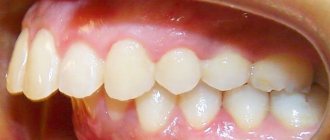
A deep bite is characterized by a strong protrusion of the upper jaw relative to the lower jaw, with the upper dentition overlapping the lower one by more than ⅓. The incisors and canines of the lower row, when closed, injure the mucous membrane of the hard palate and gums, followed by the rapid development of gingivitis.
Deep bite: photos before/after treatment
- Distal or prognathic occlusion
It is characterized by the advancement of the upper jaw relative to the lower one so that the contact of the upper and lower incisors is disrupted.
Malocclusion may be caused by:
- Excessive development of the upper jaw with normal formation of the lower jaw;
- Insufficient development of the lower jaw with a normally formed upper jaw;
- Excessive development of the upper jaw with simultaneous insufficient development of the lower jaw;
- Flattening of the upper jaw in the lateral sections, due to which it is pulled forward.
Distal bite: photos before/after treatment
- Mesial bite
A type of pathological in which the lower jaw is pushed forward relative to the upper, so that the lower incisors and canines are in front of the upper ones and may not close at all. Mesial bite is accompanied by painful sensations in the temporomandibular joint and jaws when speaking and chewing.
Mesial bite: photos before/after treatment
Visually, the chin moves forward; in profile, the face looks somewhat concave.
Characteristic appearance in profile with mesial occlusion: before/after treatment
- Crossbite
A crossbite develops as a result of uneven formation of the upper and lower jaws on the right or left side of the face.
With a crossbite, there is a pronounced asymmetrical appearance of the face, speech impairment and difficulty in chewing; while eating and talking, a person may bite his or her cheek.
At the moment of closure, the teeth of the upper and lower sections intersect with each other, the chin deviates to the side, and infection of the lips is indicated. Opening the mouth is accompanied by pain and crunching in the temporomandibular joint.
Crossbite: before/after photos
- Open bite
This is a pathological type of occlusion, characterized by a violation of the closure of teeth in the lateral (lateral bilateral open bite) or anterior sections (respectively, anterior unilateral open bite).
Types of open bite
A vertical gap is formed between the upper and lower rows of teeth. With an anterior open bite, there is difficulty in closing the lips, due to which the mouth is constantly half-open, a slanting of the chin and a vertical stretching of the lower part of the face are visually determined.
Due to breathing problems caused by an open bite - a person breathes through the mouth - dry mouth appears over time, and periodontal disease quickly develops. Due to crowded teeth, especially in the anterior section, which are difficult to free from dental plaque, the risk of developing caries increases.
Open bite: photos before/after treatment
Some terminology
The arrangement of the lower and upper teeth in relation to each other is called the bite. There is a similar concept - occlusion, which refers to the natural closure of the jaws. This is a process where the masticatory muscles, teeth, and temporomandibular joints take part. There are central, anterior and lateral clutches of the jaws. Central occlusion, in essence, is a bite. If it is correct, it is called physiological; if not, it is called abnormal or pathological. With normal occlusion, the functions of chewing and speech are not impaired. With pathology, it’s the opposite.
Causes of malocclusion
Most of the reasons for the development of malocclusion in an adult come from childhood and the period of pregnancy preceding it, when the formation of all organs and tissues occurs, including the dental system, as well as a number of other unfavorable factors:
- Hereditary predisposition realized against the background of unfavorable environmental factors;
- Rickets suffered in childhood;
- Disturbance of Ca metabolism during the formation of the dentoalveolar system and change of bite;
- Insufficient intake of vitamins and minerals during pregnancy;
- Diseases suffered during pregnancy by the mother, such as influenza, childhood infections, etc.;
- Complications during pregnancy of the mother, such as preeclampsia, toxicosis, diabetes mellitus, chronic intrauterine fetal hypoxia;
- Bad habits (taking alcohol, tobacco products and drugs) during pregnancy,
- Chronic diseases of the ENT organs: adenoiditis, deviated nasal septum, rhinitis, sinusitis, etc., as a result of the need to breathe through the mouth and not the nose, the jaws are constantly in a forced position and the process of forming a correct bite is disrupted;
- Chronic diseases of the oral cavity: periodontitis, periosteitis, gingivitis;
- Injuries to the maxillofacial apparatus, including those received during childbirth;
- Bad habits during childhood: prolonged use of pacifiers, thumb sucking, pencils and other objects, lip biting and frequent resting of the face on the hand;
- Failure to comply with the timing of introducing complementary foods in childhood;
- Insufficient amount of solid food in the diet, especially during the period of change from a temporary bite to a permanent one and the formation of a bite,
- Early tooth loss;
- Macroglossia (increased tongue size);
- Short frenulum of the tongue.
Prevention
Open bite before and after
Since most factors influence the malocclusion in childhood, it is very important for parents to pay attention to measures to prevent such pathology:
- During the period of bearing a baby, a woman should not allow a deficiency of calcium and fluoride in the body.
- Pay attention to the baby's breathing. If he breathes mainly through his mouth, then the growth of the upper jaw will slow down.
- Turn the baby from side to side. It is important that the pressure is applied alternately to one and the other jaw.
- Feed your baby correctly. If he is bottle-fed, it is necessary to make a small hole in the bottle so that the child sucks the nipple and develops his facial muscles. It is better to choose a pacifier with an orthodontic shape.
- Wean your child off sucking a pacifier by age 2.
- Wean your child off the habit of thumb sucking at the age when baby teeth appear.
- Visit a specialist regularly to examine your child’s oral cavity. It is necessary to monitor whether the child’s teeth appear on time.
Consequences of malocclusion?
- Speech impairments can manifest themselves in difficulties in pronouncing various phonemes; during a long conversation, a person with a malocclusion may experience discomfort in the dental system, as the muscles are constantly under tension;
- Breathing disorders;
- Swallowing disorders;
- Insufficient oral hygiene due to the inaccessibility of some areas of the surface of the teeth for cleaning leads to the occurrence of tartar, caries and inflammatory gum diseases;
- Malocclusions lead to disruption of the mechanical processing of food in the mouth and the formation of a food bolus, which can result in diseases of the digestive system and injuries to the esophagus from large pieces of food;
- Psychological discomfort: people with malocclusion lose self-confidence and often worry about their appearance.
How to correct malocclusion in an adult?
Correcting a bite in adults is possible with the help of braces and aligners. The treatment of malocclusion is carried out by an orthodontist. After examining the patient, he draws up a treatment plan and offers several treatment options. The patient can choose the one that suits his budget and aesthetic characteristics.
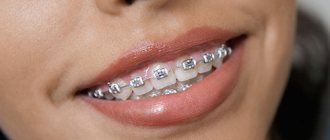
Braces are fixed orthodontic structures designed to correct pathological malocclusion. They can be made of metal (including gold), plastic, ceramics, artificially created sapphire, or combinations. Installation, adjustment and removal of braces is carried out exclusively by an orthodontist.
Types of bracket systems
The bracket system is represented by bracket-locks, which are installed on each tooth separately and an arch that unites them together. The arch bends somewhat under the influence of pathologically displaced teeth, but due to its properties it tends to acquire its original shape, as a result of which it pulls the teeth along with it, placing them in the correct position.
In the first week, painful sensations appear, which can be eliminated if necessary by taking painkillers (ibuprofen, analgin, ketarol, ketanov, ketofril, diclofenac, etc.). Pain indicates that the brace system has begun to work and the teeth have begun to occupy the desired position.
Currently, braces can be installed both on the external and internal surfaces of the dentition, making them completely invisible on the teeth and not causing psychological discomfort to the patient.
Wearing braces obliges the patient to follow certain rules for eating food and use additional tools for cleaning the oral cavity.
Bite correction using braces: before/after
Aligners are removable orthodontic structures or so-called aligners. They are made from a polymer transparent hypoallergenic material individually for each patient and are an alternative to the braces system.
Mouthguard or aligner for the upper dentition
They are invisible on the teeth, and therefore do not cause psychological or aesthetic discomfort to the patient. Do not injure the mucous membrane. They do not require special oral care or diet, since the patient has the opportunity to remove the aligner before eating and brushing their teeth.
Before treatment, a computer model of the bite is created, which will be formed after using the mouthguard, so the patient can see the result of treatment before it begins.
Surgery
If therapy with a brace system or aligner does not produce results, there are severe disturbances in the functions of chewing, swallowing, breathing, and speech; or there are congenital malformations of the dental system, especially combined; Chin dysplasia and other severe pathological disorders; bite correction is only possible through surgery. Surgical treatment is performed by an oral and maxillofacial surgeon.
Duration of treatment for malocclusion
The duration of treatment for malocclusion in adults depends on the material of the chosen structure, its serviceability, the condition of the dental system (the presence of inflammatory diseases of the oral cavity, crooked teeth and those that need to be removed) and the severity of pathological disorders in it. It should also be taken into account that, unlike children, the ligamentous-muscular framework of adults is already fully formed and because of this it is more difficult to be influenced by orthodontic structures: muscles and ligaments need more time to adapt to the newly formed bite.
Treatment of malocclusion consists of a period of wearing the orthodontic structures themselves (braces and aligners), which ranges from 6 months to several years, and a period of consolidating the result through the use of removable and non-removable retainers, it lasts 2 times longer than the first period.
