Superficial caries: photo, medical history, treatment
Destruction of tooth tissue, loss of minerals by enamel, pain - all this accompanies superficial caries. Photos, medical history, treatment - we will talk about everything related to this process today in the article.
The formation of erosion is detected during inspection, transillumination, and probing. Up to 70% of children are susceptible to the disease. In adults, the clinical picture is worse, since it is not always possible to do with small methods and only remineralization, as in childhood.
Causes of superficial caries
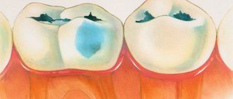
Why do tooth decay develop from a chalk stain? The process of destruction of enamel is associated with its demineralization. At the first stage, it remains intact, but only in appearance, since in fact caries is already developing. If treatment is carried out at this stage, then further growth of the defect stops.
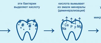
The main cause of erosion, especially in children, is considered to be the pathological activity of bacteria. It occurs due to poor oral hygiene, but there are other factors that lead to the problem:
- reduced amount of fluoride in teeth;
- heredity;
- reduced immunity;
- malnutrition;
- change in the composition of saliva.
Plaque, accumulating on a wisdom tooth or any other crown, forms a kind of plaque covering the dentin. Organic acids appear during the fermentation of leftover food, leading to a change in pH in the mouth and accelerated destruction of enamel. The appearance of a carious cavity is accelerated by wearing various orthodontic structures, dentures, somatic diseases, abnormal bite, and hyposalivation.
Symptoms
With superficial caries, there are no such striking symptoms as with deep damage to dentin. Due to the paucity of signs, a person rarely pays attention to the problem in time. In some cases, only a dentist can identify it, especially if the destruction has affected the proximal surface.
Symptoms that can help identify the problem include:
- The pain is short-term.
- Minor defects on the enamel.
- Discomfort when brushing with a toothbrush.
- Roughness of teeth.
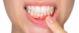
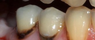
The very appearance of the affected dentin will be uneven and chalky. Caries is often preceded by the appearance of white spots, as the enamel loses its shine and begins to deteriorate. Painful sensations are more often observed when the process is localized at the neck of the tooth. If erosion appears in a convex area, then discomfort is not felt for a long time due to the growth of dentin, which is designed to protect the pulp.
A characteristic sign may be food getting stuck at the defect site. If it is located close to the gum, then it will gradually become inflamed, bleed on contact, and swelling will appear.
Photo
Kinds
> Black's disease is of the following types:
- Fissure caries affects natural cavities and depressions.
- Approximal - can spread to cutting and molar teeth. Damage is often concentrated on the edge.
- The cervical view is located at the base of the crown. This subtype is characterized by the fact that the pathological process affects several teeth at once.
> Classification by recurrence:
- primary - the area was not treated, no pathological processes were previously noted here;
- secondary - relapse in the area of the filling or under it.
> Caries can be divided according to the severity of its symptoms:
- Compensated - progress is very slow, the tooth is practically not bothered and other symptoms may be absent. This type of disease is characterized by the likelihood of a complete stop in the development of a carious cavity. In such cases, it is possible to prevent its formation by conventional hygiene methods.
- Subcompensated - although the course cannot be called sluggish, a person still feels the first signs, especially expressed in pain and discomfort, late.
- Decompensated - dentin destruction occurs rapidly, so the initial stage quickly develops into deep caries.
According to the intensity of the disease, multiple and single processes are distinguished.
Superficial caries - treatment
Superficial destruction of dentin is the initial stage of caries, so its treatment is often done without drilling. But the dentist will need to remove the already damaged layer, which is achieved by gentle grinding. A gel or application (sterile bandage soaked in a solution) with the following agents will be applied to the cleaned tooth:
- fluorine varnish;
- calcium gluconate;
- remodent;
- sodium fluoride.
Preliminary diagnosis is required to differentiate caries from other diseases affecting tooth enamel. A thorough examination and radiography are needed to determine the location of the affected crowns and decide whether it is necessary to prepare (drill) the tooth.
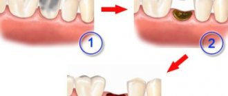
Typically, enamel restoration occurs after remineralization, but sometimes the dentist may suggest a treatment method using the Icon system. The protective layer at the defect site becomes thinner and becomes porous, so a sealant is used. Thanks to penetration into the pores, the structure of the crown is restored. The treatment process itself goes like this:
- Etching - at this stage, the dental tissue is treated with a gentle composition from the series. This procedure avoids drilling and grinding.
- Drying the surface with an alcohol solution.
- Applying the infiltrate to the affected area - after penetrating the pores of the enamel, it is polymerized by light.
The doctor monitors the condition of the tooth for some time and if the erosion continues to deepen, then a full filling is performed.
Prevention
With timely treatment and subsequent attention to high-quality oral hygiene, a favorable outcome is high. The thinning of dentin stops if you supplement regular tooth brushing with rinsing the mouth after meals, using floss and periodic examinations by the dentist.
Following preventive recommendations helps to avoid caries:
- It is better to avoid foods with strong irritants (sour, overly salty, chips). It is worth reducing the number of all kinds of carbohydrate snacks;
- if matte areas appear on the enamel, you need to contact a dentist for treatment and mineralization;
- after snacking on sweets and flour, you should brush your teeth for 10 minutes;
- regular removal of tartar and professional cleaning of plaque;
- alternating pastes with a healing effect and a cleansing effect;
- taking vitamin supplements;
- treatment of teeth with calcium and fluoride - in this case, you can adjust the diet to obtain a sufficient amount of minerals. You should drink 1-2 glasses of fluoridated water every day.
Prevention will prevent tooth decay and eliminate the need for treatment. Timely measures will protect you from unpleasant consequences such as pulpitis and prosthetics.
Video: treatment of superficial caries under a microscope.
Additional questions
> ICD-10 code
According to the international classification of diseases, the disease is coded K02.
> To treat or not?
It is imperative to treat superficial caries, otherwise it will deepen. Treatment in this case will be more expensive, but the main thing is that the process can lead to a number of dental diseases and infection of healthy teeth.
medporada.in.ua
Clinic and etiology of secondary caries
During the initial (superficial) stage, only the enamel, the outer and hardest part of the tooth, is destroyed. The disease has no symptoms yet. And that’s why people usually don’t rush to see a doctor.
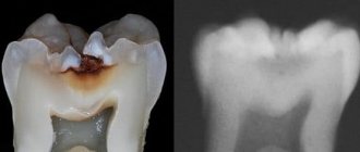
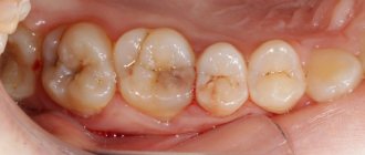
But average caries goes lower and affects dentin – the porous area of the tooth located under the enamel. Dentin is not very dense; it contains many tiny cavities that look like winding tunnels. Through these microscopic corridors, the infection quickly “gnaws” further – into the depths of the tooth.
Dentin makes up the largest part of the tooth. But it is not visible under the enamel. Many people are surprised, because they only had a small black hole in the enamel, and the dentist mercilessly drilled out half of the tooth. The fact is that the infection made its way through the enamel and began to wreak havoc in the dentin. This often happens: the hole in the enamel is small, but the carious cavity (the area of affected enamel and dentin) is very large.
By the way, if medium caries is not treated, it will inevitably lead to deep ones. Treatment will then involve removing the dental nerve. A very painful procedure. However, people usually go to the doctor precisely at the stage of middle caries. Symptoms are usually already present.
Symptoms
The main (and, in general, the only) symptom of average caries is a painful reaction to cold and hot. It's like a sudden flash of acute pain that lasts a few seconds and then disappears without a trace.
However, everything is individual. It happens that at this stage of the disease there is no pain at all.
Medium caries is usually clearly visible to the naked eye. This is an area of gray (possibly casting blue) color that is visible through the enamel. Well, it also looks like blackness on a large area of enamel (this is if it is the enamel that is severely affected).
The main (and, in general, the only) symptom of average caries is a painful reaction to cold and hot. It's like a sudden flash of acute pain that lasts a few seconds and then disappears without a trace.
As mentioned above, it is not easy to recognize secondary caries; however, there are certain symptoms, the appearance of which serves as an alarming signal and a reason for an early visit to the dentist. These include the following factors:
- The appearance of pain in individual teeth.
- Inflammation of the gums, their swelling.
- Manifestation of bleeding gums.
- The appearance of an unpleasant odor in the mouth.
You can also pay attention to some external signs, in particular, the filling will change its color either completely or along the edges, in addition, cracks or chips may appear in the enamel.
In the chronic course of secondary caries, there is a danger that, unnoticed by you, the infection penetrates deeper, undermining the dental nerve.
The first clinical signs appear 3-6 months after filling. For example, darkening of the enamel next to the filling, changing the color of the filling itself and its edges. Noticeable chips or cracks may occur.
At the stage of deep secondary caries, discomfort and aching pain occurs when brushing teeth and when eating cold, hot or sweet foods.
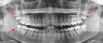
For diagnosis, a standard dentist's examination kit and an x-ray are used, since the carious lesion is hidden behind the filling, and it is difficult to determine its depth.
Secondary dental caries on x-ray
Risk factors
Factors of wear resistance of the filling:
- exposure to low and high temperatures - simultaneous intake of cold and hot food;
- eating hard, tough foods;
- bite defects, improper closing of the jaws - leads to excessive friction of the teeth;
- pathological abrasion of enamel, bruxism;
- violation of hygiene rules.
If you skimp on brushing your teeth after eating, be aware that food debris accumulates in the interdental spaces and in the cusps of your chewing teeth. A persistent bacterial plaque is formed, which will lead to secondary caries.
Possible consequences
If you ignore the emerging symptoms of the return of caries, the patient risks getting a number of problems and complications, namely:
- deep damage to bone tissue infection;
- caries damage to adjacent healthy teeth;
- the process of destruction of the root and dental canal;
- tooth loss.
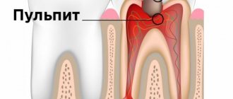
The main danger of secondary caries is that an inflammatory process develops in the pulp, which over time will lead to tissue death.
Depulpation procedure
Treatment of a superficial process that affects only the hard tissues of the tooth is limited to removing the affected part and installing a filling. Deep caries, which has spread to the soft tissues - the pulp, requires more serious intervention.
- Anesthesia (injection) is required during this procedure.
- Removing the old filling (if any).
- Drilling out hard tissues affected by caries, opening the tooth cavity.
- Removal of nerve and blood vessels from dental canals.
- Cleaning and disinfection of channels.
- Root canal filling.
- Closing the channels with a special gasket.
- Filling the cavity with filling material, grinding and polishing the filling.
Sometimes, after filling the root canals, an X-ray is taken to ensure the quality of their processing and filling. If the procedure is carried out in accordance with all requirements, the nerveless unit will no longer hurt.
Most often, chewing teeth are subjected to depulpation, this is due to the characteristics of their shape and the functions they perform. The load on them is greater than on the front ones, and care is often worse (due to inaccessibility).
Development mechanism
A secondary disease that affects healthy teeth develops in three stages:
- Microscopic gaps appear between the material used in treatment and the tooth.
- Pathogenic microorganisms begin to penetrate into these cracks and crevices.
- The proliferation of pathogenic organisms and the release of toxins that have a destructive effect on tooth enamel and installed fillings.
As a result, rejection of the material used begins.