There is a huge number of different dental diseases, among which dental hyperesthesia occupies a special place. This term is usually understood as a specific condition when people have very high sensitivity of tooth enamel. The pathology is acutely manifested by mechanical stress, as well as by ingestion of very hot, sweet or sour foods. The causes of pain may lie in injuries that occurred in the past. The development of pathology also depends on the thinning of the enamel. Other reasons may be erosion and other pathological processes. Depending on the clinical picture and the extent of the spread, dentists select the most effective treatment methods. In today’s article we will take a closer look at what dental hyperesthesia is, and also talk about how it should be treated.
Causes of dental hyperesthesia
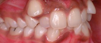
With hyperesthesia tooth sensitivity, patients may experience severe pain. Pain can occur as a result of mechanical or chemical effects. Patients often complain of discomfort when taking sour, salty, too cold or hot food and drinks. More often, this anomaly occurs in adults aged thirty to sixty years. The reason should be sought in non-carious lesions, which lead to abrasion of the enamel. An incorrectly formed bite and various wedge-shaped defects can lead to disruption of the integrity of the upper layer of the tooth. Treatment of hyperesthesia at home is ineffective. In order to properly treat the pathology, an in-person consultation and examination by a dentist is necessary. To understand how to treat dental hyperesthesia, you must first determine why it occurred. Doctors consider the following phenomena to be impact factors:
- Long and regular courses of home (and office) whitening. This can lead to thinning of the enamel, which will begin to react even to mild irritants.
- Periodontal inflammation and changes in tissue structure. When the root region is exposed, increased sensitivity develops.
- Regular use of a toothbrush with too hard bristles.
- Using highly abrasive toothpastes.
- Injury to tooth tissue.
- Hyperesthesia may be associated with disorders in the body.
- Hormonal disorders.
Hyperesthesia of tooth enamel
Hyperesthesia of tooth enamel is a lesion of the thin tissue that protects the tooth from external damage. Tooth enamel is very sensitive. A lack of vitamins and minerals, resulting from poor nutrition, disrupts the pH balance and destroys the protective layer of enamel.
- Excessive consumption of harmful foods: soda, sour foods and sweets is one of the causes of hyperesthesia of tooth enamel.
- Using hard toothbrushes and toothpastes with abrasive elements is another cause of the disease.
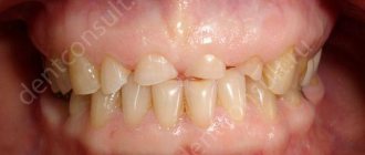
- Very often, enamel disease is accompanied by bleeding and atrophy of gum tissue.
- Failure to comply with the rules of oral hygiene, refusal to treat dental problems and visit the dentist.
- Bad habits also have a negative impact, which lead to the formation of cracks and violate the integrity of the enamel (clenching teeth, grinding teeth, biting nails, etc.).
In the absence of proper treatment, hyperesthesia of tooth enamel can provoke inflammation of the nerve and pulp of the tooth. Due to increased sensitivity, swelling of the gums occurs, which indicates an inflammatory process that requires medical treatment.
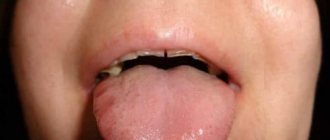
Symptoms
The first and most common symptom is the appearance of short-term pain attacks in the teeth. More often, people complain of pain after eating cold and hot foods. Pain may occur during the process or field of brushing your teeth. In the initial stages, the pain is short-term, but over time it can intensify. Often patients experience remission, at which time the irritants stop causing pain. When a period of remission begins, patients stop thinking about the problem and do not take appropriate measures. And without proper treatment, eliminating the pathology is impossible and hyperesthesia can return with renewed vigor. To eliminate dental hyperesthesia, it is necessary to establish the causes of its development.
Clinical manifestations of root caries.
The first is complaints. The patient most often complains of:
- An aesthetic defect (not beautiful) if there is caries on the vestibular surface of the front teeth.
- Discomfort while eating, pain when brushing teeth
- Symptoms of periodontal disease (bleeding gums, loose teeth, etc.)
- Short-term pain from irritants (teeth hurt from ice cream, hot tea). The pain disappears when the cause is removed.
Next, we look into the oral cavity and see the clinical picture. We will consider it taking into account the Leus classification
Along the way, root caries occurs:
- Active lesion - the edges of the cavity are undermined. The cavity is filled with softened tissues. There is a tendency towards a rapid increase in its size.
- Suspended caries - the edges of the cavity are smooth, dense, flat (not undermined). The bottom of the cavity is dense and shiny. Don't strive to get bigger.
- Secondary caries is the resumption of the process in the area of remission or after treatment (at the site of the filling).
According to the depth of the lesion:
- Without cavity formation - a white spot. Density is less than normal cement.
- With the formation of a cavity - any of those described in paragraph 1. carious defects.
Depending on what surface of the tooth it affects
Caries can affect any surface of the root, or 2 surfaces, or all surfaces in a circle (circular spread)
And lastly, according to ICD-10. There are cement caries and dentin caries.
- Cementum caries affects only the cementum of the tooth root
- Dentin caries also spreads deeper into dentin
The classification is over, and so are the clinical manifestations.
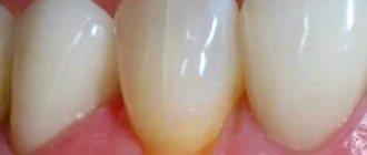
To summarize, root caries is a spot or cavity on the surface of the root of a tooth. Its edges are undermined or have smooth edges.
The cavity may be located above or below the gum.
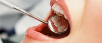
Diagnostics
The purpose of diagnostic measures is to determine and further eliminate the cause of the development of pathology. In this case, you need to consult a competent doctor. An experienced dentist will first make sure that the pain is not caused by progressive pulpitis, which has similar symptoms. It is necessary to conduct a thorough visual examination of the patient's oral cavity. After the examination, a consultation will be held, during which the dentist will take into account all the patient’s complaints. If necessary, the person will be sent for an x-ray.
Etiology and pathogenesis
Most often, hyperesthesia is observed in pathologies of dental tissues of non-carious origin, as well as in caries and periodontal diseases. It has been established that the state of permeability of dentinal tubules is a decisive factor in the development of dentin sensitivity, since a change in the flow of dental fluid in them can cause a pain reaction, which is realized due to the structural features and innervation of the dental pulp.
With caries, increased sensitivity may occur in one area. Very often, hyperesthesia is observed during abrasion of tooth tissue, when the loss of enamel reaches the dentin-enamel junction. However, not with all types of abrasion the increased sensitivity is expressed equally. Thus, with enamel erosion, hyperesthesia often occurs, while with a wedge-shaped defect it almost never occurs. Sometimes sharp sensitivity is observed even with insignificant exposure of the necks of the teeth (1-3 mm).
Theories of dental hyperesthesia
In dentistry, there are several theories of the occurrence of pain during hyperesthesia:
- Receptor - discomfort may appear as a response to irritation of the nerve in the dentin tubes.
- Nervous-reflex pain is the result of a disruption in the process of ion exchange in tissues and an increase in the perception of stimuli by the dentin receptor apparatus.
- Hydrodynamic - according to this theory, pain occurs as a result of fluid circulation in the dentin canals. It is this theory that has become most widespread.
The code for dental hyperesthesia according to ICD 10 is K03.80. In Russia, the International Classification of Diseases, 10th revision (ICD-10) has been adopted as a single regulatory document for recording morbidity. A special code assigned to dental sensitivity allows dentists to accurately diagnose and correctly select ways to treat the disease. Dental hyperesthesia ICD 10 requires the intervention of a professional dentist.
Cause of the disease
According to modern medical concepts, dental caries according to ICD 10 is considered an infectious disease. In this regard, the key cause of carious lesions is bacterial contamination of dental surfaces.
The following factors contribute to the formation of a carious defect:
Nutritional Features
Numerous clinical observations indicate that excessive consumption of sweets causes multiple dental caries.
It is very important to note that the destructive effect of carbohydrates occurs when sucrose comes into close contact with the enamel surface. In such cases, the formation of a carious cavity still requires the presence of pathological microorganisms and an insufficient level of oral hygiene.
Such gross violations of the composition of food as insufficient levels of proteins and vitamins also serve as an indirect cause of carious lesions of hard tooth tissues. This also affects the quality properties of saliva, which becomes more viscous and loses its protective properties.
One of the decisive cariogenic causes is the consistency of the food consumed. Soft foods create conditions for the formation of plaque and the formation of enamel defects.
Past illnesses
Some scientists express the opinion that some chronic diseases (tuberculosis, diabetes, etc.) have a direct impact on the severity of the carious process. This occurs due to a disruption in the blood supply to the dental pulp, which is responsible for the mineralization and strength of the enamel.
Previous systemic diseases have the most pronounced toxic effect on dental tissues during the period of change of primary teeth. At this time, the patient’s saliva composition changes significantly, which loses its protective properties.
External risk factors
Among the external cariogenic factors, ionizing effects should be noted. Thus, 6-8 weeks after completing a course of radiation therapy, a person experiences multiple chalky spots on the dental surfaces. Over time, these pigmentations turn into carious defects.
Very often, caries, this form of the disease, ends with the complete destruction of the crown. As a result, the patient requires orthopedic treatment. The pathological process is aggravated by the forced intake of soft carbohydrate foods.
In dental practice, radiation damage to enamel and dentin is usually called radiation caries.
Microbial factor
The presence of carbohydrates and pathogenic bacteria in the oral cavity is a prerequisite for the development of caries. Based on the results of numerous scientific studies, experts have found that demineralization and destruction of enamel are caused by organic acids produced by bacteria.
Problems of dental hyperesthesia and treatment methods
The problem of tooth enamel hyperesthesia is a rather serious issue, the solution of which can only be entrusted to an experienced doctor. The choice of a specific treatment method largely depends on the cause and development of the pathology. In dental practice, in addition to the ICD dental hyperesthesia code, such a concept as the dental hyperesthesia index is used. Pathology is assessed using the hypersensitivity prevalence index. The index is calculated in points and assessed according to the following indicators:
- 1 point - enamel reacts only to temperature factors;
- 2 points - hypersensitivity to temperature and chemical factors;
- 3 points - enamel reacts to temperature, chemical and mechanical influence.
Determining the hyperesthesia index allows dentists to correctly select treatment methods. In most cases, methods should be aimed at restoring dental tissue. In this regard, one of the most effective procedures is fluoridation and remineralization. Saturation of tissues with calcium and fluorine and other important elements will definitely give results. If the patient is diagnosed with the second or third stage of dental hypersensitivity, the doctor may suggest the use of special filling materials as a way to restore the shape of the crown of the tooth. When carious lesions are detected, dentists also use filling techniques. If the cause of the problem is a malformed bite, orthodontic treatment will be required. Treatment methods for dental hyperesthesia also include the use of special means:
- Pastes with a desensitizing effect - the action of these drugs is aimed at reducing hypersensitivity.
- Gels and foams are effective for strengthening tooth enamel.
- Varnishes – their function is to create a special film that will act as tooth protection.
- Electrophoresis is an effective method. It is also effective when taking special medications simultaneously.
- Traditional medicine - more often used in complex therapy or for prevention.
Hyperesthesia of hard dental tissues
Hyperesthesia of hard dental tissues is a common dental disease. Hyperesthesia is classified into several categories. There is a generalized and local form of the disease, as well as several degrees of development. Let's take a closer look at the features of hyperesthesia of hard dental tissues.
- Hyperesthesia by distribution
Painful sensitivity manifests itself both in the entire dentition and in an individual tooth. Depending on the degree of distribution of painful sensations, there is a local, that is, limited form of hyperesthesia and a generalized one.
- Local – occurs in one or several teeth at the same time. Very often, pain is associated with caries, non-carious lesions and other dental diseases of the hard tissues of the tooth. Increased sensitivity can occur due to treatment, extraction or filling of teeth.
- Generalized form - pain affects all teeth at the same time. As a rule, this form develops due to the exposure of tooth necks due to periodontal disease, tooth erosion, increased abrasion and other diseases.
- By origin
I distinguish two types of hyperesthesia, the first is associated, and the second is not associated with the loss of hard tooth tissue. If increased sensitivity arises from disruption and loss of hard tooth tissues, then this is due to the presence of carious cavities, increased abrasion of enamel and hard tooth tissues. If the disease is not associated with the loss of hard tooth tissue, then the appearance of increased sensitivity is provoked by periodontal diseases, metabolic disorders or gum recession.
- Clinical course
This category of disease has three stages. At the first stage, the tooth reacts to temperature stimuli, at the second stage, pain occurs due to temperature and chemical stimuli, and at the third stage, painful sensations appear when exposed to temperature, chemical and tactile stimuli. That is, painful sensations appear even from a light touch to the teeth.
This classification of dental hyperesthesia allows the dentist to carry out differential diagnosis and select the most effective treatment.
Prevention: what methods are effective
To protect yourself from such a problem as hypersensitivity of tooth enamel, it is necessary to periodically perform preventive procedures:
- Be careful about your diet.
- Use only optimally suitable care products.
- For teeth whitening, contact a trusted dentist.
- Visit your dentist at least twice a year and don't forget about professional cleaning.
An excellent prevention of dental hyperesthesia is regular dental examinations. Despite the fact that hyperesthesia is a serious pathological process, it can be eliminated. To eliminate the pathology, you need to contact a professional and competent doctor.
Topographic classification of dental caries
In our country, this classification is most widely used. It takes into account the depth of the lesion, which is very convenient for the practice of the dentist.
- Stage of carious stain - focal demineralization of hard tooth tissues is observed, and it can occur intensively (white spot) or slowly (brown spot).
- Superficial caries - at this stage a carious cavity appears within the enamel.
- Middle caries - at this stage, the carious defect is located within the surface layer of dentin (mantle dentin).
- Deep caries - in this case, the pathological process reaches the deep layers of dentin (peripulpal dentin).
In clinical practice, the terms “secondary caries” and “recurrent caries” are also used; let’s take a closer look at what they are:
1) Secondary caries is all new carious lesions that develop next to the filling in a previously treated tooth. Secondary caries has all the histological characteristics of a carious lesion. The reason for its occurrence is a violation of the marginal seal between the filling and the hard tissues of the tooth; microorganisms from the oral cavity penetrate into the resulting gap and optimal conditions are created for the formation of a carious defect along the edge of the filling in the enamel or dentin.
2) Relapse of caries is the resumption or progression of the pathological process if the carious lesion was not completely removed during previous treatment. Recurrence of caries is often detected under the filling during an X-ray examination or along the edge of the filling.