Tooth surfaces, dental formula
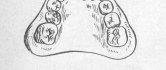
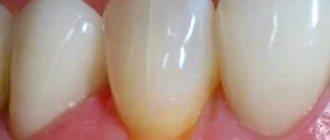
Tooth surfaces. For the convenience of describing the features of the relief or localization of pathological processes, 5 surfaces of the tooth crown are conventionally distinguished (Fig. 1).
Rice. 1. Surfaces (a), edge (b) and axis (c) of the tooth
1. The occlusal surface (fades occlusalis) faces the teeth of the opposite jaw. It is present in molars and premolars. The incisors and canines at the ends facing the antagonists have a cutting edge (margo incisalis).
2. The vestibular surface (facies vestibularis) is oriented towards the vestibule of the mouth. In the front teeth, in contact with the lips, this surface can be called labial (facies labialis), and in the back teeth, adjacent to the cheek, - buccal (facies buccalis).
The continuation of the tooth surface to the root is designated as the vestibular surface of the root, and the wall of the dental alveolus, covering the root from the vestibule of the mouth, is designated as the vestibular wall of the alveolus.
3. The lingual surface (facies lingualis) faces the oral cavity towards the tongue. For the upper teeth, the name palatal surface (facies palatinalis) is applicable. The surfaces of the root and walls of the alveoli directed into the oral cavity are also called.
4. The approximal surface (facies approximalis) is adjacent to the adjacent tooth. There are two such surfaces: the mesial surface (facies mesialis), facing the middle of the dental arch, and the distal surface (facies distalis). Similar terms are used to refer to the roots of teeth and the corresponding parts of the alveoli. On these surfaces there is a contact zone (area contingens).
Terms denoting directions in relation to the tooth are also common: medial, distal, vestibular, lingual, occlusal and apical.
When examining and describing teeth, the terms “vestibular norm”, “occlusal norm”, “lingual norm”, etc. are used. The norm is the position established during the study. For example, the vestibular norm is the position of the tooth in which its vestibular surface faces the researcher.
The crown and root of the tooth are usually divided into thirds. Thus, when dividing a tooth by horizontal planes in the crown, the occlusal, middle and cervical (cervical) thirds are distinguished, and in the root - the cervical (cervical), middle and apical (apical) thirds. Using the sagittal planes, the crown of the anterior teeth is divided into the medial, middle and distal thirds, and the frontal planes are divided into the vestibular, middle and lingual thirds.
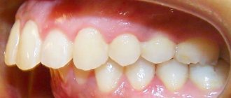
The dental system as a whole. The protruding parts of the teeth (crowns) are located in the jaws, forming dental arches (or rows): upper (arcus dentalis maxillaris (superior) and lower (arcus dentalis mandibularis (inferior). Both dental arches contain 16 teeth in adults: 4 incisors, 2 canines, 4 small molars, or premolars, and 6 large molars, or molars. The teeth of the upper and lower dental arches are in certain relationships with each other when the jaws are closed. Thus, the cusps of the molars and premolars of one jaw correspond to the depressions on the teeth of the same name in the other jaws. In a certain order, opposite incisors and canines come into contact with each other. This relationship of closed teeth of both dentitions is called occlusion (Fig. 2).
Rice. 2. The relationship between the upper and lower dentition in central occlusion:
a - direction of the teeth axes; b - diagram of the location of antagonist teeth
The contacting teeth of the upper and lower jaws are called antagonist teeth. As a rule, each tooth has two antagonists - the main and additional ones. The exceptions are the medial lower incisor and the 3rd upper molar, which usually have one antagonist each. The teeth of the same name on the right and left sides are called antimeres.
Dental formula. The order of the teeth is recorded in the form of a dental formula, in which individual teeth or their groups are written in numbers or letters and numbers. In the complete dental formula, the teeth of each half of the jaws are written in ordinal Arabic numerals. This formula for an adult looks as if the recorder is examining the teeth of the person sitting in front of him. This formula is called clinical. When examining patients, clinicians note missing teeth. If all teeth are preserved, the dentition is called complete.
Each tooth, in accordance with the full clinical formula, can be designated separately: the upper right ones - with the sign; top left ; bottom right ; lower left. For example, the left lower second molar is called , and the right upper second premolar.
The World Health Organization (WHO) has adopted a complete clinical dental formula in a different form:
Milk teeth in the full formula are designated by Roman numerals:
Individual baby teeth are pointed in the same way.
According to the WHO classification, the complete clinical dental formula for primary dentition is written as follows:
In this case, the lower left canine is designated 73, and the upper right first molar is designated 54.
There are group dental formulas that reflect the number of teeth in each group in the halves of the jaw, which can be used in anatomical studies (for example, in comparative anatomical studies). This formula is called anatomical. The group dental formulas of an adult and a child with milk teeth look like this:
This group formula of teeth means that in each half of the upper and lower jaws (or half of the dentition) there are 2 incisors, 1 canine, 2 premolars, 3 molars. Since both halves of the dental arches are symmetrical, you can write one half or a quarter of the formula.
The group dental formula can be written using the initial letters of the Latin names of teeth (I - incisors, C - canines, P - premolars, M - molars). Permanent teeth are designated in capital letters, while baby teeth are designated in lowercase letters. The dental formulas are as follows:
You can write down the complete formula of teeth using letters and numbers:
It is convenient to use such an alphanumeric formula when examining children with baby teeth whose permanent teeth have partially erupted. For example, the complete dental formula for a 10-year-old child may be as follows:
Individual teeth according to this formula are indicated with an angle sign, indicating the group of the tooth and its serial number. For example, the right upper second premolar should be written as follows: , left lower second molar: , primary right upper first molar: t1.
Human anatomy S.S. Mikhailov, A.V. Chukbar, A.G. Tsybulkin
Published by Konstantin Mokanov
Classification of mesial occlusion
Taking into account the size, as well as the position of the upper and lower jaws, the mesial occlusion is classified as follows:
- mesial bite, formed as a result of increased size of the upper, lower or both jaws (macrognathia);
- mesial bite, formed as a result of underdevelopment (reduced size) of the upper, lower or both jaws (micrognathia);
- mesial bite, formed as a result of a strong protrusion of the upper or lower jaws forward (prognathism);
- mesial bite, formed as a result of an abnormal posterior position in the skull of the lower or upper jaws (retrogression).
A combination of anomalies may also be observed, namely:
- underdevelopment of the upper jaw with increased size of the lower jaw;
- abnormal posterior position of the upper jaw in the skull with strong protrusion of the lower jaw;
- underdevelopment of the lower jaw with an increase in the size of the upper jaw;
- abnormal posterior position of the lower jaw in the skull with a strong protrusion of the upper jaw forward.
Mesial occlusion, formed as a result of lower prognathia and macrognathia, is a true progynea. False progynea is caused by the normal size of the lower jaw with underdevelopment of the upper jaw.
Violation of occlusion in the sagittal direction and the magnitude of the mandibular angle classify mesial occlusion into the following degrees:
First degree - the sagittal gap of the front teeth does not exceed two millimeters, while the first molars have irregularities in the sagittal direction of no more than five millimeters, the mandibular angle is 131 degrees.
Second degree - the sagittal gap of the front teeth does not exceed ten millimeters, while the first molars have irregularities in the sagittal direction of no more than ten millimeters, the mandibular angle is 133 degrees.
Third degree - the sagittal gap of the front teeth is more than ten millimeters, while the first molars have irregularities in the sagittal direction of about eleven to eighteen millimeters, the mandibular angle is 145 degrees.
In addition, the mesial bite can be dentoalveolar, which is characterized by an arbitrary backward displacement of the jaw located below, while achieving correct occlusion of the teeth located on the sides, as well as a gnathic mesial bite, in which displacement of the jaw is impossible.
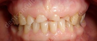
Symptoms of mesial occlusion
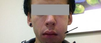
The most pronounced symptom of a mesial bite is a strongly protruding jaw in relation to the other. In addition, the patient has a violation of chewing and speech functions, diseases of the temporomandibular joint and periodontal disease. Experts note difficulties during dental prosthetics. The patient's chin has a pronounced shape, often protrudes forward, and the facial profile has a concave shape. When the jaws close, the incisors of the upper dentition are located behind the lower teeth; with normal jaw development, the position of the teeth is the opposite. The patient feels constant pain in the facial joints when opening and closing the mouth, as well as chewing food. Eating may be accompanied by a characteristic clicking and crunching of the jaws.
Experts characterize the facial expression of a patient diagnosed with mesial occlusion as “angry.”
Classification of tooth shape anomalies
Anomalies in tooth shape are usually classified as follows:
- • Fournier teeth,
- • Hutchinson's teeth,
- • Pfluger's teeth,
- • Spiked teeth,
- • Ugly shaped teeth.
The above anomalies arise as a result of insufficient development of the surface layer of tooth enamel (systemic hypoplasia).
Hutchinson's teeth have a screwdriver or barrel shape, and there is also a special hole-shaped notch on the cutting edge of the tooth. Such teeth are more often found on the upper incisors, located in the central part. Such pathology can be detected before teething begins through an X-ray examination. This pathology occurs during the formation of permanent teeth (approximately 6-7 months of intrauterine development). This anomaly is absent on baby teeth.
Fournier's teeth are screwdriver-shaped and differ from Hutchinson's teeth only in the absence of a semilunar notch on the cutting edge of the tooth. This anomaly occurs due to hereditary syphilis.
Pflueger teeth are one of the types of systemic enamel hypoplasia that occurs mainly on the first large molars. In this case, the tooth has the shape of a cone: the size of the crown of the tooth is much smaller than the size of the neck. Also, teeth with this anomaly are characterized by underdevelopment of the molar cusps. Pfluger's teeth develop due to a syphilitic infection.
Spiny teeth are characterized by a conical shape of the funnel part and resemble a spike. The reasons for the development of this anomaly are not known, but it is associated with the pathological development of the tooth buds. Quite often, spiky teeth are caused by congenital partial edentia. There is also an anomaly in the form of a splitting of the lateral incisor into two spike-shaped teeth with a cleft palate. People with this type of teeth are dissatisfied with the aesthetics of their smile, but sometimes spiky teeth can damage the oral mucosa. Another form of anomaly is twisted teeth with pronounced enamel hypoplasia. Such teeth can be observed in the anterior region of the upper jaw. A similar anomaly occurs due to the pathology of the development of the jaws and tooth buds.
Enamel hyperplasia , another type of tooth shape abnormality, is an excessive formation of hard tissue. Hyperplasia is accompanied by the appearance of enamel drops with a diameter of 1-4 mm on the chewing surface.
Anomalies are detected in rare cases in primary teeth, but most often deviations in the development of the shape of the teeth can be diagnosed after the formation of a permanent dentition.