Atypical neoplasms in the oral cavity are one of the most common dental problems. A type of such formation is exostosis, also called a bone spine. Gingival exostosis is a neoplasm formed from periodontal cartilaginous tissue. It is a benign growth and almost never degenerates into a malignant form. At the same time, it causes significant discomfort, makes it difficult to pronounce sounds, and worsens the quality of life.
Why does gum exostosis occur?
Exostosis of the gums can appear at any age, in a person of any gender. As with other bone pathologies, hereditary predisposition plays a major role in the development of exostosis. In the latter case, the tumor usually forms at an early age, and during adolescence, when the hormonal balance changes sharply, the growth begins to grow rapidly.
The main reasons for the formation of exostosis are:
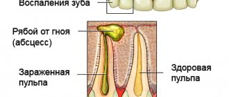
- gumboil, fistula and other purulent inflammations of periodontal tissues, accompanied by the death of the tooth root, destruction and atrophy of bone tissue;
- anomalies in the growth and development of individual teeth;
- chronic form of periostitis;
- hormonal disorders (hormones affect the qualitative and quantitative composition of bone tissue);
- syphilis;
- severe tissue damage during tooth extraction;
- fractures, dislocations and other jaw injuries.
Sometimes a lump of cartilage tissue forms in children during the period when baby teeth are replaced by molars.
Difficult tooth extraction and trauma to the maxillofacial apparatus are the most common causes of gum exostosis. In this case, active restoration of damaged tissue occurs: cells actively divide and grow in a random direction.
This situation is especially often observed when there is a refusal to apply a fixation splint to the chin, or the patient’s reluctance to monitor the immobility of the jaw at the stage of rehabilitation after a fracture.
Causes of cysts
Violation of hygiene measures - insufficient oral care - can lead to the appearance of a cyst. If you don't brush your teeth, food particles remain between them and gradually decompose. Putrefactive bacteria at the slightest injury to the gums - you may not even notice a scratch after chewing hard foods, such as crackers - penetrate into the wound. Inflammation begins.
Factors that provoke cyst growth are:
- pathology of the jaw bone as a result of traumatic influences;
- dentition disorder – congenital or acquired;
- abuse of bad habits - smoking and drinking alcoholic beverages;
- dysfunction of internal organs, exacerbation of chronic diseases.
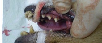
The culprit for the appearance of a growth on the gum after tooth extraction may be the individual reaction of the body and the low professionalism of the dentist.
Poorly placed fillings, gum infections during treatment, and similar manipulations lead to the appearance of a cyst.
In addition to purulent fistulous neoplasms, bone growths may appear on the gums. The reasons for their occurrence are also dental errors during tooth extraction and jaw injuries. The development of hard growths on the gums is influenced by hereditary factors.
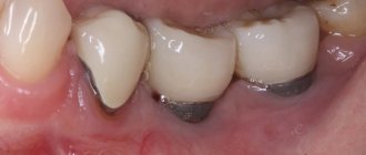
When cysts with purulent contents appear in the mouth, swelling appears, touching the mucous membrane is painful, and the general condition worsens. If, after tooth extraction, exostosis appears on the gums—the so-called hard bone growths—then it is possible to detect them in the initial stage only by palpating the problem area with the tongue.
Symptoms
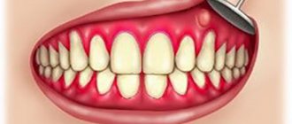
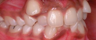
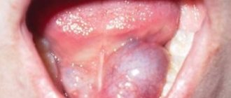
Exostosis looks like a round bump or sharp peak. Most often it is localized at the base of the fangs or incisors. Patients with a genetic predisposition almost always develop multiple growths located symmetrically after injury.
The formation of the growth is asymptomatic. A small, slowly growing lump forms under the gum. At the initial stages it may be invisible, and in most cases it is discovered by chance at a dentist’s appointment.
In advanced stages, the following symptoms allow one to suspect the development of pathology:
- a rounded bump (less often a sharp peak) clearly visible under the mucous membrane, hard to the touch;
- light, white gums in the area of the growth;
- feeling of constant presence of a foreign body in the mouth;
- difficulty chewing;
- speech disorder;
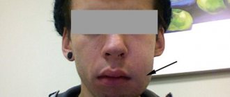
- facial asymmetry due to exostosis.
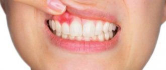
Exostosis usually does not hurt (except for cases when the tumor puts pressure on the roots of the tooth), but often provokes inflammatory processes: the inner surface of the lip or cheek constantly rubs against the bump and is damaged. When an infection enters the wound, inflammation begins. The lump turns red, the inflamed area swells, ichor or purulent contents begin to be released, and bad breath appears.
In dentistry, there are 2 types of exostosis:
- Develops after an injury to the maxillary sinus and does not affect the roots of the tooth. It can cause various complications in the nasopharynx.
- Formed from the cartilage tissue of the joint connecting the upper and lower jaws, it limits the range of motion. The patient cannot open his mouth completely, has difficulty smiling and chewing food. Because of this, over time, aching pain appears in the cheekbones, which causes discomfort to the patient and interferes with proper rest.
Exostosis - what it is and the reasons for its appearance
Exostosis of the jaw (osteophyte) is an area of hardened cartilage tissue that stands out on the surface of the jaw and appears as a lump on the mucosa. The cause of this phenomenon is most often a complex tooth extraction or other injury. If the tumor is small in size, it usually does not cause any discomfort. As a rule, this phenomenon is localized in the area of the median palatal suture or on the gum in the area of the alveolar process of the lower jaw.
Exostosis - the defect can be easily detected by touch
If the defect is not too pronounced, it does not in any way affect the functionality of the jaw system and the condition of the soft tissues. However, if the growth continues to increase in size, then the mucous membrane becomes thinner and is subject to constant trauma. There are difficulties opening the mouth, and when you press the protrusion, painful sensations appear.
On a note! One of the reasons for the formation of such bone growths is heredity. In this case, the neoplasm appears at an early age and begins to rapidly progress during puberty. In some situations, the growth resolves on its own over time.
Among other possible prerequisites for the appearance of exostoses, experts identify the following reasons:
- infections and inflammatory processes leading to the gradual destruction of bone tissue and its atrophy. These include flux and fistulas with purulent processes, cystic neoplasms, chronic syphilis,
- mechanical trauma – damage to the integrity of individual areas of the maxillofacial apparatus can cause fragmented destruction of osseous tissue,
- anomalies in the growth and development of individual teeth, causing curvature of areas of bone tissue and the formation of nodular hardenings,
- problems with the endocrine system - hormonal imbalance affects the qualitative composition of bone tissue, as a result of which it loses density and begins to gradually atrophy, which creates favorable conditions for the formation of bone formations.
Untreated gumboil can cause bone growth
The most common cause of exostosis is the difficult removal of a wisdom tooth. A bone tumor often forms in the socket of an extracted tooth. The culprit may be a violation of the technology of performing the procedure or the peculiarities of the clinical picture.
Diagnostics
If the development of exostosis is suspected, the dentist must conduct a differential diagnosis to distinguish it from a cyst, supragingival (epulis), or malignant tumor. The doctor takes a panoramic x-ray of the jaw, with the help of which he establishes the exact location, size, and shape of the exostosis. The diagnostic procedure also helps to identify:
- destruction of the tooth root and purulent inflammation near the growth;
- cracks and other hidden damage after impact, injury, tooth extraction;
- thickening of bone tissue, the initial formation of another exostosis.
If it is necessary to exclude the possibility of developing an oncological tumor, a biopsy or cytological examination is performed.
If necessary, the patient is sent for consultation to specialists in related specialties.
Characteristic symptoms
Exostosis in an adult is formed not only in the oral cavity; growths also appear on the feet, hands and collarbone. Only the bones of the skull are not susceptible to this disease. A person may develop several growths at once, their shapes and sizes can vary dramatically. Sometimes the neoplasm looks more like a thorn; often it has the shape of a mushroom with a narrow base and a wide surface.
The clinical picture largely depends on the location of the growth, its shape and size. Some patients have difficulty detecting small growths in their mouth, often only becoming aware of the problem after receiving x-rays. Sometimes the osteophyte acquires enormous dimensions; its shape may resemble a round orange.
At the initial stage, the disease is asymptomatic. At this stage, the disease can only be diagnosed in a clinical setting. The dentist will determine the location of the growth and determine the cause of its appearance.
As the disease progresses, the symptoms become more pronounced:
- A raised area forms on the gum. It is covered with a mucous membrane.
- The neoplasm gradually increases in size. If a small growth did not cause discomfort, then the presence of a large lump in the mouth interferes with the correct position of the tongue. The patient constantly experiences the sensation of the presence of a foreign object in the oral cavity.
- The patient begins to experience pain (of varying intensity).
- The mucous membranes covering the growth change color - they become bright pink.
- Occlusion occurs. The movement of blood through the capillaries and vessels in the affected area slows down.
- Partial dysfunction of the lower jaw is observed.
The difference between exostosis of the jaw and inflammatory diseases is the absence of itching, burning and elevated body temperature. The formation of a bone growth is not a disease, but a pathological process that requires constant monitoring.
READ ALSO: what to do if a growth forms on the gum after tooth extraction and its photo
Treatment of gum exostosis
If the cause of exostosis is endocrine disorders, and the diameter of the lump does not exceed 2-3 mm, they resort to wait-and-see tactics. After normalization of endocrine disorders, the tumor may disappear spontaneously.
In other cases, surgical removal of the tumor is performed. Indications for surgery are: rapid growth of a bone lump, pain, aesthetic discomfort, severe difficulties in eating and speech disorders, planned implantation and prosthetics.
Problems with the functioning of the adrenal glands, endocrine pathologies, blood clotting disorders, and diabetes mellitus are contraindications for surgery.
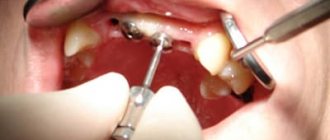
The standard removal procedure is carried out as follows:
- local anesthesia is administered, the oral cavity is treated with an antiseptic;
- the gum is carefully cut off, opening access to the exostosis;
- the cartilaginous growth is cut down using a special drill attachment, the bone base is ground to a smooth state;
- if there was a crack under the growth, a special plate is placed on it;
- The gum tissue is sutured, and the wound is covered with a sterile bandage.
An alternative treatment method is laser removal of exostosis. The laser beam heats and melts the pathological cartilage tissue. This eliminates the risk of bleeding and infection, shortens and facilitates the rehabilitation period.
Laser removal lasts about 60 minutes, the standard procedure requires a little more - 1.5-2 hours.
Rehabilitation
Immediately after surgery, pain and swelling may occur, and body temperature rises to subfebrile levels. This is a normal reaction of the body to surgery.
The main recovery period lasts about a week, but complete rehabilitation is completed in 3-4 weeks, when the soft tissues are completely healed and the gums grow together.
At the rehabilitation stage it is recommended:
- taking painkillers and anti-inflammatory drugs (Ketanov, Nimesil) in the first few days after surgery;
- rinsing the mouth with antiseptic solutions (Chlorhexidine, Chlorophyllipt, Rotokan) to prevent infectious complications;
- application to the wound area of applications with drugs that stimulate regeneration (Solcoseryl) in order to accelerate healing;
- internal use of antibiotics (Doxycycline, Tetracycline, Lincomycin) in cases where the growth of exostosis was accompanied by purulent inflammation.
During the recovery period, it is important to follow a number of rules and recommendations:
- eat only soft foods;
- do not consume too hot or cold food or drinks;
- do not touch the wound area with your tongue or hands;
- during the first 2-3 days after surgery, exclude any physical activity;
- stop smoking and drinking alcohol (tobacco smoke and alcohol irritate the mucous membranes and slow down the regeneration processes).
Possible complications
Failure to comply with medical recommendations during the recovery stage can lead to the development of complications, including:
- suture dehiscence caused by too intense chewing load or damage to the wound by solid food particles;
- inflammation that occurs as a result of unsatisfactory care of the wound and oral cavity, which is accompanied by swelling and redness of the mucous membrane, purulent processes.
The development of any complications requires immediate consultation with a doctor.
Gum exostosis is not a dangerous, but rather unpleasant problem. The appearance of a bone growth makes it difficult to speak and eat; if large, it injures the buccal mucosa, causing inflammatory processes. It is impossible to completely prevent the formation of exostosis. However, you can reduce the likelihood of a defect forming. To do this, it is important to monitor oral hygiene, avoid injuries, undergo preventive examinations with a dentist every six months and promptly treat identified pathologies of teeth and gums.
Indications for removal of exostosis
- rapid growth or large size,
- pain in neighboring teeth,
- pronounced cosmetic defect (when a growth forms on the outside of the jaw),
- the danger of changes in bite, especially in a child (due to the pressure of exostosis on adjacent teeth),
- preparing the patient to wear orthopedic or orthodontic structures.
Good to know! Most often, bone protrusions on the upper jaw grow onto the outer surface, giving a negative cosmetic effect, but on the lower jaw - on the inner surface, causing discomfort when chewing and speaking.