Infectious and inflammatory damage to bone tissue or osteomyelitis is a serious disease, often leading to disability or even death of the patient. A favorable outcome of the disease depends on the state of the patient’s immunity, the presence of pathologies of internal organs and the characteristics of the inflammatory process. But the main thing is the correct treatment of osteomyelitis. How quickly the body will clear the infection, how much bone tissue will be destroyed, whether the disease will become chronic and will not cause serious complications depends only on well-chosen methods of therapy.
Features of the purulent process in the bone
Osteomyelitis is one of the diseases that is very difficult to treat. This occurs due to the characteristics of the inflammatory process in bone tissue. When microorganisms multiply, the volume of bone marrow and spongy substance increases. This leads to compression of the blood vessels and disruption of the blood supply to the bone. Deprived of nutrition, large areas of bone tissue die, which creates favorable conditions for the proliferation of bacteria. Sometimes the purulent process affects nearby joints, muscles, ligaments and skin.
Another feature of osteomyelitis is that its causative agent is most often Staphylococcus aureus, which is very difficult to treat with antibiotics. Sometimes the causes of inflammation are hemolytic streptococcus, Escherichia coli or Pseudomonas aeruginosa. When treating, it is very important to identify the pathogen, otherwise incorrectly chosen antibiotics will lead to even greater bacterial resistance.
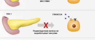
Pathogenic microorganisms are often present in the human body, but they are not always the cause of osteomyelitis. The seriousness of the disease and the complexity of its treatment are due to the fact that it occurs against the background of a weakened immune system, viral or other infectious diseases, diabetes mellitus, tumors, and pathological processes in internal organs. Therefore, osteomyelitis develops most often in weakened patients, elderly people or children.
The last feature of the disease that explains why it is difficult to cure is the difficulty in making a timely diagnosis. In the initial stages, bone inflammation shows almost no effect, especially if the infection has entered it from the inside through the bloodstream. Therefore, it happens that the patient does not consult a doctor on time, so the time required for successful treatment of osteomyelitis is lost - the inflammation becomes chronic. In addition, in some cases the disease develops rapidly: with high fever, severe pain, symptoms of intoxication, and disruption of the functioning of internal organs. Without medical care, this course of osteomyelitis can lead to the death of the patient.
Competent medical care and correct diagnosis are the main factors for successful treatment of the disease.
Causes
Jaw osteomyelitis occurs due to the penetration of pathogenic bacteria or viruses into the body. The further course of the disease depends on the state of the person’s immunity. If he is fine, then the disease enters the chronic stage or does not develop at all. But with immunodeficiency, the inflammatory process will begin to progress.
What diseases provoke osteomyelitis?
All causes of the development of osteomyelitis can be divided into several types:
| Name of the group of causes of the disease | Description |
| Odontogenic | The source of infection is a diseased tooth. Harmful substances enter the bone through the pulp when:
|
| Hematogenous | Pathogenic microorganisms enter the skeletal system from the primary source of inflammation:
|
| Traumatic |
|
| Radiation | Associated with oncological formations in the maxillofacial area and subsequent chemical and radiation therapy. The cells can develop into cancer cells. |
| Toxic | While taking narcotic drugs. |
Treatment tactics depend on accurate diagnosis, but with any type of pathology, you first need to destroy the source of inflammation, and only then act on the organs and systems that have been infected for the second time. If the patient suffers from diabetes, pathology of the hematopoietic organs or cancer, the prognosis worsens.
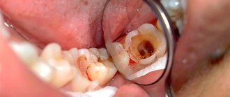
Osteomyelitis after tooth extraction
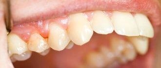
Osteomyelitis can occur after tooth extraction. In this case, the infection enters the hole due to insufficient quality of treatment of the oral cavity. From the hole, pathogenic microorganisms penetrate into the bone tissue. One of the main signs of the disease is increased mobility of the teeth located next to the socket.
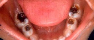
Osteomyelitis in the presence of milk teeth
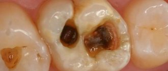
A carious cavity in a baby tooth that is not treated in time can also cause the development of pathology. Moreover, the infection often destroys the rudiments of the main tooth, so if any problems appear with baby teeth, they must be treated quickly, and not hope that they will fall out soon. Milk teeth are also removed if they have become a source of inflammation in the body.
What determines the effectiveness of treatment?
Only a specialist can determine how to treat osteomyelitis correctly in each specific case. Therefore, the effectiveness of treatment depends primarily on the patient’s timely seeking medical help. Based on the examination and complaints of the patient, the doctor can make a preliminary diagnosis. This is especially easy to do in case of post-traumatic osteomyelitis, when signs of tissue damage and the presence of pus are visible. But to confirm the diagnosis, it is very important to conduct an examination. It includes blood and urine tests, as well as instrumental methods.
Modern equipment for hardware diagnostics makes it possible to determine the degree of bone destruction, the presence of purulent tracts, and their location. For this purpose, radiography, computed tomography, magnetic resonance imaging, scintigraphy, densitometry, ultrasound and other methods are prescribed. To choose the right antibacterial drug, it is necessary to examine the purulent contents for pathogens.
Diagnostics
Diagnosis of osteomyelitis of the jaw and subsequent treatment is carried out based on the following examinations:
- radiography;
- biochemical blood test;
- general urinalysis;
- bacteriological culture to identify the type of pathogenic microflora.
A biochemical blood test is performed.
Important! In the acute form of the disease, the results of X-ray examination are uninformative or completely ineffective due to the absence of structural changes in the bone.
Subacute and chronic types of pathology are easy to establish based on radiography. The images will show foci of osteosclerosis, osteoporosis and sequesters - multiple or single.
Objectives of osteomyelitis treatment
To more effectively stop the inflammatory process, it is necessary to immobilize the infected limb. This is especially important when the tibia is affected, which is often subject to deformation. For this, corsets, splints or a plaster splint are used. The patient needs to remain in bed, this will help him maintain strength to fight the disease, and also protect his legs from overload and bone deformation.
In any form of the disease, it is very important to remove the pus from the bone so that it does not spread throughout the body. This is done by surgically opening the bone, cleaning it and rinsing it with antiseptics. If this is not done on time, blood poisoning, sepsis and death are possible. Such complications occur especially often when the bones near the shoulder joint become infected.
For successful treatment, it is very important to open purulent foci in time and remove pus
Therefore, in severe cases of acute infectious inflammation, detoxification therapy is very important to cleanse the blood of toxins. These are, for example, plasma transfusion, hyperbaric oxygenation or hemosorption, ultraviolet irradiation or laser therapy.
Complex treatment of osteomyelitis must fulfill one more task - restoring the integrity of the affected bone. This is done after the inflammatory process has subsided and the infection has been destroyed.
During the entire treatment, it is necessary to maintain the patient's strength to fight the disease. For this purpose, various immunomodulatory drugs, means to improve metabolic processes, vitamin complexes, dietary supplements and folk remedies are used.
Symptoms of osteomyelitis
The course of osteomyelitis depends significantly on the form. The severity of general symptoms, the degree of bone destruction, and the rate of progression of the process are determined by this factor. Therefore, it is advisable to consider different types of disease independently of each other.
Symptoms of acute hematogenous osteomyelitis
This form is characterized by the most severe course, due to the presence of severe general symptoms. National recommendations distinguish three variants of the course of hematogenous osteomyelitis:
Adynamic (toxic)
It is characterized by an unfavorable course, as it is accompanied by endotoxic shock. This is a deadly condition that develops when large amounts of bacterial toxins enter the blood. The patient's health suddenly deteriorates:
- Blood pressure drops;
- Consciousness is depressed (coma or stupor may develop);
- Temperature 40-41oC;
- Shortness of breath, without signs of lung damage;
- Children often develop seizures due to high fever and depression of brain function.
This leads to rapid development of heart failure and death. Even timely adequate treatment is not a guarantee of cure.
Local symptoms of bone osteomyelitis often go unnoticed due to the severity of general reactions. The patient cannot complain of pain in the affected bone due to depression of consciousness, and special diagnostic procedures (directed X-ray/CT) are not indicated for shock. Therefore, often its cause is revealed only at autopsy.
In rare cases (about 20%), upon examination it is possible to detect the following signs of acute osteomyelitis of the limb:
- moderate or mild swelling;
- redness and increased temperature in the projection of bone damage;
- contracture (difficulty in passive movements) of the joint located next to the infected bone;
- contouring of superficial veins (asymmetrical).
The detection of these symptoms does not significantly affect medical tactics, since first of all it is necessary to stabilize the patient’s condition. Only after this is surgical intervention indicated for osteomyelitis possible.
It should be noted that this is the rarest variant of the disease, which most often develops in children 14-17 years old.
Septic-pyemic
Also a severe form of osteomyelitis, which is more common than adynamic. General symptoms occur when the microbe enters the bloodstream, and not its toxin. It is characterized by fever up to 40°C, confusion, dull pressing headache, low blood pressure, severe sweating, and dehydration.
The peculiarity of this form is that local signs are expressed equally with the general ones, which is manifested:
- Severe cutting/bursting pain in the pathological focus. Typically, the patient can pinpoint the location of the lesion;
- Swelling of soft tissues and their redness over the affected bone;
- Contracture of the adjacent joint is possible.
A frequent complication of septicopyemic osteomyelitis is infection of other organs by microorganisms, with the formation of their failure. At the same time, one of the forms of the infectious process develops in them: inflammation of the organ (myocarditis, pneumonia, pyelonephritis, and so on) or a limited purulent focus (abscess, cyst).
It ends in death in 40-52% of cases, even with adequate treatment. With late presentation, the prognosis is much worse (mortality up to 70%).
Local
The most favorable option. With it, intoxication of the body is also observed, but less pronounced than with other forms. It is limited to the following symptoms:
- Fever 38-39oC;
- Decreased/lack of appetite;
- Sweating;
- Weakness;
- A headache of a dull nature, of medium or low intensity.
Local symptoms are extremely disturbing to the patient and reduce his quality of life. He complains of sharp, bursting pain in the projection of the infectious focus. NSAIDs (Ketorol, Ibuprofen, Analgin) and antispasmodics (Drotaverine/No-Shpa) do not have a significant effect on it. Upon examination, you can detect other local signs of osteomyelitis (swelling, redness, contouring of veins, contracture).
Symptoms of subacute osteomyelitis
As a rule, this is a transitional condition that ends in the formation of chronic osteomyelitis. General and local signs of the disease are less pronounced than in the acute form. The clinical picture is most often limited to the following symptoms:
General:
- Fever up to 37.6°C (low-grade);
- Slight weakness;
- No/minor headache.
Local:
- The intensity of pain decreases. It becomes dull, erased, and intensifies with load on the limb;
- Swelling is absent or insignificant on the affected limb;
- There may be contracture of the joint adjacent to the infected bone.
The main criterion for transition to the chronic stage is a persistent change in the affected bone.
Symptoms of chronic osteomyelitis
The patient's well-being with chronic osteomyelitis is much better than with other forms. Signs of a general illness disappear or low-grade fever remains. The pain in the limb becomes insignificant, aching in nature. Minor swelling usually persists with this form.
A distinctive feature of chronic osteomyelitis is the formation of pathological changes in bone tissue. These include:
- A purulent fistula is a canal that is connected to the infected bone on one side, and the exit hole is formed on the skin. Often several fistulas form, forming a kind of infected network;
- Curvature of the limb (usually shortening);
- Sequestration (separation of destroyed parts) of bone. This is an x-ray sign that is not reflected in symptoms.
The course of chronic osteomyelitis is staged. There is an alternation of phases of remission (disappearance of symptoms) and relapse/exacerbation. In the second phase, the clinical picture is similar to that of acute osteomyelitis.
The chronic multifocal form has a similar course. The difference lies in the prevalence of local symptoms (they appear on several bones) and the absence of purulent fistulas.
Osteomyelitis of the maxillofacial region
It should be noted that the symptoms of osteomyelitis of the jaws (upper/lower) also depend on the form of the disease. The peculiarity of the clinic is the possible appearance of a number of additional symptoms: pain during swallowing, contracture of the temporomandibular joint (the patient cannot open his mouth), severe swelling of the face. General symptoms do not have any particularities.
It is recommended to treat a disease of this localization with an oral and maxillofacial surgeon, since he has sufficient knowledge to timely identify complications (cellulitis of cellular spaces, purulent leaks, and so on) and has the ability to perform specific surgical interventions.
Symptoms and course of atypical forms
Currently, three atypical forms of chronic osteomyelitis have been described. The reason for their development is unknown, since the role of bacteria has not been confirmed. Specific bone changes and some clinical features made it possible to isolate them into a separate group. Highlight:
- Brodie's abscess is the formation of a small cavity in the bone containing purulent/serous fluid. It can occur latently or with unexpressed local symptoms (pulling pain, periodic swelling of tissues). Limb deformity is rare;
- Gare's osteomyelitis (sclerosing) - symptoms resemble classic subacute osteomyelitis. Features: increased symptoms at night, the bone thickens at the site of the lesion, more often observed in men under 30 years of age;
- Ollier osteomyelitis (albuminous) - occurs as chronic osteomyelitis. Isolated in a separate form, since pus does not form in the lesion. Instead, a serous fluid enriched with proteins is secreted.
Diagnosis of these forms is carried out using instrumental and invasive (violating the integrity of the skin) methods.
Basic methods of treating the disease
Osteomyelitis is a serious disease that affects the entire body. Therefore, its treatment must be comprehensive. Only by influencing the source of inflammation using different methods can the infection be dealt with. Therefore, a set of measures is most often used, which includes:
- drug therapy;
- dietary nutrition;
- local treatment with ointments and antiseptic solutions;
- physiotherapeutic treatment;
- traditional methods;
- surgical intervention.
Drug therapy
The main goal of treating osteomyelitis is to destroy the infection. This is best treated with medication. Various groups of drugs are prescribed. But to destroy the infection, which is the main goal of treatment, antibiotics are needed. First, you can start therapy with broad-spectrum drugs, then, after identifying the causative agent of the infection, special agents are prescribed. For osteomyelitis, the following drugs are especially effective: Cefazolin, Vancomycin, Fusidin, Kefzol, Linkamycin, Ceftriaxone, Co-Trimoxazole. Sometimes, in severe cases, a combination of two antibiotics is required. This therapy continues for at least a month, often even longer.
To eliminate the effects of intoxication and the consequences of antibiotic use, detoxification therapy is prescribed. This may be the administration of saline solution, washing the blood with Ringer's solution, Trisol or Normosol, as well as the use of diuretics.
Immunomodulators must be used in complex treatment. They are needed to strengthen the body's defenses and increase its resistance. This could be Timolin, Amiksin, Thymogen. It is important to restore the intestinal microflora, which will help strengthen the immune system. For this you can use Linex, Bifiform, Acipol, Hilak Forte and other drugs.
Physiotherapeutic methods help speed up recovery
Theory of the mechanism of development of the disease
The transition of acute osteomyelitis to chronic is evidenced by the lack of effectiveness of therapy within 30–45 days. The patient's condition may improve, but the results of diagnostic procedures indicate further development of destructive processes. In children, the chronic form of the pathology is usually hematogenous in nature.
The slow nature of the development of the inflammatory process is characteristic of various forms of pathology. The disease is accompanied by the formation of fistulas and voids in the bones. If bone destruction begins due to postoperative complications, purulent fistulas form in the area of the sutures.
Hematogenous osteomyelitis promotes the spread of exudate affecting the entire bone. Often inflammation affects connective and cartilaginous tissues. This alternation of phases can take quite a long time. An exacerbation can happen at any time. It can be triggered by:
- infectious and colds;
- excessive physical activity;
- weakening of the body's defenses.
The fistula closes and pus begins to accumulate in the bone cavity.
The exacerbation begins abruptly: the temperature rises, signs of intoxication intensify. The patient complains of general weakness, headaches, and fever. Redness and swelling occur in the affected area, and severe pain is observed. These symptoms disappear when the fistulas open and the purulent masses come out.
The pathogenesis of osteomyelitis has not been fully revealed, but doctors have identified the following 3 theories according to which the disease develops:
- Nervous-reflex. Negative mental states or post-traumatic stress lead to vasospasm in bone tissue. If you do not use antispasmodic drugs, regular strong contraction of blood vessels provokes tissue necrosis. In combination with the penetration of bacteria into the body, osteomyelitis occurs.
- Vascular. It is characterized by a decrease in the rate of blood circulation, which creates a risk of blood clots and death of bone tissue. If there are foci of infection, bacteria travel through the bloodstream throughout the body and cause suppuration.
- Allergic. The body's high sensitivity to irritants can cause an allergic reaction. Due to this reason, inflammation develops in the perivascular spaces. The pathological condition leads to compression of blood vessels, blood supply is suspended, and bone tissue lacks oxygen. Swelling negatively affects surrounding tissues, affecting healthy areas. If a microorganism gets into the focus of the inflammatory process, osteomyelitis develops.
Physiotherapeutic treatment
After the patient’s condition has stabilized, the temperature has dropped and the pain has subsided, physiotherapeutic methods are prescribed. They are designed to stimulate blood circulation and tissue regeneration processes, prevent the development of contractures and restore limb mobility. First of all, this is physical therapy. You can start exercising shortly after your condition improves. But the affected limb can be loaded no earlier than 20 days after surgical treatment was performed.
Laser therapy for bone inflammation is now considered the most effective. In this case, special substances are introduced into the blood, which quickly penetrate into the purulent focus and linger there. After this, they are burned out by laser radiation along with the cells affected by the infection. This method is often used for osteomyelitis of the calcaneus.
Drug electrophoresis, UHF therapy, ultraviolet and ultrasound irradiation, mud applications, paraffin treatment, ozokerite and other methods are also effective for treatment.
Symptomatology and clinical picture
The toxic (adynamic) form of hematogenous osteomyelitis is characterized by extremely rapid (lightning-fast) development and an extremely severe course. The clinical picture of the disease is dominated by general and septic phenomena. The picture of the toxic form, which is unclear for typical forms of osteomyelitis, usually presents significant difficulties for diagnosis.
In this regard, the treatment of such patients, and sometimes the section, is carried out under a different diagnosis. The disease either occurs suddenly or is preceded by a prodromal period, manifested by mild malaise, which is usually visible. The prodromal period lasts on average from 1 to 4 days. In the toxic form of osteomyelitis, the patient’s temperature immediately rises to very high numbers (39-40ºC) and, unlike other forms, remains at this level until the child’s death.
Most of these patients die within 1-3 days. The child soon falls into an unconscious state and at times becomes delirious. Such patients do not experience violent chills; the child’s face is hyperemic, he is covered in sweat, and there is a toxic exanthema on the skin. The patient usually lies motionless; at times, convulsions occur. With the most careful examination of the patient, it is usually not possible to identify an area of pain on the limb.
The septicopyemic (severe) form is observed in 40% of patients. At the onset of the disease, general phenomena predominate, indicating a severe septic condition of the patient. The disease begins suddenly with a sharp chill with an increase in temperature to 39^ and above. There is general weakness, weakness, headache, vomiting, drowsiness, but the child does not lose consciousness.
Appetite disappears, the tongue is coated and dry, the face becomes pale, lips and mucous membranes are cyanotic, blood pressure is reduced, heart sounds are muffled, the pulse is rapid, weakly filled and corresponds to the temperature, breathing is rapid and shallow. The liver and spleen are enlarged and painful on palpation. Little urine. There is protein and casts in the urine.
The temperature in the first days remains at the level of 39-40ºС; as suppuration develops, it takes on a remitting character with large fluctuations throughout the day.
In the blood test, the changes are significantly greater than with the fulminant-toxic form. There is pronounced leukocytosis, a sharp shift in the formula to the left and the appearance of young forms of leukocytes. Anemia develops quickly.
Complaints of acute pain in the affected limb are typical already on the first day of the disease. Due to reflexive muscle contraction, the limb assumes a semi-bent position. Over the next 1-2 days, changes are detected in the form of painful swelling of soft tissues, moderate redness of the skin, swelling, and a local increase in temperature.
By the end of the second day of the disease, a subperiosteal abscess forms in the affected area. Subsequently, the pus perforates the periosteum and reaches the subcutaneous tissue from the depths. At this point (5-7 days) fluctuation appears. With the development of intermuscular phlegmon, the patient's condition improves somewhat.
For the purpose of early detection of acute hematogenous osteomyelitis, special importance should be given to the details of the disease history, which can be established with the help of the child’s mother. When examining a child, it is advisable to conduct the examination while he is sleeping. If necessary, you can resort to medicated sleep.
In young children, with a rapid course of the process, early radiological signs of osteomyelitis appear after 3-5, and in older children - 12-15 days after the onset of the disease. An early diagnostic symptom of osteomyelitis is detachment of the periosteum. Bone changes begin in the metaphysis and are expressed in blurring of the bone beams.
Local (mild) form. A milder onset and further course of the disease are observed. Local inflammatory phenomena prevail over general ones. The inflammatory process does not always resolve with suppuration. Sometimes, under the detached periosteum in the affected area, not pus is found, but an accumulation of mucous-protein fluid mixed with hemorrhagic exudate.
Diet therapy
For a patient with osteomyelitis, it is very important to adjust your diet. During illness, the body needs a large amount of vitamins and minerals. Therefore, you need to include foods rich in calcium, iron, magnesium, and phosphorus in your diet. B vitamins are also needed. You need to eat more vegetables and fruits, drink at least 2 liters of water. And every morning drink a mixture of beet juice (2 parts) and carrot juice (5 parts).
In the diet of a patient with osteomyelitis, it is important to replenish protein losses. Therefore, it is necessary to include lean meat and fish, liver, eggs, and dairy products in your diet.
Herbal baths and compresses are effective in the complex treatment of osteomyelitis
Local treatment
The outcomes of acute hematogenous osteomyelitis depend on the timeliness and adequacy of treatment, in particular, on surgical intervention.
Doran and Brown figuratively wrote back in 1925 that if the time from the onset of the disease to surgery is measured in hours, then the patient’s recovery is measured in weeks, but if the time before surgery is measured in days, then recovery is delayed for months and years.
The impact on the macroorganism is aimed primarily at relieving intoxication, eliminating acute disturbances of vital functions, and preparing the patient for surgery.
Correction of water and electrolyte balance is carried out by intravenous infusion of solutions: glucose with insulin, plasma, albumin, polyglucin, rheopolyglucin, hemodez. Electrolyte disturbances are corrected by administering Ringer-Locke solutions, lactosol, etc.
1) pulse, arterial and central venous pressure;
2) hemoglobin and hematocrit levels;
4) the content of potassium and sodium in the blood plasma.
In order to detoxify the body, the method of forced diuresis, hemosorption, etc. is used.
With hyperthermia, t.s. If the body temperature rises above 38.5ºC, they resort to giving humidified oxygen through a nasal catheter, rubbing the skin with alcohol until reddened, blowing with a fan, applying an ice pack to the head and groin areas, intravenous administration of a 5% glucose solution cooled to 4°C. Medications such as analgin, amidopyrine should be used less frequently.
In cases of hypoxia, it is advisable to use hyperbaric oxygenation. Pain relief is achieved by administering non-narcotic analgesics. The use of protease inhibitors—trasylol, contrical, etc.—is indicated. Allergization of the body is relieved by prescribing desensitizing drugs.
The energy balance is maintained by the administration of glucose, fructose, protein and fat preparations. B vitamins and vitamin C are needed.
Nonspecific immunotherapy: pentoxyl, potassium orotate. prodigiosan, levamisole, thymalin, etc.
Specific immunotherapy: staphylococcal vaccine, toxoid, gammaglobulin, hyperimmune plasma (targeted action).
Puncture method. A special needle is used to puncture the bone at an angle of 60-70º, followed by the introduction of antibiotics through it. The method is indicated only in the early stage of the disease, in the first 2 days.
Method of decompressive osteoperforation. Applying small burr holes and leaving them near the distal hole of the needle for administering antibiotics. Usually 2-3 holes are enough. The distance between the holes is 2-3 cm, the diameter is from 3 to 7 mm, depending on the age of the patient and the caliber of the bone.
Recently, laser osteoperforation followed by wound treatment with low-frequency ultrasound and a pulsating jet of antiseptic has been successfully used.
The operation is completed by draining the wound using silicone tubes and preparing for intraosseous lavage. Lavage is carried out with 0.1% hydrochloric acid. 0.1-0.2% solution of chlorhexidine digluconate (hibitan), 0.1% solution of rivanol, etc. Antibiotics and enzymes (trypsin, chemotrypsin, chymopsin) must be introduced into the bone cavity.
Intraosseous lavage is carried out continuously or more often in fractions of 4-6 hours for 5 to 10 days or more.
Recently, ultraviolet irradiation of blood, as well as endovasal irradiation of blood with a laser beam, have been used with good effect.
Treatment in a controlled abacterial environment is used.
The outcome of treatment of acute hematogenous osteomyelitis: currently, mortality ranges from 1 to 7%; transition to the chronic stage in 10 to 20% of cases.
Traditional methods of treatment
In the complex treatment of the disease, after consultation with a doctor, any methods can be used to alleviate the patient’s condition. Therefore, treatment of osteomyelitis with folk remedies is also often used. But it should not replace therapy prescribed by a doctor.
All traditional methods are used only as additional treatment:
- It is recommended to drink a tablespoon of fish oil and a raw home-made egg in the morning;
- pour 200 g of walnut partitions into 0.5 liters of vodka and leave for 2 weeks, then drink a tablespoon of the tincture three times a day;
- in the chronic course of the disease, you can apply compresses of chopped onions mixed with laundry soap to the affected area at night;
- for osteomyelitis of the foot, baths with ash from aspen logs are effective;
- an infusion of lilac flowers with vodka is used for compresses and drunk 2 drops per day;
- An application made from this dough draws out pus well and promotes wound healing: mix liquid honey, rye flour, butter and yolks;
- Wash wounds after traumatic osteomyelitis with juice from fresh nettle leaves or chamomile decoction;
- apply a paste of crushed aloe leaves, this will help cleanse the lesion from pus.
To prevent complications, it is important to remove pus from the osteomyelitic cavity in a timely manner.
Surgery
Surgical treatment is necessary for osteomyelitis to cleanse the bone tissue of pus. This is easiest to do when all parts of the bone are affected. And when an infection develops from the inside, it is necessary to open the periosteum and expose the bone marrow. After removing the purulent focus and washing the cavity with antiseptics, a drainage tube is inserted into the bone to drain fluid and pus. Through it, the osteomyelitic cavity will be subsequently treated with antibiotics, accumulated pus will be removed and washed. This operation is called osteoperforation.
With chronic osteomyelitis, there is often a need for repeated surgery. This happens if conservative treatment is ineffective, frequent relapses occur, many fistulas and sequestration are formed, and purulent lesions of soft tissues occur. In such cases, operations are more traumatic. For example, sequesternecrectomy is performed to remove all dead and necrotic areas. Sometimes it is also necessary to perform a resection - complete removal of the affected area of the bone and connecting its remaining ends with grafts. For this purpose, artificial materials or the patient’s own tissues are used.
Forms
According to the nature of the course, osteomyelitis of the jaws is divided into several forms:
- Spicy. It develops rapidly and is characterized by a vivid manifestation of symptoms: fever, chills, severe pain at the site of the lesion, which soon begins to radiate to the ear, temple, or orbit.
- I'll sharpen it up. It is a transitional form between acute and chronic osteomyelitis. The symptoms subside, the patient’s general condition improves, but the infection continues to spread. Often the “quiet period” is facilitated by the spontaneous discharge of purulent exudate through the fistula.
- Chronic. It is diagnosed if the disease lasts more than 1.5 months. Symptoms are moderate, there are structural changes in bone tissue, and the formation of sequesters - isolated dead areas.
The acute form develops rapidly, accompanied by elevated body temperature and chills.
Important! Chronic osteomyelitis of the jaw can develop as a complication of acute and subacute types, or as an independent form.
Features of the treatment of post-traumatic osteomyelitis
If bone inflammation began after an injury, its treatment has some features. Post-traumatic osteomyelitis is characterized by the presence of a wound through which it is convenient to wash the purulent focus. In addition, fistulas are usually located around the affected area. Therefore, proper timely antiseptic treatment will help avoid the development of the inflammatory process.
After gunshot wounds and complex comminuted fractures, osteomyelitis develops most often. Treatment in such cases should be surgical. It is imperative to remove all bone fragments, dead tissue and foreign objects from the wound. The affected area is pricked around with antibiotics, and a drainage tube is inserted into the cavity. After this, treatment methods are the same as for other forms of osteomyelitis.
There are also special features in the treatment of chronic post-traumatic osteomyelitis. With a long purulent process, the bone is deformed and joint mobility is impaired. To restore limb function, osteosynthesis and replacement of lost areas with grafts are performed.
Acute hematogenous osteomyelitis
Hematogenous osteomyelitis is a common disease. It affects mainly children and adolescents in 90% of cases. Boys get sick 2-3 times more often than girls.
up to 5 years - 20-30%, from 5 to 14 years - 70-75%.
Among the bones of the skeleton, the femur and tibia are most often affected (80%), the patella, shoulder blades, ribs, vertebrae, and skull bones are rarely affected.
Regarding the zones of primary inflammation of long tubular bones, it should be noted that when the femur is affected, the process is most often localized in the area of the lower metaphysis, and when the tibia is affected - in the upper metaphysis, which, as is known, corresponds to the zones of maximum growth of these bones in length.
Thus, of all the diaphyseal bones of the limb, the bone bearing the greatest functional load is most often affected, and along their length the inflammatory process, as a rule, develops near the most active growth plate. This pattern must be taken into account in the complex of causes and conditions that explain the pathogenesis of hematogenous osteomyelitis, and first of all requires special study.
Treatment of odontogenic osteomyelitis
One of the most common forms of the disease is inflammation of the jaw bones. It often develops in children under 12 years of age. If treated incorrectly, this leads to facial deformation, loss of teeth and other dangerous consequences. Therefore, it is very important to choose the most effective methods of treatment for odontogenic osteomyelitis.
Treatment should be carried out in a hospital. The main method is surgical removal of affected tissues and teeth, washing the source of infection. It is necessary to create conditions for the outflow of pus and prevent further proliferation of pathogenic flora. For this purpose, antibiotic therapy, rinsing with antiseptics, and laser irradiation are used.
Incorrect treatment can lead to patient disability
Kinds
Pathology is classified according to several criteria. Depending on the location, osteomyelitis of the lower jaw or upper jaw is distinguished. The first develops in 80% of clinical cases. The second occurs several times less - it is diagnosed in 30% of patients.
According to the degree of prevalence, limited and diffuse types of the disease are distinguished. In the first case, the inflammatory process is local in nature and affects the area of the jaw body and alveolar processes ranging from 2 to 4 teeth. The diffuse type covers all or most of the jaw.
According to the nature of occurrence, pathology is divided into:
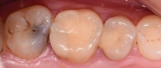
- Odontogenic. The cause of the lesion is always an infected tooth. Pathogenic microorganisms that multiply in the inflamed or necrotic pulp (neurovascular bundle) penetrate into the bone tissue through the apical foramen of the root.
- Hematogenous. The infection enters the bone through the bloodstream from another source of inflammation. This species is characterized by the reverse method of development: first the body of the jaw is affected, and then the tooth at the site of inflammation.
- Traumatic. Bacteria and microbes are brought inside during mechanical damage to tissues.
Important! Odontogenic osteomyelitis occurs most often - up to 80% of cases. Less common are hematogenous (11%) and traumatic (9%) forms of the disease.
Complications of the disease
If treatment is not started in a timely manner or if the treatment is chosen incorrectly, osteomyelitis can cause serious complications. This happens especially often in patients who are weakened, elderly, or suffering from pathologies of internal organs. All complications of the disease are very serious and therefore require immediate response.
What consequences can osteomyelitis cause:
- purulent lesion of soft tissues – abscess or phlegmon;
- infectious arthritis;
- muscle contractures and joint ankylosis – loss of mobility;
- spontaneous bone fractures;
- malignant tumors;
- blood poisoning;
- severe kidney damage;
- anemia.
Prevention of osteomyelitis
This serious disease is very difficult to cure. Therefore, everything possible must be done to prevent the bone from becoming infected. To do this, you need to strengthen your immune system, eat right, give up bad habits and exercise. After all, bacteria multiply best in a weakened body. It is necessary to treat all chronic diseases in a timely manner, as well as eliminate foci of infection: caries, sinusitis, sinusitis, sore throat, purulent wounds. If skin damage or injury occurs, it is necessary to treat the wound with an antiseptic, such as hydrogen peroxide or Chlorhexidine. In case of a fracture, you must immediately contact a medical facility.
Osteomyelitis is a dangerous infectious disease that can cause considerable harm to human health. If not treated correctly, it often leads to loss of performance and disability, and in some cases ends in death due to damage to the kidneys, liver or other organs. Properly selected therapy is the main way to quickly restore the functions of the affected limb.