1166
Many people suffer from various inflammatory diseases occurring in the oral cavity. Some of them leak inside the gum tissue and are completely invisible during normal visual inspection. One of these diseases is chronic fibrous periodontitis.
What is
This disease is an inflammation that develops in the periodontium. These are very thin (up to 0.25 mm) tissues that are located in the slit-like space between the cementum of the dental root and the alveolar plate. Blood and lymphatic vessels and nerve fibers pass through them. Periodontitis compensates and redistributes the loads that occur during chewing.
We will be told in more detail about the concept of “periodontitis” in the following video:
Inflammation in these tissues is usually chronic and occurs without complications. The disease is characterized by a strong decrease in the number of “native” cells and their replacement by others. Their place is taken by cells of coarse fibrous tissue. Namely, this process is characteristic of fibrous periodontitis.
Important: pathology, depending on the type of changes occurring in periodontal tissues, has several forms. Fibrous periodontitis is the most harmless form of the disease for humans.
Causes
The appearance of the disease is associated with two groups of causes: local and general. The first group includes the following factors:
- infection that developed after the filling was reabsorbed;
- infection of tissues by pathogenic microorganisms due to untreated caries or pulpitis;
- incorrectly selected or installed orthopedic systems;
- the presence of a foreign element in the periodontal tissues - a filling mass taken in large volumes, an exposed edge of the pin;
- accidental damage to periodontal tissues by the placed filling (this often happens when the height of occlusion increases);
- effective therapy for granulomatous/granulous periodontitis , when these pathologies pass into a safe fibrous stage.
The most common causes belonging to the second group are diseases:
- nervous;
- circulatory (heart and blood vessels);
- endocrine systems.
Fibrous periodontitis often develops with reduced immunity.
Antibiotics
Antibiotics for periodontitis are prescribed to prevent the development of the inflammatory process. Most often they are prescribed for purulent acute periodontitis, when there is a danger of pus entering the patient’s blood. In this case, the doctor, after medicinal treatment and filling of the tooth root canals, prescribes antibiotics to the patient to localize the inflammatory process and prevent its spread to surrounding tissues.
Drug therapy involves the use of not only antibiotics, but also other medications and vitamins, including painkillers. Every practicing dentist has modern and effective drugs in his arsenal that help cope with the disease.
Drugs for the treatment of periodontitis help not only to eliminate the source of inflammation, but also promote the regeneration of damaged periodontal tissue and restoration of bone tissue, which is especially important in the treatment of granulating and granulomatous periodontitis.
Symptoms
Under the influence of these reasons, structural changes occur in the periodontal tissues over a long period of time: their ligaments become significantly thicker, and the cells are replaced by fibrous ones. Because of this, the tissues themselves become scarred, thickened and roughened.
But a person does not feel such changes in any way, since the disease has a sluggish and almost inexpressive course. Symptoms are poor: in isolated cases, slight pain may occur from strong pressure on the gum or from tapping on the tooth.
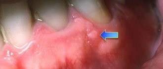
Signs characteristic of most dental diseases (severe pain, sensitivity to temperature changes, fever, swelling of the mucous membrane and tissues) are absent. In rare cases, the gums in the affected area change color.
We'll cover the pros and cons of getting In-Ovation braces in our next article. And here you can find out what the cost of installing retainers is and what it depends on.
Symptoms of acute periodontitis
In acute cases, the patient actively complains about his well-being. At the same time, permanent changes have not yet occurred in the soft tissues, despite the actively developing inflammation. If adequate dental intervention is performed at this stage, there is a high probability of complete elimination of the pathology.
Serous
Apical periodontitis, which occurs after injury or an allergic reaction to dental medications, most often develops as a serous type. It manifests itself only by local symptoms of damage to the dentofacial apparatus, such as:
- The pain is aching or throbbing, quite intense. It weakens somewhat after taking anti-inflammatory drugs (Ketorol, Ibuprofen, Citramon, Nise, etc.). May increase when palpating the affected area with the tongue or finger;
- Excessive mobility - due to the production of fluid in the gum pocket, the tooth becomes more mobile. Even when exposed to the tongue, a slight displacement and discomfort is felt;
- Unpleasant sensations when “biting” - any load on the inflamed periodontium leads to pain.
By examining the place where signs of the disease occur, you can detect an area of redness of the gums, local swelling and smoothing of folds on the mucous membrane. This form is more favorable for the patient than the purulent form, since there is no infectious process in the dentofacial apparatus. Complications of periodontitis with the serous variant are extremely rare.
Purulent
Bacterial infection leads to the development of a purulent process. It differs from serous in the following ways:
- Intoxication of the body. Microbes have the ability to produce toxic substances and penetrate into the blood. This leads to the formation of a general inflammatory reaction, manifested by fever (37-38°C), headache, decreased performance, decreased appetite;
- Formation of cyst/phlegmon.
Pus is a biologically and chemically active substance that can corrode normal tissue. If the body manages to limit it, a cyst is formed (a cavity with walls made of dense fibers). Externally, it is defined as an elastic volumetric formation in the gum area. With purulent periodontitis occurring against the background of weakened immunity, the development of phlegmon - a diffuse inflammatory process - is possible. In this case, there is pronounced swelling of the soft tissue and skin, its redness, soreness and an increase in local temperature; - High probability of complications. The activity of bacteria and the lack of timely treatment can lead to the spread of infection to the lower/upper jaw (osteomyelitis), tonsils (bacterial tonsillitis), maxillary sinus (sinusitis) and throughout the body (sepsis).
If you suspect the presence of a purulent process, you should not delay treatment of periodontitis. It must be done as early as possible to avoid serious complications.
Diagnostics
Since the symptoms of the pathology are very mild, fibrous periodontitis is very difficult to diagnose. The patient is misled by the absence of any of its manifestations, and is revealed only with a full examination for other diseases of the gums and teeth.
The only method that allows timely and accurate diagnosis of periodontitis is radiography . The x-ray shows that the periodontal fissure has expanded evenly and slightly, sometimes hypercementosis (overgrowth of dental cement) is observed.
Considering that the pathology does not have specific signs and outwardly it can easily be mistaken for a chronic form of pulpitis, deep caries or gangrenous periodontitis, it is important for the dentist to correctly carry out a differential diagnosis and distinguish fibrous periodontitis from these diseases:
- with caries there is always sensitivity to temperature changes;
- the chronic form of pulpitis is accompanied by an unpleasant putrefactive odor and bleeding upon probing;
- with gangrenous periodontitis, differences are visible only on an x-ray.
In order to accurately make a diagnosis and decide on a treatment method, fibrous periodontitis must be correctly differentiated from conditions with similar symptoms.
Treatment
The specificity of fibrous periodontitis is that in some cases there may be no treatment. Since the disease is characterized by a closed and limited course of inflammation, its therapy is not required if:
- pathology develops inside a tooth with well-sealed canals;
- the disease arose as a result of treatment of its acute form, caries or pulpitis;
- there are no complaints or deterioration in the person’s general condition.
In dental conditions
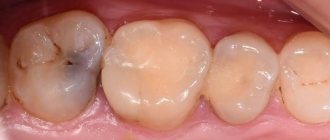
If defects are noted in a filling that was placed long ago or there is no filling at all, treatment is mandatory. It consists of 2 stages of visiting a specialist.
Initial visit to the dentist , during which he performs the following procedures:
- X-rays are performed to make an accurate diagnosis;
- local anesthesia is performed (if necessary);
- plaque and all deposits are removed;
- if the color of the dentin is greatly changed, the layer affected by inflammation is removed;
- The roots of the affected unit and the pulp must also be removed;
- the shape and depth of the channels change (if necessary);
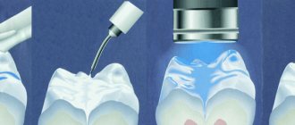
- the enlarged canal is treated with an antiseptic to prevent the recurrence of inflammation;
- A temporary filling is placed in the cavity, which has previously been enriched with calcium.
Second visit to the dental clinic . Usually prescribed 2-4 days after the first. Depending on the general well-being of the patient, further treatment can take place in two ways:
- If the patient has no pain in the unit with a temporary filling and no foreign taste, this filling is removed, and the canals are treated with an antiseptic. Then the usual filling of the cavity is carried out, fluoroscopy is done and the issue of subsequent restoration of part of the tooth is decided (if necessary).
- If within 2-4 days after placing the filling the patient experiences discomfort and the manifestation of pain increases, the dentist removes the filling and leaves the cavity completely open for 2-4 days. During this time, antibacterial therapy is carried out.
Another treatment option is also possible: the periosteum is dissected, drainage is placed and antibacterial therapy is prescribed. After the inflammation has subsided, the doctor places a permanent filling.
Important: treatment of this form of periodontitis is a simple procedure (when compared with other forms of the disease). The effectiveness of cavity cleaning and subsequent filling is high, and re-inflammation does not develop in the treated unit.
The total cost of the course of treatment includes all procedures performed: radiography, canal cleaning, antibacterial therapy, filling.
The approximate cost of the entire course is in the range of 6-9 thousand rubles. The final figure depends on the extent of the lesion and the number of canals.
Drug therapy
Well-sealed root canals are not prepared when fibrous periodontitis is detected. Typically, in this case, specialists prescribe two groups of medications:
- antiseptics;
- antibiotics (rarely).
Antiseptic drugs . Since the main reason for the development of inflammation is infection of dental tissues by pathogenic organisms, prescribed antiseptics are applied topically in the form of rinses or irrigations of the oral cavity. The dentist may prescribe:
- "Aminoflurid" with fluorine;
- "Vagotil";
- "Chlorhexidine";
- "Miramistin";
- "Iodopirone".
Antibiotics . They are prescribed in exceptional cases when there is an urgent need to neutralize a certain type of microorganism. Such drugs include:
- "Biseptol";
- "Ciprofloxacin";
- "Lincomycin";
- "Metronidazole";
- "Doxycycline."
It is necessary to understand that, by destroying pathogenic flora, antibiotics negatively affect the body’s protective properties, upset the balance of microflora in the mouth and create the preconditions for the re-development of the disease.
All of the medications listed are prescribed individually for each patient and only after a complete examination.
Therapy at home
Traditional medicine recipes are not able to completely cope with the inflammatory process. They are able to temporarily slow down the development of the disease if it is impossible to immediately consult a doctor.
For fibrous chronic periodontitis, you can try the following recipes:
- Rinse your mouth with saline solution : dissolve 1 tsp well in 200 ml of warm water. salt and use as directed up to 4 times a day. The liquid has an anti-inflammatory and antiseptic effect.
- Wipe the problem area with honey . It has a strong anti-inflammatory and antibacterial effect, so it can be used for rubbing twice a day. You can add a little sea salt to the honey, but the mixture should be rubbed into the gums carefully, without pressing, also 2 times a day. The product strengthens gums and reduces inflammation.
- Lemon applications . Prepare a mixture of freshly squeezed juice of half a lemon and 2 tsp. salts with large crystals. After the salt has completely dissolved in the juice, moisten a cotton pad in it and apply it to the problem area. After 3-5 minutes, remove the disc and rinse your mouth with water. This mixture also has a strong anti-inflammatory effect.
- Rinsing with infusions of medicinal herbs . To prepare infusions, you can use any plant that has a pronounced anti-inflammatory effect: calendula, yarrow, chamomile, nettle. For this, 2-3 tbsp. l. any plant, pour 500 ml of boiling water, leave for about an hour in a closed container, then strain and rinse your mouth 3-4 times a day.
We characterized the Victory bracket system in a special review. And here you can read reviews about the T4k trainer.
At this address: https://dentist-pro.ru/ortodontiya/prikus/effektivnye-metodiki-korrekcii-perekrestnogo.html you will find out whether crossbite can be corrected with braces.
All these remedies have only a short-term effect and cannot replace professional therapy. You should seek medical help as soon as possible.
Conservative surgical methods for the treatment of chronic periodontitis
Conservative surgical methods for the treatment of chronic periodontitis . Resection of the root apex. In recent years, this method has been used extremely rarely. It is now resorted to only in cases where there is a progressive destructive process in the bone, especially if there is a fistulous tract, cystogranuloma and other methods are not successful, and the tooth remains stable.
Replantation of teeth. With the introduction of antibiotics into dental practice, reports about the successful use of this method in the treatment of chronic periodontitis again appeared in periodicals. Data from V. A. Marchenko (1967), V. A. Kozlov (1974) and others indicate that the method of tooth replantation should be used for certain indications in the treatment of chronic periodontitis in all dental institutions, especially since the method helps preserve tooth, prevents atrophy of the alveolar process, restores the bite, relieves patients from additional, often painful manipulations.
It should be especially recommended to carry out replantation of teeth in the lower jaw, where the preservation of each tooth is the prevention of bone tissue atrophy and guarantees good fixation of the dentures. If, from a practical point of view, dental replantation is quite well developed, is available to all doctors and can be carried out in outpatient settings, then in terms of studying the causes of resorption of roots and replanted teeth and ultimately their rejection, this issue requires further development. The study of immunological reactions during dental replantation, as well as the influence of various medications under the protection of which the specified surgical treatment is carried out (antibiotics, vitamins, etc.) deserves the most serious attention. After replantation, drugs that stimulate immune reactions should not be prescribed: streptomycin, neomycin, nucleic acid preparations, vitamins A, B, C, PP, dibazol, pyrogenal, etc. (Petrov R.V., 1969). Of the antibiotics, penicillin or tetracycline should be preferred, since they have immunosuppressive properties.
Other methods of conservative surgical treatment of periodontitis are hemisection, root amputation, coronary-radicular separation.
Hemisection (the term was first introduced by Curson, 1966) refers to the removal of one of the roots of a multi-rooted tooth (usually the medial root of lower molars) with the adjacent coronal part of the tooth and the subsequent use of the remaining distal root for a bridge.
Root amputation involves removing the entire root up to the root origin (furcation) without removing the coronal portion. Hemisection is carried out on double-rooted teeth - molars of the lower jaw and premolars of the upper jaw, and root amputation - on molars of the upper jaw, although, in essence, both operations are identical and differ only in the technique of execution.
Corona-radicular separation consists of cutting the tooth into parts and then reuniting it in one common crown. The choice of one or another treatment method depends on the relevant indications and contraindications. Indications for hemisection and root amputation are:
1) the presence of deep intraosseous pockets in the area of one of the roots of the lower molar, one or two buccal roots or the palatal root of the upper molar; 2) exposure of the entire length of the root due to bone loss in teeth with periodontal damage; 3) significant damage to the cement and dentin of one of the roots by caries; 4) perforation in the area of bifurcation or the wall of the root canal; 5) obstruction of the root canals of teeth with chronic periodontitis; 6) obliteration of the root canal after previous treatment with significant loss of bone tissue in the area of the root apex; 7) cases where preservation of one of the roots may contribute to the further spread of the pathological process to adjacent teeth and roots, despite well-performed endodontic therapy; when a tooth has already been used or will be used as a support for a fixed bridge and the removal of which would lead to the need for a removable denture.
Corona-radicular separation is carried out in case of inflammatory and destructive changes in the area of bifurcation or trifurcation of multi-rooted teeth; with perforations of the area of the bottom of the tooth cavity, idiopathic resorption with destruction of the root branching area.
Contraindications include:
1) significant loss of bone tissue in all roots, which leads to an unfavorable clinical crown-to-root ratio, which prevents the teeth from withstanding normal loads during the act of chewing; 2) the presence of fused roots that cannot be separated; 3) the presence of impassable channels in the roots that need to be preserved; 4) when the furcation is so close to the apices of the roots that an attempt to expose it, necessary to divide and create a normal contour, would lead to the need to remove a large part of the bone in the remaining bed; 5) when endodontic, periodontal and orthopedic procedures are harmful to a weakened patient.
There are various options for amputating the roots of upper molars. These include: 1) removal of both buccal roots while preserving the palatal root; 2) removal of one of the buccal roots while preserving the palatal and other buccal roots; 3) removal of the palatal root while preserving both buccal roots; 4) cutting off one of the roots of the upper premolar while maintaining the functioning of the other.
Of the listed options, the most appropriate is to remove one of the buccal roots while preserving the palatal root and the other buccal one, or to remove both buccal roots while preserving the palatal root. The opinion of Basaraba (1967) is correct, who believes that preserving both buccal roots when removing the palatal root significantly reduces the useful life of such teeth, since the vertical loads falling on the restored crown on such roots will not be distributed in the axial direction. A preventive measure that eliminates mobility of the remaining segment and the possibility of secondary occlusal injury is temporary stabilization, carried out in most cases after surgery. It can be carried out optionally: using wire and quick-hardening plastic, orthodontic devices or amalgam. After hemisection, the treated tooth is splinted.
A necessary step prior to hemisection and root amputation is endodontic treatment. It consists of careful instrumentation and sterilization of all root canals, followed by filling them with phosphate cement, zinc-vinol cement and applying a filling.
Hemisection technique. The operation can be performed in two ways: without tilting the mucoperiosteal flap or with folding it.
The first method is the simplest. After filling the canal and applying a filling, the crown of the tooth is cut into two halves under anesthesia, exactly at the furcation site, using a long cone-shaped diamond bur, after which the second half of the root is removed. Separation and removal are carried out, if possible, carefully so as not to damage the interradicular septum and adjacent teeth, and also not to destroy the bone tissue surrounding the remaining segment.
The operation according to the second method begins with the formation and detachment of a mucoperiosteal flap, after which the alveolar ridge bone is cut with a diamond bur along the length of the root using a turbine unit. The roots are separated with a diamond bur or disk, and removal is carried out using an elevator or forceps. The flap is placed in place and secured with sutures. Sometimes the entire root is removed, preserving the crown of the tooth. However, this operation is associated with certain technical difficulties, and the remaining overhanging part of the tooth is a kind of cantilever, which is justified only when removing the root located under the supporting crown of the bridge.
Root amputation technique. Amputation of the roots of the maxillary molars can be carried out in two ways, with or without tilting the mucoperiosteal flap. In the first case, if one or two buccal roots of the upper molar are subject to amputation, after detachment of the mucoperiosteal flap on the buccal surface, they are exposed by removing the outer wall of the alveolus. Then a long fissure or diamond bur is inserted into the area where the roots emerge, the root is separated exactly along the enamel-cement border, after which it is removed using a straight thin elevator or forceps. Next, curettage is performed, the mucoperiosteal flap is placed in place and sutures are applied. If the palatal root is to be removed, then two incisions are made on the palatal surface, parallel to the long axis of the tooth, 2-3 mm medial and distal to the root; the incisions can be oval or trapezoidal.
The second method, proposed by Kirchoff and Gerstein in 1969, is the simplest and most atraumatic. It avoids cutting out a mucoperiosteal flap and removing a large part of the bone surface.
The method consists in a peculiar reshaping of the anatomical crown of the tooth according to the root to be amputated. To do this, a wedge-shaped excision of a small part of the tooth crown is performed using some cutting tool (mill, diamond or fissure bur). Thanks to this, a cross-section of the root to be removed is exposed in the surgical field. After this, the root is easily removed using an elevator or forceps. The chewing surface of the tooth crown remains intact, and the tooth crown must be shaped in the way required by the remaining roots.
When performing surgical hemisection and root amputation, it is necessary to carry out preoperative and postoperative radiological monitoring to identify hard tissue overhangs in the separation area and carefully smooth the separation surface in the presence of any protruding area.
Orthopedic treatment of molars treated by hemisection or root amputation depends on a number of conditions and is also the choice of the dentist. Most teeth treated with this method require covering with an artificial crown and can serve for a long time as the main support for a permanent bridge.
Corona-radicular separation technique. The operation is quite simple and usually does not cause complications. Under anesthesia with a diamond bur, the tooth is cut in half, the overhangs are removed, then a thorough curettage is carried out in the furcation area and a gingival bandage is applied. Subsequently, crowns are made for each of the roots, welded together. Preparing the roots for crowns and taking impressions can be done during surgery.
The currently available arsenal of tools and methods allows us to treat all single-rooted and most multi-rooted teeth with passable canals.
In conclusion, it should be noted that despite the recent successes achieved in the treatment of chronic periodontitis, many issues of their treatment still remain unresolved. These include: a significant number of exacerbations, high labor intensity and incomplete effect of treatment of multi-rooted teeth, a lack of drugs aimed at enhancing repair processes in periodoitis. In addition, the degree of influence of the periapical lesion and its treatment on the state of the body remains unclear in all aspects (Ovrutsky G.D., 1966), despite some successes in the study of these issues.
What is meant by a source of infection? The source of infection is currently considered not only as an accumulation of microbes, their metabolic products and destruction of organic matter of connective tissue, which are antigens, but also as a source of irritation of the autonomic nervous system, through which it is transmitted to higher nerve centers, and from here in the form of pathological impulses to the periphery. As for the ways of influence of odontogenic infection, they can be different: purely bacterial (happens relatively rarely); toxic - products of tissue decay; antigenic with the development of immune reactions (Burkov T. et al., 1969; Rabinovich A. S., 1971; Rudzit K·, 1973; Türk, 1973; Vera, 1974, etc.).
Recent years have brought some advances in testing and studying periodontitis as a source of chronic infection. But the electrical tests currently used (Zicher, Battner, 1965; Battner, 1965; Burkov T. et al., 1969; Heuser, 1971, etc.), the histamine-conjunctival test according to Remki et al. to identify and characterize foci of odontogenic infections (chronic periodontitis, periodontal disease, periodontitis) can only be reliable when combined with other data: x-ray examination of the alveolar processes of the jaws, examination of peripheral blood (number of leukocytes, monocytes and lymphocytes), protein fractions of blood serum, study of capillary resistance, skin - allergy tests with streptococcal allergens, dynamic reaction indicators using antistreptolysin-O, rheumatoid factor and other immunological studies.
Considering the influence of infectious periodontal foci on the development and course of the rheumatic process (Zhukova I. I., 1964; Chepulis S. P. et al., 1968, etc.) and their potential role in the pathogenesis of other autoimmune diseases (streptococcal glomerulonephritis, systemic red lupus, rheumatoid arthritis, scleroderma, periarteritis nodosa, dermatomyositis, blood diseases, hepatitis), multi-rooted teeth with chronic granulating and granulomatous forms of periodontitis should be removed. Chronic infectious foci in the area of single-rooted teeth must be eliminated surgically using resection of the root apices under the cover of antibiotics and steroids. Such a radical approach should take place in similar cases and with deep pathological gingival and bone pockets (periodontal disease, periodontitis).
The most common complications, their causes and methods of elimination are presented in Table 6.
Features of treatment in children
The method of treating the disease in children depends entirely on their age and the condition of the affected area. Temporary units are subject to deletion if:
- tooth mobility has developed as a result of illness;
- there are no more than 2 years left before their replacement;
- there was already inflammation or damage to more than half of the root.
In tooth-preserving treatment, special attention is paid to stopping the infection in order to prevent recurrence of inflammation.
Important: the specificity of fibrous periodontitis is considered to be safe for babies and children who are changing teeth. This is explained by the fact that the disease develops further only around fully formed tooth roots.
Features of treatment in pregnant women
When carrying a child in the first and third trimesters, any dental intervention is undesirable. A woman may receive medical care only in an emergency.
X-rays are also prohibited - it is prohibited to do an X-ray examination of the dentofacial apparatus at any stage of pregnancy.
Treatment of the disease can only be carried out in the second trimester . Usually, when fibrous periodontitis is detected, the affected tooth is removed. When selecting anesthesia, preference is given to drugs with a very low rate of passage through the placenta.
Causes
In most patients, the development of the disease is associated with an infectious process. Bacteria or their toxins, passing through the root, enter the adjacent tissue and cause inflammation. This situation often occurs when pulpitis and caries are treated untimely or incompletely. Also, periodontal infection can occur from other parts of the dentofacial apparatus (sockets or gums), if there is a pathological process in them.
In addition to infection, the cause of acute periodontitis can be trauma to the jaw (blow, bad fall) or the tooth itself. For example, when biting hard objects or playing musical instruments with a mouthpiece (saxophone, recorder, etc.). As a rule, patients ignore unpleasant sensations, attributing them to a natural reaction of the body. This attitude often leads to a protracted course of the disease.
Periodontitis after dental intervention is also a common occurrence. The reason may be the installation of an oversized filling, the use of low-quality equipment, medications, or the patient’s allergic reactions to medications. To reduce the risk of pathology, you must contact only licensed clinics whose level of care meets modern standards.
Forecast
Of all types of periodontitis, only fibrous is the most stable and has the most favorable treatment outcome. Its timely detection guarantees a complete cure of the disease within a few days if it appears and develops as an independent pathology. If the disease is a consequence of therapy for other types of periodontitis, the presence of fibrous periodontitis is ignored.
Prescribed antibacterial treatment guarantees the death of pathogenic microflora in the root canals, and a hermetically sealed filling prevents the possibility of relapse of the disease due to infection of periodontal tissues.
If treatment for fibrous periodontitis is not started at the initial stage or if you try to cope with it by self-medication, it easily turns into other, more severe and dangerous forms: granulomatous and granulating, leading to exacerbation of tissue inflammation.
Possible complications
Any inflammation in the soft tissue of the gums or in the tooth is considered a factor that complicates the treatment of periodontitis. Lesions cannot always be prevented. Sometimes this happens due to the patient’s fault due to late contact with the dentist. Carrying out surgical therapy or filling, regardless of the stage of treatment, does not always give a positive result. In some cases, complications are possible:
- Pain of varying intensity.
- Enlargement of the inflammatory focus.
- Temperature increase.
- Severe swelling and redness of soft tissues.
The occurrence of pain is related to how periodontitis is treated in a clinical setting. The fact is that the dentist’s instrument can damage tissue during root canal treatment, when the inflammatory secretion is removed. Otherwise, pain after therapy occurs due to the filling material getting into the bone tissue.
In some cases of periodontitis, the causes of pain after the installation of a filling are difficult to determine. In this case, the patient does not experience additional symptoms of inflammation. In this case, doctors recommend rinsing with antiseptic solutions. After 5 days, the discomfort disappears.
Pain syndrome after therapy can persist for several weeks. Discomfort occurs only when food gets on the tooth or when the jaw is clenched. To eliminate these sensations, the patient is advised to reduce the load on the affected area.
In some cases, symptoms of general malaise may occur after treatment. The patient needs to see a doctor for additional treatment for periodontitis. Antibacterial agents are prescribed from medications. To reduce pain, they carry out: UHF, microwave, laser therapy.
Some time after treatment of periodontitis, the focus of inflammation may increase. In most cases, this goes away without further symptoms. In this case, an operation is prescribed and part of the root is removed. When suppuration resumes, the infection can enter the bloodstream.
Due to constant circulation, inflammatory viruses can spread to other organs. Therefore, if periodontitis is not treated in a timely manner or the patient is inattentive to relapse, this can lead to serious complications: diseases of the liver, kidneys, and heart .
Prevention
The main method of preventing fibrous periodontitis is considered to be timely relief of dental diseases, especially severe forms of the disease itself.
In addition, dentists advise following the following recommendations to prevent the onset of the disease:
- Perform regular and high-quality oral hygiene;
- promptly, at the first signs of any dental pathology, seek medical help;
- do not try to cope with diseases on your own and only with folk remedies;
- regularly sanitize the oral cavity.
Forecast and prevention of chronic periodontitis
The course and prognosis of chronic periodontitis, of course, depend on the timeliness of visiting a dentist and the quality of the treatment he provides. With effective root canal treatment, the area of damaged bone is restored, and the tooth retains its functional properties. If the patient does not apply in a timely manner or treatment is unsuccessful, there is a high probability of tooth loss. Complications of chronic periodontitis can pose a serious threat to health and life. That is why patients need to improve their dental culture in matters of oral care, undergo regular examinations with a doctor and promptly treat odontogenic foci of infection.