home
Diseases
Cancer
Salivary gland cancer: symptoms, diagnosis, treatment
The liquid that is found in the mouth is called saliva. It performs particularly important functions in the body: it moistens the mucous membranes, the food consumed, helps swallowing, greatly facilitates articulation, and protects against the appearance and proliferation of all kinds of harmful bacteria. This fluid is produced by the salivary glands. They are divided into two main groups:
- large (located under the jaw, near the ears and under the tongue);
- small (characterized by a microscopic structure, scattered throughout the entire oral cavity).
Note that there are more than a hundred small glands.
Oncology can develop in all these glands. Salivary gland cancer in most cases originates in the parotid location. If you look at the statistics, they show that this happens in 7 out of 10 cases. Most rarely, salivary gland cancer forms and develops in the sublingual or small salivary glands. Approximately 1-2 tumors out of 10 cases occur in the glands that are located under the jaw.
Causes of salivary gland cancer
- A change in a normal salivary gland cell due to the formation of a certain set of mutations.
- Mutations in oncogenes or suppressor genes (they repair “damaged” DNA, activate or suppress cell reproduction).
- Age. Salivary gland cancer depends on age. This is due to an increase in mutated cells in the human body. Over the years they accumulate. Most often they are in a period of rest, but certain external or internal factors can provoke their activity. Such factors include exposure to harmful chemicals, stress, as well as various infectious diseases that a person has suffered from.
- Sexual characteristic. Men are a special risk group, as they are most predisposed to developing salivary gland cancer.
- Irradiation of the head. The disease can appear if a person has previously undergone a course of radiation therapy or has been exposed to ionizing radiation in another place (for example, at work).
- Professional hazards. The cause of the appearance and development of salivary gland cancer can be professional activities that involve contact with harmful chemicals. Contact with nickel alloy dust and asbestos is especially dangerous. Oncology of the salivary glands is often diagnosed in people working at rubber manufacturing plants. Woodworking industries are also dangerous.
- Heredity (but this is the most minimal, insignificant percentage).
- There is an opinion that exposure to mobile phones affects the formation of cancer of the salivary glands. Moreover, this is not just an assumption, but specific data that was obtained by scientists during a series of studies.
Turban tumor or skin cylindroma
Skin cylindroma is a fairly rare benign disease, which is most often localized on the scalp.
More often, tumors are multiple; they merge with each other, forming growths that resemble a fancy headdress. That is why this tumor is called turban. Cylindroma develops more often in women; the disease can also manifest itself in childhood. There are frequent cases of turban tumor formation in blood relatives.
Reasons for the development of the disease
Scientists believe that this tumor may develop from apocrine glands.
The causes and mechanism of development of cylindroma have not yet been elucidated.
Even in determining the histogenesis of the tumor there is still uncertainty.
Most scientists consider cylindroma as an eccrine tumor, however, there is an opinion that a neoplasm develops from apocrine glands or from hair structures.
Since turban tumor is often observed in blood relatives, we can assume that this disease is inherited in an autosomal dominant manner. The likelihood of developing the disease in children born from sick parents is not precisely established.
Clinical picture
The main symptom of cylindroma is the formation of many solitary nodes localized on the skin of the head and neck. One of the characteristic features of this disease is that turban tumor often acts as one of the symptoms of complex cutaneous hamartomas. Combinations can be very different. The most common diagnoses are:
- Cylinder - adenoma of the parotid glands - trichoepithelioma.
- Cylinder - adenoma of the parotid glands - eccrine spirpdenoma.
- Cylindroma – milium – trichoepithelioma, etc.
Clinical manifestations of cylindroma consist in the appearance of a large number of tumor-like, nodular formations with a smooth surface. The color of the formations is usually pink, but can have different shades. Consistency – dense, elastic. Rarely, a tumor with a cylindroma contains a cystic component, which gives it a bluish tint.
New growths with a turban tumor are always multiple, sometimes the growths occupy almost the entire surface of the head and spread to the face and neck.
In cylindroma, the tumor is located in the subcutaneous fatty tissue and dermis. The layer of epidermis above the tumor is thinned, the interpapillary processes are smoothed. The tumor consists of several lobes of round shape and different sizes.
Cylindroma is characterized by rather slow growth of nodes that have a rounded shape and protrude above the surface of the skin. Hair does not grow on the surface of the tumor. Large and long-standing cylindroma nodules may be riddled with telangiectasia.
A characteristic symptom of cylindroma is a gradual and progressive course. Periods of active growth of old nodes and the formation of new ones alternate with periods of relative stability.
It is likely that hormonal fluctuations influence the growth of tumors, since the most active development of cylindroma is observed in women during adolescence, during pregnancy and during menopause.
The rest of the time, the tumor may not undergo any changes for years.
Malignant degeneration for cylindroma is atypical; however, the literature describes cases in which both solitary nodes and multiple growths were subjected to malignancy. When cylindroma degenerates, regional metastasis may occur.
Diagnostic methods
Diagnosis of cylindroma is carried out on the basis of studying the clinical picture and conducting histological studies. It is necessary to distinguish cylindroma from lipoma, atheroma, basal cell carcinoma, dermatofibrosarcoma protuberans, and eccrine spiradenoma.
When conducting histological studies of cylindroma tissue in the dermis, one can observe numerous nests and papillomas of cells whose structure is similar to the cells of the basal layer. Within the tumor islands there are two types of cells: in the center - with light-colored nuclei, at the edges - with dark-colored nuclei.
Cylindroma belongs to the category of heterogeneous tumors; microstudies can reveal four types of the disease.
- Trichoepitheliomatous;
- Hydroadenamatous;
- Mixed;
- Undifferentiated.
In the undifferentiated type of cylindroma, the tumor cells have intensely stained nuclei and are small in size. Cells are arranged in the form of cells that are surrounded by membranes.
A distinctive feature of the hidradenomatous type of cylindroma is the presence of cavities between the tumor cells that resemble the ducts of the sweat glands.
The trichoepithelial type of cylindroma is characterized by the presence of structures that indicate the involvement of hair follicles in the process.
With a mixed form of cylindroma, a picture can be observed that is characteristic of any of the types of tumor described above.
Treatment
Treatment tactics for cylindroma are selected depending on the shape of the tumor, as well as the number of nodes, their size and location.
In most cases, skin grafting is used to treat cylindroma.
In case of single nodes, they are surgically removed. But, in most cases, due to the large area of excision, it is necessary to resort to skin grafting.
With multiple growths, attempts to remove especially large conglomerates do not always yield positive results. Firstly, there is a high risk of the formation of pronounced scars, which neutralize the cosmetic effect of the operation. Secondly, cylindroma often recurs when incompletely removed. Radiation therapy is sometimes used to target the area where the cylindroma is located.
For small cylindroma nodes, it is possible to use gentle surgical methods for removing tumors - diathermocoagulation, laser treatment.
Treatment with traditional methods
It is impossible to completely get rid of cylindroma with the help of medicinal plants, however, using traditional medicine methods it is possible to slow down the growth of nodes.
For cylindroma, it is recommended to take a decoction of oak bark. The healing remedy is prepared as follows: you need to take 200 grams of oak bark, 100 grams of dried pomegranate peels, 150 grams of viburnum branches. Brew daily by taking a tablespoon of the mixture in a glass of water.
Boil the broth for 7 minutes over low heat, then let it cool, covering with a lid. Drink during cylindroma, dividing the prepared decoction into three parts. Take the product until the prepared mixture of medicinal raw materials runs out.
Then take a month's break and repeat the course of treatment.
Prevention and prognosis
Prevention of cylindroma has not been developed. The prognosis for life, in most cases, is favorable, but for complete recovery is doubtful.
Cylindroma has a long, slowly progressive course; periods of tumor growth alternate with periods of stabilization.
Cylindroma extremely rarely degenerates into a malignant disease, however, this possibility cannot be excluded. Therefore, patients should be monitored by an oncologist. If suspicious symptoms appear (sharp changes in the size of tumors, bleeding, formation of ulcers, etc.), it is necessary to conduct a histological examination of the tumor tissue.
Source: https://dermalatlas.ru/opuxoli-kozhi/tyurbannaya-opuxol-ili-cilindroma-kozhi/
Symptoms and signs of the disease
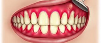
- Feelings of compaction or swelling in the mouth are the first signals that indicate an urgent need to consult a doctor. This must also be done if there are seals in the neck, cheeks, or jaw.
- Facial asymmetry. If you notice certain changes in the shape of your face, be sure to undergo a medical examination.
- A sign of salivary gland cancer is pain in the mouth and other locations (neck, ear, jaw, cheeks).
- Numbness in a certain part of the face is another reason to go for examination to a specialist.
- A symptom of salivary gland cancer can also be weakness in the facial muscles. Because of this, one part of the face may noticeably appear lower.
- If you cannot open your mouth wide, then this may also be one of the symptoms of salivary gland cancer.
- Difficulty swallowing.
Symptoms
In the early stages, the tumor practically does not manifest itself, since pain does not develop.
In the future, the following symptoms can be noted:
- compaction of the tumor structure;
- the neoplasm has no clear boundaries;
- the tumor is immobile;
- pain with further growth;
- damage to the masticatory muscle;
- facial nerve paresis;
- skin hyperemia.
Due to damage to the facial nerve and tissue proliferation, jaw mobility is impaired, the patient feels discomfort when chewing, severe pain and swelling. The tumor most often affects the salivary gland on one side.
ul
When to see a doctor
At the first symptoms of salivary gland cancer, you should consult an oncologist. The earlier the disease is diagnosed, the more effective the prescribed course of treatment will be. If you are looking for a highly qualified doctor in Moscow, then contact the oncological JSC "Medicine" (clinic of Academician Roitberg), which is located at 2nd Tverskoy-Yamskaya lane 10 (Mayakovskaya metro station).
The first symptoms of salivary gland cancer:
- bumps or swelling in the neck, cheeks, jaw, ears;
- muscle weakness;
- “drooping” of the face and other signs, which were discussed in more detail above.
Stages and grades of salivary gland cancer
All malignant tumors have a stage and degree of development, as well as their own classification, and salivary gland cancer is no exception.
There are five main stages:
- "zero". This means that salivary gland cancer is “in place.” The tumor has not yet spread, but is located within the upper layer of cells;
- first. Cancer of the salivary gland has not spread beyond its limits, its size is no more than 2 cm;
- stage II. The tumor has become larger than 2 cm, but still does not exceed 4 cm;
- stage III. The cancerous tumor exceeds 4 cm and has begun to spread to neighboring organs and tissues;
- stage IV. The tumor began to develop rapidly. It is actively spreading to other locations.
Before treatment for salivary gland cancer begins, the extent of its spread should be determined. There are only three of them:
- grade I. Characterized by low malignancy. It grows slowly. The prognosis for salivary gland cancer is most favorable. Tumor tissue is very similar to normal tissue;
- degree II. Refers to a moderately differentiated tumor. Moreover, tumor tissue differs significantly from normal tissue;
- degree III. This is a poorly differentiated tumor. Cancer is particularly aggressive.
Classification
- The most common type of cancer is mucoepidermoid tumor. She is rarely aggressive.
- Adenoid cystic carcinoma rarely metastasizes and grows slowly.
- Adenocarcinoma.
There are also types of tumors that rarely become malignant. This:
- squamous cell carcinoma;
- epithelial-myoepithelial carcinoma;
- anaplastic small cell carcinoma;
- undifferentiated carcinomas.
The stage of salivary gland cancer, its degree and type determines how effective the treatment will be.
Determination of stage (staging)
The stage of a cancer describes the size and spread of the tumor beyond its original site. Knowing the type and stage of the tumor, the doctor can choose the most appropriate treatment method for the given case.
Classification according to the TNM system
Most often, when determining the stage of cancer, the TNM classification system is used, where:
- Category T means tumor size
- Category N means the presence or absence of cancer in the lymph nodes
- Category M means the presence or absence of metastases in distant organs
Each category has numerical values that describe the tumor in more detail. For example, category T1 means that the tumor is very small and limited to one layer of tissue, while tumors in category T4 are large and spread to multiple layers of tissue.
The specific meanings of the T, N, and M categories depend on the type of cancer.
Numerical classification of cancer
In addition to classification using the TNM system, specialists also use a numerical classification of cancer. Typically, it involves three or four stages for each type of tumor.
Stage 1 corresponds to the early stages of cancer development, when the tumor is still very small and has not spread. Stage 4 describes advanced disease in which the cancer has metastasized to other organs. Stages 2 and 3 are intermediate.
The numerical stage is a combination of different categories according to the TNM system. Thus, a stage 1 tumor can be described as T1, N0, M0 or T2, N0, M0.
The numerical stage can also be divided into substages, each of which describes the size and extent of the tumor in more detail. For example, stage 3 cancer can be divided into stage 3a, stage 3b and stage 3c. However, stage 3b differs from stage 3a either in the size of the tumor or in the presence of metastases in the lymph nodes.
Talking to your doctor about the stage of your cancer
Over the past few years, the cancer staging system has become very complex. Now the classification of cancer by stage allows us to describe in great detail the size and prevalence of various tumors. Cancer staging greatly facilitates the choice of treatment and allows one to determine the prognosis of the disease.
However, when talking to patients, doctors tend to greatly simplify information about the stage of cancer. The doctor may use words such as "early" or "local" if the tumor has not spread. If the cancer spreads to surrounding tissue or nearby lymph nodes, the doctor refers to the tumor as “locally advanced.” When the cancer has spread to distant organs, it is called "advanced" or "advanced." In each specific case, the doctor discusses with the patient the specifics of determining the stage of cancer.
ul
Life expectancy by type of cancer
Today, the survival of patients after thyroid cancer is assessed by the patients reaching the five- and ten-year mark. These indicators are usually calculated from the moment an accurate diagnosis is made until death occurs. The five-year survival rate for patients with thyroid cancer depends on the stage and type of pathological process that develops in the thyroid gland.
Papillary
About 80% of neoplasms in the thyroid gland occur in the papillary form of the disease.
- In the initial stages (stages 1 and 2) of the disease, almost 100% of patients reach the five-year mark of life expectancy.
- At the third stage, 90-95% of patients achieve complete recovery after surgery.
- At the late stage (stage 4) of malignant tumor growth, the postoperative survival rate reaches only 45-51%.
Follicular
On average, this form of the disease accounts for about 10-15% of clinical cases.
- Almost 100% of patients whose final diagnosis was made at stage 1 or 2 of the disease overcome the five-year life expectancy mark.
- The third stage of malignant tumor growth accounts for only 80% of patients who survive to the five-year mark.
- At stage 4 of carcinogenesis, this figure is usually no higher than 45-50%.
Modular
This form of thyroid cancer occurs only in 6-10% of all cases.
- The survival rate is 100% for those patients diagnosed at stage 1 of the disease.
- 98% of operated patients live at stage 2 for at least five years.
- Patients who reach stage 3 can expect a survival rate of 75-81%.
- The survival rate at the terminal stages (stage 4) is significantly reduced, it is only 24-28%.
Anaplastic
This form is the rarest and most aggressive, accounting for about 2-4% of cases.
The postoperative survival rate is only 7%, and it is not uncommon for patients to survive only 4-6 months after a final diagnosis.
ul
Treatment of salivary gland cancer
There are different types of treatment for salivary gland cancer.
- Surgical intervention. The only true method for tumor resectability. But if it has a significant degree and stage, then they resort to other methods of treatment.
- Radiation therapy. This method is used if it is not possible to perform surgery for certain indications. Often such therapy is prescribed after surgery.
- Chemotherapy. This method is used most rarely for cancer of the salivary glands, but still occurs.
- Rehabilitation. This method is aimed at completely restoring the body.
Treatment
The patient is treated by a team of doctors, which may include: a clinical oncologist, an ENT doctor, an oncologist-surgeon, an oral and maxillofacial surgeon, a chemotherapist, a radiotherapist, etc. The treatment program is determined by the stage of cancer, the histological type of the tumor, its location (which gland is affected) , age, general condition and concomitant diseases of the patient.
Surgery
If the tumor has not grown much into the surrounding tissue, then it is resectable, that is, it can be removed surgically. The surgeon’s task is to excise the tumor while capturing the surrounding tissue so that there are no cancer cells left on the cut line, that is, to ensure a negative resection margin. If tumor cells have spread to the lymph nodes, or a biopsy reveals aggressive cancer, the lymph nodes are also removed.
For parotid salivary gland cancer, surgery presents certain difficulties, because the facial nerve passes through the gland, which controls the work of facial muscles. If the tumor affects only the superficial lobe of the gland, you can remove it separately - perform a superficial parotidectomy. There is no risk of damaging the facial nerve. In some cases, it is necessary to remove the entire gland, and if the tumor has grown into the facial nerve, then it too.
For cancer of the sublingual and submandibular gland, the surgeon removes the gland itself and some of the tissue located around it, including, possibly, bone tissue. In some cases, it is necessary to excise the nerves that control sensitivity, movements in the lower part of the face, in the tongue, and the sense of taste.
For cancer of small glands, the affected gland and part of the surrounding tissue are removed. The extent of the operation depends on the size and location of the tumor.
Radiation therapy
Indications for the use of radiation therapy for malignant tumors of the salivary glands:
- To combat malignant tumors that cannot be removed surgically due to their location or size. Sometimes radiation is supplemented with courses of chemotherapy.
- After surgical treatment. This type of radiation therapy is called adjuvant and is sometimes combined with chemotherapy. Radiation after surgery helps destroy remaining cancer cells and prevent recurrence.
- For advanced cancer. In this case, radiation therapy is aimed at combating pain, difficulty swallowing, bleeding and other symptoms.
Radiation is typically given five days a week for 6–7 weeks. If radiation therapy is used for palliative purposes, the course will be shorter.
Chemotherapy
Chemotherapy is used quite rarely for malignant neoplasms of the salivary glands. Anticancer drugs can reduce the size of the tumor, but are not able to completely destroy it. They are most often prescribed for advanced cancer as palliative treatment or in addition to radiation therapy.
Depending on the type and other characteristics of the cancer, the doctor may prescribe combinations of different chemotherapy drugs: carboplatin, cisplatin, 4-fluorouracil, doxorubicin, paclitaxel, cyclophosphamide, vinorelbine, docetaxel, methotrexate. Chemotherapy for cancer is always given in cycles. The patient is administered the drug, then takes a “break” for several days. The course of treatment may consist of several cycles.
Rehabilitation
After treatment, some problems associated with nerve damage may persist: dysfunction of the facial muscles, speech disorders, swallowing, and cosmetic defects. Some side effects of chemotherapy and radiation therapy go away after treatment is completed, while others persist for a long time. In such cases, rehabilitation courses are indicated. The doctor draws up a rehabilitation treatment program individually, depending on the severity and nature of the disorders.