What it is
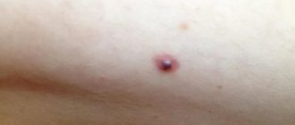
Fibroma in the mouth is a tumor of a soft structure that is benign in nature. A neoplasm is formed from soft mucous layers and has an unchanged shell.
On this topic
- Oral cavity
Complications after removal of a dental cyst
- Olga Vladimirovna Khazova
- December 5, 2020
Unlike many other formations, for example, papillomas, fibroma of the oral mucosa does not have an upper keratinized layer.
The shape of the tumor is spherical, the surface is smooth. If the fibroma is located in a part of the oral cavity where it is not subject to mechanical damage, the tumor does not change its size for a long period of time.
Kinds
Oral fibroids are divided into several types, depending on their consistency, nature and shape.
Solid tumor
This type of fibroma is dense to the touch and consists mainly of connective tissue. It is most often found on the upper palate or gums.
Soft tumor
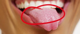
A soft fibroma is formed from connective tissue with multiple nuclei. Location: on the cheek, on the tongue.
Irritable fibroma
This type of tumor occurs due to constant mechanical or thermal irritation of the oral mucosa. It is a papule, the color is pink.
Fibroma, which occurs as a result of frequent irritation of soft structures in the oral cavity, is not a full-fledged tumor; it is tissue hyperplasia, which occurs as a reaction to constant irritation, for example, frequent biting of the inside of the cheek.
If this fibroid is constantly bitten, it may bleed, but, as a rule, there is no risk of infection.
Lobed type
Lobulated fibroma has a bumpy surface. The cause is permanent trauma to the gums due to the installed removable denture.
Symmetrical tumor
I consider this type of formation to be one of the subtypes of oral fibroma, although it is not one.
On this topic
- Oral cavity
Laser removal of dental cyst
- Natalya Gennadievna Butsyk
- December 5, 2020
Symmetrical fibroma is a pathological growth of soft tissue of the gums.
A distinctive feature is that the shape of the tumor is similar to a bean. Localization - gums.
Types of oral fibromas
There are several types of fibroids:
- Dense (hard) fibroma - consists of coarse connective tissue fibers tightly adjacent to each other. Due to this, the tumor is characterized by a dense consistency. Typically, such fibroids develop in the gum area and on the hard palate.
Soft fibroma - formed from thin, loosely fitting connective tissue fibers with a significant number of nuclei. Often the location is the mucous membrane of the cheeks and tongue.
Irritation fibroma is one of the most common neoplasms in the oral cavity. Despite its name, this type of fibroma is not a true tumor, but merely the result of reactive hyperplasia. It is a pale pink papule with clearly defined boundaries, which gradually turns into a nodule. It is usually localized on the mucous membrane of the cheeks, gums, lips or tongue.
Fibroma of the tongue. This type of fibroma most often forms under the mucous membrane of the tongue, growing from connective tissue fibers. It grows very slowly and forms dense, painless nodules with a non-ulcerated surface. Mainly soft fibromas are formed on the mucous membrane of the tongue; dense fibromas can develop in the soft tissues of the tongue. They are easily removed after dissecting the mucous membrane by peeling out the fibroma and then applying sutures.
Gum fibroma. When a gum fibroma forms, the patient feels a dense formation with a smooth surface in this place. During a medical examination, you can notice that the color of the mucous membrane has not changed and there is no pain on palpation. On the gums, mostly hard fibroids develop, characterized by very slow growth.
Symmetrical fibromas form on the palatal part of the gum, in the area of the third molars (teeth). They have a dense consistency and a rounded elongated shape. However, this is not a true tumor. The formations represent only the growth of the gums as a result of reactive hyperplasia. When the gums are chronically injured by removable dentures, lobular fibroma develops. It is characterized by a lumpy surface with traces of scar changes.
Causes
In a child, fibroma in the oral cavity occurs due to the following factors:
- mechanical injuries to the mucous membrane with hands, solid food;
- thermal irritation from too hot food or drinks;
- poor oral hygiene
- inflammatory gum diseases;
- gingivitis;
- stomatitis.
In adult patients, fibroma can occur as a result of untreated multiple caries or wearing removable dentures, the careless installation of which can severely injure the delicate mucous membrane of the gums.
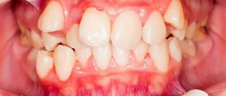
The likelihood of fibroids forming in the oral cavity increases in people with malocclusion and a genetic predisposition to oral fibroids.
Diagnostics
Considering the characteristic symptomatic picture of fibroma, making a diagnosis is not difficult, and the presence of a pathological formation is determined by the dentist during a visual examination of the patient’s oral cavity.
To clarify the degree of development of pathology and the depth of penetration of fibroid cells, an ultrasound examination of the oral cavity is performed.
Rarely, a fibroid grows or becomes more painful; in this case, a biopsy is required, during which a sample of fibroid tissue is taken and sent for histological examination.
On this topic
- Oral cavity
Removal of tooth granuloma
- Natalya Gennadievna Butsyk
- December 4, 2020
An important role in diagnosing fibroma is also played by determining the causes of the appearance of the pathological formation. For this purpose, X-rays, radiovisiography, and periodontograms are performed.
Differential diagnostics are also carried out, the purpose of which is to distinguish fibroma from similar formations such as warts, neuromas, and benign oncological tumors on the tongue.
Collagenous fibroma of the hard palate: description of a rare clinical case
Introduction
Collagenous fibroma is a benign soft tissue lesion first described by Evans in 1995. It develops from a cell with a fibroblast phenotype and is characterized by significant accumulation of collagen. The disease is quite rare and does not have a characteristic anatomical location. The etiology of the disease is unknown; it is assumed that it is based on either a reactive process or a true neoplastic process. One of the existing hypotheses suggests that the basis of the disease is a chromosomal rearrangement (11q 12).
Clinically, the disease manifests itself as a slowly growing painless formation. Localization can be different: the formation can be located on the arms, shoulder girdle, feet or legs, thighs and hands. However, only a few cases of localization on the head and neck are described in the literature: two on the hard palate and one in the parotid gland.
Microscopic examination reveals a well-defined soft tissue formation consisting predominantly of a collagenous extracellular matrix with rare dispersed spindle-shaped fibroblasts. It can vary in size from two millimeters to several centimeters.
As a rule, the disease is asymptomatic, with the exception of secondary traumatic ulceration of the surface. The standard of treatment is complete surgical excision of the formation. Recurs rarely.
This article describes a clinical case of collagenous fibroma of the hard palate, located from the upper second premolar on the right to the upper second molar on the right. There was no history of mucosal trauma or irritation.
Description of a clinical case
A 28-year-old woman was admitted to the periodontal department with a complaint of unaesthetic appearance of her teeth and a desire to whiten her teeth. During a general examination, no developmental anomalies or external signs of the disease were noted. Dental history, life history without any peculiarities. Denies systematic use of medications. Lymph nodes are not palpable.
Rice. 1. Photograph of the hard palate of the oral cavity before surgery.
Upon examination of the oral cavity, a single hard, non-displaceable, pedunculated formation measuring 4x2 cm was discovered, located on the right side of the palatal plate from the second right upper premolar to the second upper right molar (see Fig. 1). The affected tooth is asymptomatic, without pathology. The surface of the formation is not ulcerated, there is no pathological bleeding. The color of the formation corresponds to the adjacent mucous membrane of the hard palate. Periapical radiographic examination revealed no pathology (see Fig. 2), although occlusal radiography was not performed.
Rice. 2. X-ray of the affected area before surgery.
According to the patient's questioning, the formation appeared about one year ago. There was no history of trauma or irritation of the mucous membrane. Education reached its current size in one year. No other pathology of the oral cavity was detected during visual examination. Based on the results of the examination, a preliminary diagnosis of fibroma was made.
Fig 3. Removed tissue.
Laboratory data: hemoglobin 97 g/l, bleeding time 3 minutes, blood clotting time 5 minutes 30 seconds.
Rice. 4. Photograph of the affected area of the hard palate 1 week after surgery.
After careful treatment and sanitation of the oral cavity, complete surgical removal of the formation was performed under local anesthesia; the tissue sample was sent for histological examination. A periodontal surgical dressing was applied and recommendations were given. The postoperative period passed without complications (see Fig. 4).
Rice. 5. Microscopic examination of a sample of removed tissue.
Histopathological examination reveals cell-poor soft tissue with clear boundaries, consisting predominantly of fibrous tissue nodules (see Fig. 5). Mitotic figures, areas of necrosis, bone formation from periosteum, and calcification were not observed.
The patient was observed regularly for 2 years, once every 3 months; no recurrence of the disease was observed.
Discussion
Soft tissue cancers are relatively common, but diagnosing them often presents certain difficulties. The diagnosis of collagenous fibroma is made primarily on the basis of morphological data, since immunohistochemical studies are not informative enough.
Rice. 6. Photograph of the hard palate 2 years after surgery.
However, in the differential diagnosis, it is wise to exclude other diseases, including inflammatory fibroma, traumatic fibroma, giant cell fibroma, and neurofibroma. Only a few cases of collagenous fibroma localized on the head or neck have been described in the literature (two cases with fibroma localized on the hard palate and one case localized in the parotid gland). The etiology of the disease is unknown; it is assumed that it is based on either a reactive process or a true neoplastic process. The hypothesis of post-traumatic development of the disease is discussed in the literature. There is also a hypothesis about the development of the disease due to chromosomal rearrangement (11q 12).
Conclusion
This article describes a clinical case of the development of collagenous fibroma on the hard palate, in the absence of a history of trauma or irritation of the oral mucosa. There was no recurrence of the disease after treatment over a two-year period. The prognosis of the disease is good.
Treatment
The only effective treatment for fibroids in the oral cavity is surgical removal. Drug therapy is carried out only to relieve the symptomatic picture if the pathological formation is painful or bleeding.
Before removing fibroids, it is necessary to treat concomitant diseases in the oral cavity in order to prevent future relapses.
To remove oral fibroids, predominantly minimally invasive methods are used, for example, laser or radio wave removal.
Minimally invasive removal of fibroids in the mouth is performed under local anesthesia. Radio wave and laser resection are preferred, since such techniques are bloodless, the process of soft tissue regeneration after surgery is much faster, and there are no risks of complications.
In the case where the fibroma has a stalk, a small incision is made on both sides to completely remove the pathological formation.
If the fibroma is large, after its removal, the soft tissues of the oral cavity are sutured using the patch closure method to prevent the tumor from re-forming. Adjacent soft tissue is used to form a V-shaped flap.
An open method of surgical removal of fibroma in the oral cavity, with small incisions of soft structures, is used in cases where the pathological formation is located on the inside of the lip, on the tongue, or on the cheek.
When using radio wave or laser removal, no scars are left; if surgical resection of a fibroid with a stalk was performed, an incision was made in the soft tissues, a small scar may remain after the operation.
Postoperative recovery
In order to prevent infection of the wound that will remain after removal of the fibroid, the patient is prescribed regular rinsing of the mouth with an antiseptic solution - Chlorhexidine.
It is important to maintain good hygiene and rinse your mouth after every meal. For 1-2 weeks, it is forbidden to consume hot foods and drinks so as not to provoke bleeding.
Diagnosis and treatment of oral fibroma
Diagnostics includes mandatory and additional procedures:
- The examination is always based on bimanual palpation. This technique is enough for the dentist to determine the type of formation.
- A consultation with an oncologist is also required.
- To determine the depth of germination, it is recommended to undergo an ultrasound.
- If ulcerated areas are observed at the location of the fibroma, a biopsy is indicated. Histological diagnosis is carried out after tumor removal.
- In the presence of inflammatory processes, the patient is given an x-ray.
If a person uses dentures, a consultation with an orthopedic dentist will be required.
Open removal
Treatment of oral fibroma is only possible with surgery. At the first stage, the factor that caused the pathology is eliminated: inflammation is removed, infection is eliminated. Excision of the formation is carried out under local anesthesia. The operation consists of the following manipulations:
- If the nodule is on the lip, make a transverse incision perpendicular to the bordering muscle structures.
- When localized on the cheek or tongue, the excision is performed in an arcuate manner.
- The edges of the wound are placed in the mucous layer and sutured.
If the formation is large, the incision is made sagittal. A large nodule in the molar area or on the palate is removed using converging excisions. The wound is then covered with a flap and sutured. This is necessary in order to avoid further deformation of the shell.
For multiple fibromas, genetic Gardner's syndrome, which affects the tongue, incisions are made in a direction longitudinal to the axis of the back.
Minimally invasive methods of treating fibroids
Fibroids can also be removed using radio waves or a laser. These methods have a number of advantages:
- the technique is bloodless;
- regeneration is accelerated;
- no scars remain.
After the operation, the patient is prescribed medications for rapid healing and medications to prevent the development of infection. Antiseptics used for rinsing: Chlorhexidine or Fukortsin. At first, it is recommended to avoid toothpastes containing abrasive or whitening components. The causes that caused the formation are eliminated.
Even if oral fibroids are not growing or causing concern, it is worth consulting with your dentist about surgery. With constant trauma, it can degenerate into a malignant tumor.
The prognosis after removal is favorable in most cases. Complications are rare and are usually associated with poor hygiene or failure to follow doctor's instructions.
Possible complications
Fibroma in the oral cavity rarely causes pain and may not increase in size for many years; however, it carries risks of complications. Rarely, with constant mechanical damage to fibroids from rough food, the formation can degenerate into a malignant tumor.
The most common complication is infection. This happens if the fibroma often bleeds, and there are infected lesions in the oral cavity - caries, gingivitis and other diseases, or if a person does not maintain hygiene.
The likelihood of re-formation of fibroma in the mouth is present in cases where the patient neglected the need for careful hygiene, or the diseases - the root causes of the development of fibroma - remained untreated.
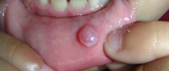
Signs of oral fibroma
Fibroma looks like a pink hemispherical formation, rising above the general surface of the mucous membrane and having a wide, strong base or thin stalk. Fibroids do not cause pain. Its surface is smooth and does not have any growths, unlike papilloma. As a rule, no changes in tissues and mucous membranes are observed in the area of the fibroma, but in some cases ulcerations may appear over the neoplasm. In this case, an infection develops followed by inflammation, expressed in redness, swelling and pain in the area where the fibroma is located.
A standard fibroma in the oral cavity grows slowly, almost unnoticeably. And if it is constantly exposed to trauma, then the growth of the tumor may slow down, and the tumor itself will be within the initial stage of development. It is worth considering that constant injuries lead to complications: the tumor degenerates into a malignant one.