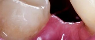
The structure of the alveoli in the mouth
Alveola is translated as cell. The term is used in pulmonology, dentistry and other medical fields. The alveoli have a spongy structure penetrated by:
- a network of nerve endings that ensure their sensitivity;
- a network of blood vessels that supply the alveolar processes with nutrients.
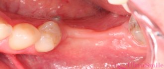
The walls of the dental alveoli are divided into internal, located closer to the throat, external, located on the side of the lips, and interdental. From the inside, the sockets are separated by thin bone partitions, the location of which depends on the structure of the roots of the teeth. Their inside is covered with a spongy plate, the size of which exactly matches the size of the tooth located in the socket. The outer edge is covered by a cortical jaw plate.
Photo: alveoli in the mouth
Elastic fibers predominate in the composition of alveolar tissue. The main function of the remaining cells is the constant restoration and renewal of bone tissue. The balance between the processes of its destruction and growth depends on them.
Bone tissue contains both organic and inorganic particles. Its main components are:
- proteoglycans;
- osteoclasts;
- collagen;
- osteocytes;
- osteoblasts.
If a tooth falls out or is removed, the hole heals quite quickly. A few weeks after extraction, the gums heal, and after a few months, a new gum cover is completely formed.
Location and structure
Dental alveoli (sometimes also called alveolar sockets or cells) are depressions located in the alveolar bone of the upper and lower jaw. Alveolar bone is represented by two components:
- alveolar process (anatomical part that forms the so-called “dental bed” to which all the teeth of the upper jaw are attached);
- alveolar part (anatomical bone surface intended for attaching teeth to the lower jaw).
Dental alveoli
The alveolar processes are filled with bone spongy substance, osteons (structural units) of which form the walls of the alveolar socket. Inside, the dental cells are separated by bone partitions, that is, in one hole there can be from one to three or four recesses in which the dental roots are located, surrounded by periodontal tissues. In rare cases, the number of cavities can reach five if a person is diagnosed with the eruption of abnormally modified “eights” (“wisdom teeth”) with a complex root system.
The relationship between dental alveoli and nutrition
The structure of the alveolar cells is porous (due to a large amount of loose connective tissue). Nutrition of the alveolar bone is carried out by diffusion through blood vessels located in periodontal and periodontal fibers, as well as cancellous bone substance. The structure of the alveoli also contains nerve plexuses and lymphatic vessels.
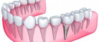
Supporting apparatus of the tooth
Functions of alveolar cells
The alveoli are designed to securely attach the teeth to the jaw. Their structure ensures the stable position of the teeth and prevents them from moving and falling out.
Thanks to dental alveoli, people can chew food without the risk of incisors, canines and molars becoming loose or falling out, unable to withstand the load.
Between the sockets and teeth there are connective fibers of periodontal tissue. Penetrating into the upper layers of the bone tissue of the tooth and the walls of the cells, periodontal tissue firmly binds them, which contributes to the correct position of the tooth in the socket. Additionally, the periodontium acts as a shock absorber, reducing the load on the dentition and slowing down its destruction.
Post-extraction alveolitis
The main cause of inflammation of the tissues of the alveolar cell is the displacement of the blood clot, which should protect the periosteum and nerve plexus from injury and infection. The prevalence of alveolitis is about 40% of all types of post-extraction complications (complications that arise after the removal of a tooth and its root).
Causes
Almost 90% of all alveolitis have an infectious etiology, so after tooth extraction, doctors often prescribe antimicrobial and antiprotozoal agents, as well as antibiotics, to the patient. Failure to comply with these recommendations and prescriptions, as well as insufficient or improper hygiene, can cause infectious and inflammatory changes in the injured socket.
Recommendations after tooth extraction
Dentists also name a number of factors that increase the risk of post-extraction complications. These include:
- smoking (if a person cannot give up his habit for several days, it is necessary to at least carefully rinse his mouth with warm water after each cigarette smoked);
- consumption of spicy, fatty, smoked, pickled foods and alcoholic products;
- using a toothbrush with stiff bristles until the alveoli are tightened;
- incomplete removal of bone fragments, fragments of cysts, granulomas and other pathological tissues during surgery;
- failure to comply with aseptic measures in the dental office and operating room (in case of complex removal);
- active rinsing of the mouth on the first day after extraction.
Tooth extraction
Chronic diseases leading to persistent weakening of the immune system, as well as the complex nature of the removal (for example, the complex removal of the eighth molar) can contribute to the development of pathogenic flora and infection of the socket.
Wisdom tooth extraction
Clinical features
One of the main and most clinically significant signs of post-extraction alveolitis is severe toothache in the place where the extracted tooth was located. The intensity of pain during alveolitis is usually high, difficult to relieve with analgesics. In some cases, the patient cannot calmly eat, sleep and perform household and work duties, so one of the main tasks of treating alveolitis is to relieve the inflammatory process and reduce pain.
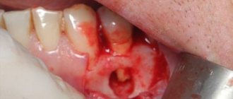
Normal socket healing
An inflammatory process after dental extraction can also be suspected based on other symptoms that appear during the first three days after surgery. These signs include:
- a strong smell of decay that does not go away after brushing your teeth;
- systemic hyperthermia (up to 38°-39° C);
- grayish coating on the surface of the alveoli;
- discharge of purulent or serous exudate from the injured socket;
- severe intoxication syndrome (headache, nausea, severe weakness, lethargy);
- inflammation, swelling and hyperemia of the soft tissues around the affected hole.
Headache
In some cases, the patient may have enlarged lymph nodes in the neck and under the lower jaw.
Treatment
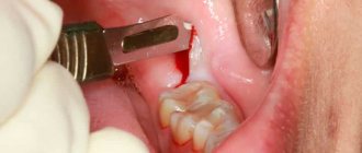
Post-extraction alveolitis almost always has an acute course and can be easily treated if you consult a dentist in a timely manner (it is better to contact the specialist who performed the extraction). First, the doctor will conduct a visual examination, after which he will perform curettage of the alveolar socket and clean it of remnants of a blood clot, granulations, food and infiltrate. If necessary, drainage is carried out followed by treatment with antiseptics. After all the manipulations, a tampon soaked in an antibacterial solution is placed in the hole.
Curettage - photo
Treatment at home after emergency dental procedures is usually carried out according to the following scheme:
- antibacterial (amoxicillin 500 mg 3 times a day) or antimicrobial (metronidazole 250-500 mg 2-3 times a day) therapy;
- NSAIDs to reduce inflammation, eliminate intoxication and fever syndrome and pain;
- local treatment (rinsing) with antiseptics, for example, hydrogen peroxide 3% or chlorhexidine.
Chlorhexidine bigluconate
Acute pain after starting therapy usually disappears by the end of the third day, but moderate and mild pain can persist for up to 15-20 days. If necessary, the patient can receive a certificate of temporary incapacity for work, the duration of which is usually 3-5 days.
Certificate of incapacity for work
Important! The main prevention of alveolitis is to maintain the integrity and location of the blood clot, as well as providing antiseptic treatment and the necessary hygienic care. A gentle regimen should be followed for 3 days after surgery.
Video - Pain in the socket
The alveoli in the mouth, which are commonly called dental sockets, are an important anatomical element of the jaw, necessary for speech (conversation) function and the ability to chew food normally without shifting the dentition. Pathologies of the dental alveoli occur mainly after the removal of permanent teeth, but in rare cases they can occur even before the birth of a person against the background of impaired embryogenesis. Prevention of alveolitis (the main disease of the tooth socket) consists of following medical recommendations regarding the nutritional and hygienic regime, as well as timely antiseptic treatment with disinfectant solutions and antibacterial ointments.
Formation of dental cells
Photo: alveolar process
The formation of alveoli begins in the perinatal period. When the tooth germs are separated from the jaw plate, bone crossbars are formed around them - the future walls of the alveoli. The cells are completely formed during the eruption of dental units.
The formation of the alveolar process occurs in infancy under the influence of the restructuring of the alveolar bone caused by tissue changes that accompany the eruption of primary incisors, canines and molars. Subsequently, the bony outgrowths fuse together and form sockets surrounding each tooth.
In adulthood, the structure of the alveoli of the teeth undergoes the opposite changes: atrophic processes are activated in the bone tissue and pulp, as a result of which the elasticity of the collagen fibers in the socket decreases. As the condition of the alveolar tissue deteriorates, the risk of loosening and displacement of incisors, canines and molars increases.
The rate of development of atrophic processes depends on the state of the body and bone tissue, the quality of oral hygiene and nutrition. The solution to the problem should be approached comprehensively: attention must be paid to all factors that may affect the condition of the cells.
Dentists evaluate the condition of the alveolar sockets based on the clarity of diction and how firmly the teeth stand in the dentition.
Structure
Varies depending on the shape and functionality of each individual tooth. For example, the alveoli of multi-rooted teeth have interradicular septa, which are absent for single-rooted teeth. One is separated from the other by a special bone interdental septum. Both septa are similar in structure and purpose, the only difference is the length: the interroot septa are shorter, they are significantly less than the length of the root.
The alveolar process consists of:
- outer wall (depending on the location, it can be labial or buccal);
- internal wall (regardless of location, it can only be lingual);
- a special spongy substance in which the alveoli with teeth are directly located.
The walls of the alveoli are strong and consist of bone plates that are completely penetrated by periodontal fibers. In this case, the walls are porous and have small microscopic holes in which blood vessels and nerve fibers are located. This is why the alveoli are so sensitive.
The alveolar process is close to bone tissue and consists of inorganic and organic substances. All the spaces that inevitably form between the alveoli and the process are filled with the so-called spongy substance, from which the interdental and interroot septa are also made.
Over time, the structure of the alveoli undergoes some age-related changes, since mobility increases with age, the walls show signs of resorption, which are most noticeable on the medial side of the alveoli, because it is in this direction that the tooth moves when it moves to the side when chewing food. The opposite wall, on the contrary, experiences some tension.
The same changes in the structure of the alveoli can be detected during orthodontic treatment associated with movements in the dentition. The wall in the direction of tooth movement experiences increased pressure, while the opposite wall experiences tension, as a result of which new bone formations may appear, which is normal in this case.
When you hear the word “alveolus,” the first thing that comes to mind is the structure of the lung tissue. But alveoli are not only found in the lungs. Dentists also use this term. Alveoli are the sockets in which the roots of the teeth are located. The structure of dental alveolar cells, their functions and possible pathologies will be discussed below.
What are the features of the upper alveoli of the teeth?
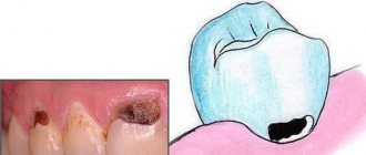
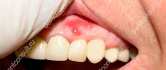
Photo: alveolitis
Regardless of which jaw the alveoli are located on, there are no significant differences in their structure. The only peculiarity of the upper alveoli of the teeth is that their structure affects diction and intelligibility of speech , which is due to the close location of the alveolar process and the palate.
The alveoli are susceptible to a number of dental diseases, the most dangerous of which is alveolitis. The disease can cause relaxation of the alveolar tissue, which can cause the tooth to shift, become loose, or even fall out. If you suspect that your teeth have begun to shift, you should immediately contact the dentist.