Osteomyelitis is usually a purulent infection affecting the bone marrow (myelitis), bone tissue (ostitis), and the periosteum around the bone (periostitis). When it first occurs, it is called acute. If it lasts for a long time with remissions and exacerbations, it is classified as chronic.
The etiology of osteomyelitis is:
- specific,
- nonspecific,
- contact,
- postoperative,
- hematogenous.
Treatment mainly consists of opening and destroying pus, fistulas and other things. Women are less susceptible to this disease than men. The elderly and children suffer from it more often than young people and middle-aged people. The main risk group is boys. What is dangerous about osteomyelitis in this group is its transition to a purulent systemic infection. The disease can be fatal. Complications in adults are rare and local.
Causes
If osteomyelitis has developed, its only causes are the ingress of pathogenic bacteria, leading to purulent inflammation. It is usually caused by Staphylococcus aureus. In some cases, the reason is:
- intestinal, Pseudomonas aeruginosa,
- intraosseous invasion of Proteus,
- hemolytic streptococcus.
The bone can become infected from the inside. In such a situation, bacteria end up in the tissue with the blood carried through the vessels. Osteomyelitis of this hematogenous type usually occurs in young people and children, including infants.
Exogenous causes of osteomyelitis - when microorganisms penetrate from the outside. A contact analogue occurs when the inflammatory process moves from the nearest soft tissue to the bone. The main causative agents of hematogenous osteomyelitis are streptococci and staphylococci. Often many different microorganisms are found at once.
The causes of osteomyelitis (acute hematogenous type) may be due to one of the following infections:
- angina,
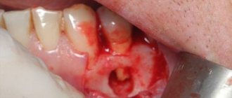
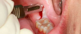
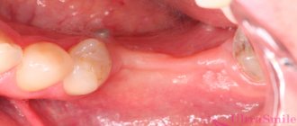
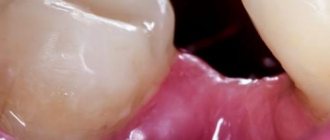
- a festering tooth
- inflammation in the middle ear,
- furunculosis,
- furuncle,
- felon,
- purulent skin diseases,
- inflamed navel ring,
- pneumonia,
- scarlet fever,
- measles,
- other infectious diseases.
The lesion also develops due to:
- trauma surgery, especially with the use of metal screws, pins or plates,
- gunshot wound,
- open fracture,
- wide injuries with contamination of soft tissues.
At risk are people who abuse smoking, alcohol, or taking drugs (through the veins). Low weight, poor nutrition, and advanced age can also lead to the disease. The disease is sometimes a complication due to other health problems:
- immunity disorders,
- vascular atherosclerosis,
- varicose and venous deviations,
- diabetes,
- functional liver, renal disorders,
- malignant tumors,
- removal of the spleen.
The pathogens that led to this lesion can be mono- or mixed cultures. Sometimes the pathogen does not grow on nutrient media. There are a number of triggering or predisposing reasons for a microbe in the intraosseous capillaries to lead to inflammation. As a rule, they predispose:
- weakened immunity,
- hidden infectious foci,
- fasting for a long time,
- vulnerability to allergies,
- any other depletion of the body.
The development of the disease can be triggered by:
- burn,
- injury,
- ARVI,
- frostbite,
- stress and so on.
This disease is also recorded in newborns. There is an opinion that they receive it due to the presence of hidden infectious foci in mothers during pregnancy. However, microbes are practically unable to “get” through the umbilical cord to the fetus.
According to scientists, most likely, against the background of infectious foci, a woman during pregnancy finds herself in a state of allergization. The number of reasons for the proliferation of lymphocytes with immunoglobulins is increasing. And these substances already penetrate the umbilical cord with the blood, increasing the allergic background of the fetus. Because of this, when the umbilical cord is cut, it can become inflamed. From the resulting focus of pus, microbes migrate into the bone, causing osteomyelitis.
Contraindications and complications
Sometimes the patient's condition does not allow surgical intervention. In this case, the doctor selects a different therapy method. Contraindications to surgical treatment are:
- general serious condition of the patient;
- severe pathologies of the upper respiratory tract;
- diabetes;
- period of bearing a child;
- thrombosis.
Provided that the intervention is carried out correctly and there are no parallel pathologies, after the operation the person can live as before. However, if the algorithm is violated or the patient does not follow the doctor’s instructions, there is a risk of developing the following complications:
Anemia in a patient is a possible consequence of violating the doctor’s recommendations.
- soft tissue abscess and phlegmon;
- involuntary fractures;
- contractures;
- sepsis;
- anemia;
- amyloidosis.
Osteomyelitis is a severe pathology that requires a quick response. With timely treatment and compliance with the doctor’s instructions, the disease will pass without leaving a trace. The main thing is to consult a doctor in a timely manner and follow his instructions. In addition, it is important to take measures to prevent illness and strengthen the body’s protective properties.
Symptoms
The course of osteomyelitis and its symptoms greatly depend on the form of the disease. For example, the hematogenous form is characterized by a severe course. The toxic variant is especially unfavorable. It is accompanied by a deadly endotoxic shock associated with the entry of many bacterial toxins into the blood.
A person’s blood pressure drops, consciousness is depressed - even to the point of coma, shortness of breath appears, and the temperature exceeds 40 degrees. Babies may have seizures. The result is heart failure and death. Symptoms of the underlying disease are often not noticed due to severe general reactions. Depressed consciousness prevents patients from complaining of bone pain. And the reasons are revealed only at autopsy.
In one case out of five, examination reveals:
- edema,
- redness and increased temperature in the affected area,
- joint contracture,
- asymmetry of superficial veins.
In this case, stabilization is necessary, and only then surgery is possible. Such an extreme course occurs most rarely. It usually occurs in adolescents aged 14-17 years.
The septic-pyemic form is more common and is accompanied by:
- low pressure,
- fever with a temperature up to 40 degrees,
- dull, pressing headache
- dehydration and sweating,
- confusion.
Local symptoms of osteomyelitis are also present. In the affected area there is a cutting or bursting, but invariably severe pain. The patient usually cannot indicate the exact location of the pain. The soft tissue over this lesion swells and turns red. Contracture occurs in the adjacent joint.
There are also complications in such cases: other organs become infected with the same microorganisms. An infectious process develops in the form of a purulent focus or inflammation of the entire organ. Even with timely, high-quality treatment, approximately half of patients die.
The local option is the most favorable. Intoxication also occurs, but it is much less pronounced, accompanied by:
- temperature 38-39 degrees,
- not very intense dull headache,
- weakness,
- sweating,
- decreased appetite.
When examining the patient, the doctor observes local signs of osteomyelitis: redness, swelling, contracture, contouring of the veins located next to the affected bone.
Subacute form is transitional to chronic. Signs of the disease, both local and general, are not as pronounced as in the acute form. Are common:
- temperature within 37.6 degrees,
- minor (or no) headache,
- slight weakness.
At the site, the pain becomes less intense, erased and dull, intensifying with exercise. There is no swelling or it is almost invisible. Contracture of the joint adjacent to the damaged bone is quite likely. The bone lesion becomes permanently altered.
With chronic osteomyelitis, the patient becomes much better. General signs disappear except for a slight increase in temperature. The pain in the damaged area is weak and aching. There is swelling, but it is moderate.
Pathological changes form in the affected bone, for example, purulent fistulas, channels connecting the infected point with the skin, with the formation of an outlet. Limbs become bent and shortened. The destroyed areas of bones are separated. Phases of exacerbation and remission alternate.
If the jaws are affected, additional symptoms appear:
- pronounced swelling,
- contracture of the joint of the temple and lower jaw,
- pain when swallowing.
There are also atypical forms, which will be discussed below, that do not have pronounced, obvious symptoms.
Symptoms and stages
Bone marrow disease is difficult to cure. For this reason, you should pay attention to any changes occurring in the body. The general symptoms of osteomyelitis can be confused with manifestations of a cold. The first signs of inflammation:
- fever accompanied by chills;
- arrhythmia.
After a few days (on days 2 or 3), local symptoms appear.
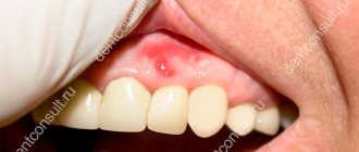
Swelling develops in the affected area, pain is felt, stiffness of movement occurs, and the skin turns red. The most commonly affected are the femur and tibia of the lower extremities or the radius, ulna, and humerus of the upper extremities. Rarely, the disease develops on the bone tissues of the feet, hands, pelvis, ribs, spine, shoulder blades and collarbones. To understand what the inflamed area looks like, you need to study the photo. When infected with harmful microorganisms, intoxication often develops; it can manifest itself as follows:
- pain in the heart area;
- hypotension;
- convulsions;
- loss of consciousness;
- dizziness;
- changes in the skin: yellowness, pallor.
Stages of development of osteomyelitis:
- acute;
- chronic.
In the first case, the pathological condition manifests itself clearly: the temperature rises (up to 40 ° C), the patient shudders, the intensity of pain increases during movement, the affected area swells, and the skin turns red. In the chronic form of the pathological condition, the symptoms will be different: the temperature is lower (37.5...38.5 °C) than during the acute course of the disease, sleep is disturbed, the pain is constant, but not severe. Fistulas form in the affected area, and pus comes out through them.
Classification of osteomyelitis
Bone osteomyelitis is divided into specific and nonspecific.
Specific osteomyelitis occurs when:
- syphilis,
- brucellosis,
- articular, bone tuberculosis.
The second type is associated with:
- coli,
- streptococcus,
- occasionally fungi.
There is also a classification of osteomyelitis according to the ways microbes enter the bone: exogenous and endogenous. In the first case, the infection occurs during surgery, injury, or from adjacent soft tissues and organs. In the second - through blood from a destroyed lesion: an abrasion, a boil, a carious tooth, and so on. At the start, their manifestations are different; at a further stage of osteomyelitis, the differences are smoothed out.
The following forms of osteomyelitis are also distinguished:
- contact,
- post-traumatic,
- postoperative,
- firearm.
The course of the disease at the beginning is usually acute, then with recovery or transition to a chronic form. There are atypical forms of osteomyelitis that do not have an acute phase. Some types of osteomyelitis associated with infectious diseases are also primarily chronic.
Hematogenous osteomyelitis
This type of disease is associated with the introduction of pathogenic microorganisms into the bone vessels with the subsequent appearance of an inflammatory focus. Children aged 3-14 years and men are at particular risk of developing hematogenous disease. With outbreaks of viral infections, the number of cases of this disease is also increasing. Useful article on the topic of acute hematogenous osteomyelitis.
Most often it is excited by Staphylococcus aureus. Less common:
- Proteus,
- intestinal, Pseudomonas aeruginosa,
- hemolytic streptococcus.
It is usually localized in the femur, humerus or tibia. There are several options for its course.
The precipitous type is the most favorable. The body’s reaction is pronounced, recovery is intense. You can fully recover in two to three months. The protracted type is accompanied by a long subacute course. The patient's immune status is low and recovery processes are weak. However, recovery can still be expected - in a little more than six months.
The fulminant type is the most dangerous and rapid, appearing most often due to staphylococcus. Bacteria are released into the blood quickly. The microbe is destroyed, releasing aggressive endotoxin. Result: blood pressure drops to zero and brain death six minutes later. A patient can only be saved by providing massive medical assistance.
The chronic course lasts more than six months, accompanied by relapses and remissions. Areas of dead tissue appear that support inflammation. Connective tissue forms around the fistulas, sometimes causing scars and muscle atrophy. Neighboring internal organs may also be affected.
Post-traumatic osteomyelitis
Traumatic osteomyelitis occurs with open fractures. The reason is contamination of the wound due to injury. The more complex the fracture, the greater the chance of developing this disease. Usually all bone parts are affected.
If the fracture is linear, then the site of injury becomes inflamed; if it is splintered, the purulent process can spread. Accompanying:
- severe intoxication,
- hectic fever,
- increase in ESR,
- leukocytosis,
- anemia.
The wound area is swollen and very painful, with a lot of pus coming out of it.
Gunshot osteomyelitis
The appearance of gunshot osteomyelitis is promoted by widespread lesions of soft tissues and bones, coupled with stress, reduced ability to resist, and poor wound treatment. General symptoms are similar to the previous type, local symptoms are smoothed out.
The development of the disease can be determined by changes in the surface of the wound: a dull tint and a grayish coating. Inflammation then spreads to all bone layers. Despite infection, the bone will heal over time if the damage was not too severe. Foci of pus enter the callus.
Postoperative osteomyelitis
This is a type of post-traumatic osteomyelitis that occurs after operations performed on bones. Appears due to the high traumatic nature of the procedure and failure to follow the rules of asepsis.
Contact osteomyelitis
Contact osteomyelitis spreads from a purulent process in nearby soft tissues. At the same time, swelling increases, pain in the damaged areas intensifies, and fistulas form.
Osteomyelitis: classification, symptoms, treatment methods
Osteomyelitis is a process of infectious progression that penetrates the bone and causes inflammation.
Many people think that only the bone is affected, however, the bone marrow as a whole also suffers. There are both acute osteomyelitis and its chronic form. There are certain statistics that indicate that after injuries and operations, osteomyelitis appears in only 7% of all diseases that are associated with the musculoskeletal organs. The types of osteomyelitis are different, for example, depending on the etiology, there are specific and nonspecific osteomyelitis, hematogenous, post-traumatic, contact and others. The picture of the disease directly depends on the type and form of osteomyelitis, which shows the further course of treatment of this disease.
Diagnostics
When osteomyelitis is suspected, diagnostics are carried out with instruments, as well as paraclinical and laboratory diagnostics. In a general blood test, the leukocyte formula shifts to the left, band neutrophils exceed the norm from two to five times. In this case, hemolytic anemia develops, hemoglobin, reticulocytes and red blood cells are reduced, and platelets are increased. The rate at which red blood cells settle is also taken into account. The amount of protein in the urine increases, the relative density decreases, casts, erythrocytes, and leukocytes appear.
In a biochemical blood test, signs of inflammation appear, and sometimes indicators of acute liver or kidney failure. Depending on the pathogen, glucose levels increase or decrease. The values of chlorine, sodium and potassium decrease, phosphorus and calcium - increase.
Ultrasound helps to examine intermuscular phlegmon and fistulous tract. Also, the diagnosis of acute osteomyelitis is based on infrared scanning. In this way, areas with elevated temperatures are identified.
Bone puncture is not only a diagnosis and examination for osteomyelitis, but also treatment. After collecting the pus, the pathogen is identified. Treatment is carried out by reducing the pressure inside the bone and forming an artificial fistula. In this case, inflammation stops progressing. It is used only for children whose bone tissue is relatively soft.
Osteomyelitis is a disease whose diagnosis is most often made on the basis of radiography. Pictures are taken on a pair of projections. At the same time, bone necrosis is localized, its extent and severity are determined. The procedure is repeated to control the dynamics.
A CT scan, or computed tomography scan, is best for visualizing signs of osteomyelitis. It provides all the X-ray data as well as a three-dimensional reconstruction of the entire affected area. The examination helps to distinguish it from other bone lesions.
Also, at all stages, phases, stages of osteomyelitis, radionuclide diagnostics are carried out, based on the use of radioactive pharmaceuticals. They accumulate in a special way in the inflamed area. The research is high-quality, but very expensive, requiring special premises and sophisticated equipment.
Diet for osteomyelitis
With this disease, the intestinal microflora changes, immunity decreases, and the functioning of the digestive tract is disrupted. To reduce the load on these organs, you should follow a diet. Basic recommendations:
- Avoid fatty and fried foods, as well as smoked foods. It is not recommended to consume coffee, chocolate, spices.
- Products that contain large amounts of vitamins and microelements are introduced. With osteomyelitis, calcium, iron, magnesium, and phosphorus are especially important, so you need to eat meat (only lean), dairy products, eggs, fish and liver.
- The basis of the diet should be fiber: vegetables, fruits.
- It is recommended to drink at least 2 liters of water per day. This does not include soups, teas and other drinks.
- To support your immune system, it is recommended to drink fresh beet and carrot juice every day. A combined drink is made from these root vegetables in a ratio of 2:5.
Treatment
When osteomyelitis is diagnosed, treatment must be timely and comprehensive. The unpredictability of the flow forces us to constantly monitor the dynamics. Ideally, you should combine methods:
- surgical,
- medicinal,
- physiotherapeutic.
Treatment of osteomyelitis with medications is carried out exclusively in combination with surgery. Combinations of antibiotics are selected that most effectively destroy the infection inside the bone. They are administered intraosseously.
During and after surgery, different antibiotic therapy regimens are used. Having identified the causative agent of the disease, the set of medications is reviewed. Subspecialty specialists prescribe medications that correct the functioning of the systems and organs affected by the disease.
At home, treatment of osteomyelitis is almost impossible - surgery is needed in almost all cases. A concomitant decompensated chronic disease may become a contraindication - if complications from the surgeon’s intervention may be more serious than osteomyelitis itself. In the acute stage, surgery is performed to force the disease to become chronic. As soon as the inflammation subsides, they begin to sanitize the lesion.
Surgical treatment of osteomyelitis consists of removing the purulent focus. Surgery is resorted to only when purulent formations are accurately identified. In the chronic stage, inflammation and scar changes are removed. The operation is performed under general anesthesia in a septic operating room. The bone is cleaned and washed.
A drainage tube is left in the bone marrow canal for a week, through which it is then washed with special antibiotics. Then the drainage is left only in the soft tissues. When the wound heals, it is also removed. They do not put a plaster on the wound, but a splint, so that the wound does not rot, and it is more convenient for doctors to control the healing process.
When osteomyelitis is treated, rehabilitation is also carried out. A dietician, together with the attending physician, selects a diet rich in iron, calcium and protein. Patients eat fractionally: five to six times a day in small portions. Be sure to consume:
- meat,
- cottage cheese,
- eggs,
- milk,
- bananas, apples and other fruits.
You should drink at least two and a half liters daily.
Physiotherapy procedures are also carried out, the most effective are:
- UV irradiation,
- electrophoresis with moderate antibiotic solutions,
- UHF.
Exercise therapy is also indicated. The specialist selects exercises in accordance with the current condition of the patient. Bone osteomyelitis is treated if a set of therapy and rehabilitation measures is selected and carried out competently.
Indications for surgery
Since osteomyelitis of the jaw is an irreversible purulent process that involves an infectious agent, surgery cannot be avoided. Lack of surgical intervention leads to further osteomalacia and necrosis. Acute process and exacerbation of chronic, diffuse and localized osteomyelitis - all these are indications for urgent treatment. Even if it is possible to avoid partial removal of the jaw bone, access to the purulent focus is still required to remove the bacterial exudate and wash the resulting cavity, which is called osteoperforation, with antiseptics.
Prevention
Prevention of osteomyelitis allows you to avoid the disease, timely diagnosis – complications and transition to a chronic condition. You should not self-medicate - when the purulent focus is in the bones, only surgery will help.
Osteomyelitis is too serious for prevention to be underestimated. It is important to consult a doctor promptly if symptoms are detected. All diseases leading to osteomyelitis are treated with special attention. It is important to avoid the presence of chronic foci of infection, as well as foci of inflammation.
Prognosis and prevention
It is difficult to say what the threat of osteomyelitis is in each specific case, since the course of the disease is influenced by a number of factors: the characteristics of the patient’s immunity, age, condition of the affected tissues, and the body’s response to drug therapy. Successful treatment is indicated if there are no relapses of this pathological condition over the next 2–3 years. The prognosis is positive in most cases.
Sometimes the inflammatory process leads to irreversible changes in the musculoskeletal system. Then the patient is registered as disabled.
Prevention of osteomyelitis comes down to careful treatment of wounds and deformed areas in case of any injuries and surgical intervention. It is important to prevent the development of postoperative inflammation of bone tissue. In this case, a purulent focus and fistula may appear at the incision site. The main reason is insufficient quality of cleaning the canal from infection.
Frequently asked questions from patients
Osteomyelitis is a dangerous disease that can lead to disability or death. It is important to diagnose the disease in a timely manner, carry out proper treatment and undergo rehabilitation.
Which doctor treats you?
If there are signs of osteomyelitis, you should seek help from an orthopedist. In public medical institutions, you must first visit a surgeon, who will give a referral to a specialist.
Can it be cured completely?
Cases of complete recovery of patients with osteomyelitis are not an exception, but a fairly common occurrence. This is possible with timely diagnosis and proper treatment. Even when the disease develops in newborns, doctors give favorable prognoses.
Is a child given disability after suffering from osteomyelitis?
You can get a disability if the pathology is severe, the bone is deformed, and the patient has lost the ability to lead an active life. For example, if the lower limb becomes shorter, then the child is indicated for disability. In other cases, he is exempt from physical education for 6-12 months.
Is osteomyelitis contagious to others?
When treated in a hospital, the patient is placed in a general ward. The pathology is not contagious, although it has an infectious etiology. With chronic osteomyelitis, the patient leads a normal lifestyle, but with some limitation of physical activity.
Acute osteomyelitis
The manifestations of this form of the disease depend on the following factors:
- general condition of the body;
- route of infection;
- the extent of traumatic damage to surrounding soft tissues and bone.
The acute form of osteomyelitis is characterized by the fact that even 2-3 weeks after the onset of the disease, changes are visible on the x-ray. There are several varieties of this pathology.
Hematogenous
Develops in childhood, most often before 1 year. Affects the femur and tibia. In some cases, multiple bone lesions are observed. Microbes enter the bloodstream from an infected wound, phlegmon, or soft tissue abscess, and then spread throughout the body.
Long tibias have a wide network of capillaries with reduced blood flow. Infectious agents settle in the spongy bone. If unfavorable conditions arise, such as weakened immunity or hypothermia, microbes begin to actively reproduce, and hematogenous osteomyelitis occurs.
The pathology is accompanied by the following symptoms:
- increase in body temperature to +39°C…+40°C;
- vomit;
- headache;
- chills;
- hemolytic jaundice;
- convulsions;
- rave;
- loss of consciousness;
- increased heart rate;
- decrease in pressure;
- bursting, boring or sharp pain in the affected area.
Post-traumatic
It develops when there is an open bone fracture as a result of infection getting into the wound. The likelihood of pathology occurring with a comminuted fracture, vascular insufficiency, extensive soft tissue damage, and weakened immunity increases. The disease can affect all elements of the bone.
With this pathology, severe intoxication, fever, leukocytosis, anemia, and increased ESR are observed. In the area of the fracture, the tissue swells, turns red, and sharp pain occurs. A large amount of pus is released from the wound.
Firearm
This is a suppurative process in the area of a gunshot fracture, which occurs with blind shrapnel wounds. In this case, severe destruction of the bone occurs, which is accompanied by the formation of fragments and the formation of necrosis along the edge of the wound and in the bone. Most often, pathology develops with injuries to long tubular bones. The symptoms are the same as for post-traumatic osteomyelitis.
Postoperative
This is inflammation of bone tissue, which occurs as a purulent complication after surgical interventions for osteosynthesis of closed fractures, insertion of wires when applying skeletal tension or compression-distraction devices (wire osteomyelitis). The disease develops due to excessive trauma of the operation or non-compliance with antiseptic rules.
Contact
It occurs if a purulent-inflammatory process has developed next to an intact bone: bedsores, chronic ulcers, purulent wounds, etc. The pathological process affects the periosteum, which is the source of nutrition for the bone, leading to its death. Because of this, the blood supply to the bone is disrupted, the infection begins to actively multiply, and osteomyelitis develops. In this case, there is an increase in swelling, increased pain and the formation of fistulas.
Osteomyelitis of the bone structure
The clinical picture and therapeutic actions for infection of the bone structure are determined by the localization of the source of inflammation. The mildest course is observed with inflammation of the bone in the heel area, since only a small area of the limb is affected. So, even with swelling and problems with motor functions, installing drainage to drain liquid contents and treating with antibiotics helps to quickly cope with the disease.
If the lower leg is affected, the tibia is involved in the process, which causes severe disturbances in the functionality of the leg in the form of:
- severe pain;
- swelling of the lower leg;
- inability to move;
- increasing signs of poisoning;
- hectic fever.
If only the fibula is affected by the inflammatory process, the severity of pain is lower. But surgical care in this case is more difficult to provide. If this disease is accompanied by problems with blood supply to the legs, the healing process is often slowed down.
If the femur is affected, severe intoxication develops and often immobilizes the patient. Simple trepanation in this case is insufficient, since there are a large amount of muscles around. So in such a situation, open surgery is performed, which is why the recovery period and the final cure of the person will take longer.
With osteomyelitis of the ischial bone structure, sharp pain appears in the lumbar region. Rarely, due to paresis of the limbs, the patient cannot move at all. Open surgery is used in rare cases; trepanation and conservative treatment are usually used.
If any bone in the lower extremities is affected, you can resort to alternative medicine. A compress with herbal decoctions or tinctures applied to the affected area helps to quickly get rid of swelling and enhance the effect of antibiotic therapy. But when installing open drainage, such means will not help, since foreign plant substances can accelerate bacterial growth in the wound surface.
Diagnosis of osteomyelitis
The main method for diagnosing osteomyelitis is x-ray examination. It is mandatory to perform radiography of the affected segment in several projections. However, for assessing the prevalence of the destructive process in soft tissues, the capabilities of this method are very limited. To fully characterize the pathological process, conventional radiography data are usually not sufficient. In most cases, when examining large joints and bones, computed tomography and MRI are necessary to identify or clarify the prevalence of destructive changes and sequestration.
Radioisotope scintigraphy is used for early detection of the disease and allows one to determine the approximate size and location of the lesion.
To identify purulent leaks and paraosseous phlegmon, it is necessary to perform an ultrasound examination to assess the volume and extent of the purulent-necrotic focus of soft tissue.
In the presence of fistulas, fistulography is mandatory. This method makes it possible to clarify the prevalence of fistula tracts in soft tissues, bone structures and the volume of the pathological cavity for an adequate choice of surgical approach.
Bacteriological research serves to assess the qualitative and quantitative microbial composition of a purulent focus.