general information
Normally, the jaw bones have a dense structure that ensures the correct performance of the supporting and chewing function. When exposed to aggressive factors, the quality of bone tissue is impaired and foci of inflammation appear. Dead jaw syndrome is a noticeable loss or complete destruction of bone.
The essence of pathological changes:
- with osteonecrosis, the blood supply to the bone is disrupted;
- with a lack of nutrients, microcracks appear;
- tissue density is gradually disrupted, the structure deteriorates;
- teeth often fall out;
- in advanced cases, the infection affects a wide area, penetrates deep into the tissues, and destroys the bone marrow.
Description of osteonecrosis of the jaw
What is osteonecrosis of the jaw?
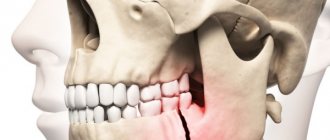
This anomaly is known as “dead jaw syndrome”; in medicine the concepts of aseptic or avascular necrosis are used. This is a serious disease of the upper or lower jaw, as a result of which the life of the cells of the skull bones (osteocytes) fades away while the main substance is preserved. This leads to reduction or destruction of tissues and disrupts the process of supplying them with blood. Pathology causes cracks, leads to bone damage and weakening, and loss of teeth. Unfortunately, the disease does not manifest itself in the early stages, so it is often detected accidentally during dental procedures and x-rays. She suddenly announces herself with sharp pain in the affected area. With avascular necrosis in the initial stage, symptoms may appear:
- a feeling of numbness in one or more areas of the face;
- severe swelling of tissues, pain in the gum area;
- when pressing on the teeth, their high mobility is revealed;
- the jawbone is exposed (an abscess may develop);
- the inflammatory process of the mucous membrane progresses and is difficult to treat.
Once a person develops this condition, symptoms may not appear for months. It will only become obvious when the following signs appear:
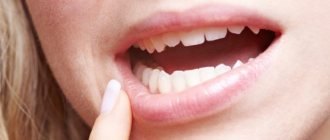
- acute pain, swelling of the gums and infection in the mucous membrane;
- gums cannot be cured with the use of strong medications;
- teeth are loose;
- exposed bone is visible through missing gum tissue;
- heaviness or numbness of part of the face appears.
The development of atrophy can lead to a deterioration in the general condition of the body. A person feels weak, his sleep is disturbed, headaches often appear, and his body temperature rises greatly.
Causes of tissue destruction
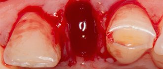
Problems with teeth and gums are one of the main provoking factors. When a tooth is removed, an infection enters the wound. Pathological changes begin if the bone remains exposed after extraction of the affected unit/dental operation.
With a decrease in immunity, negative processes develop over a short period; if the body is sufficiently strong, the asymptomatic course lasts for weeks or months.
Find out the instructions for using the drug Scandonest in dentistry.
Which teeth are better to insert: plastic or ceramic? Read the answer on this page.
There are other risk factors that increase the likelihood of osteonecrosis:
- chemotherapy;
- radiation therapy to the head and neck area;
- penetration of infectious agents - intestinal and pseudomonas bacteria, Klebsiella, staphylococcus;
- viral infections;
- cancer;
- severe pathologies of the hematopoietic system;
- the formation of gum pockets filled with purulent masses, inflammation of the gums;
- poor oral hygiene;
- poor nutrition, lack of vitamins;
- steroid therapy;
- mechanical damage to the jaw (injury with infection);
- excessive drinking of alcohol, smoking;
- problems with blood clotting.
Bisphosphonates and osteonecrosis
Many scientists tend to believe that the use of potent drugs that maintain bone density in cancer has side effects in the form of osteonecrosis. In some cancer treatment regimens, bisphosphonate therapy is a mandatory element.
Simultaneous observation by an oral surgeon and an oncologist will help reduce the likelihood of complete bone destruction. Doctors will develop a program to monitor bone condition and adjust the dosage and form of taking the potent drug in a timely manner.
Thermal form
With severe burns (IV degree), pathological processes develop in the tissues. The cortical plate of the upper or lower jaw is rejected.
Chemical necrosis
Bone condition worsens among workers in hazardous industries. The reason is a violation of safety regulations, exposure to toxic substances, including aggressive vapors.
Some patients experience problems after visiting the dental office. The reason is the incorrect use of paste containing arsenic compounds in the treatment of pulpitis.
Prevention
Prevention of osteonecrosis involves a special method of treating oral wounds during surgical procedures. These wounds are potential trigger points for the onset of the necrotic process.
Author: Roman Kutsenko, maxillofacial surgeon, Ph.D.
Rating: Select ratingVote: 1/5Vote: 2/5Vote: 3/5Vote: 4/5Vote: 5/5Average: 2.7(3 ratings)
Video
Diagnosis of dizziness 12/24/2018 All videos
Reviews
Alexander Moscow
Thank you Dr. Novozhilov! A rare combination of high professionalism, intelligence and positive attitude.
All reviews
Treatment stories
Fracture of the inferior wall of the orbit
For one of our patients, a simple fall resulted in a fracture of the lower wall of the orbit. Outwardly, this manifested itself only as swelling of the eyelids and “bruising,” but the internal damage turned out to be much more serious. The patient was concerned about double vision that appeared after an injury. He addressed this complaint to the EMC. Most
05.23.2019 All
Classification
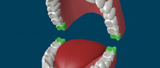
According to the degree of bone damage, they are distinguished:
- partial osteonecrosis. Tissue death occurs in certain areas;
- full. Necrotization of the entire bone is observed.
Localization:
- deep. Deep layers of hard tissue are affected;
- superficial. Necrotic changes are visible on the surface of the bone.
Depending on the amount of tissue necrosis, jaw surgeons distinguish four stages of pathology:
- I. The lesion reaches 10% of the structure, pain occurs periodically, bone mobility is normal;
- II. Microcracks appear, severe pain is noted, mobility deteriorates;
- III. Bone necrosis reaches a third, sometimes half of its volume. The patient constantly feels severe pain;
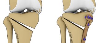
- IV. The bone is completely destroyed, excruciating, unbearable pain develops. Surgery is required to remove dead areas.
Symptoms
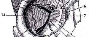
The first sign of osteonecrosis of the jaw is facial pain. Then there are inflammatory phenomena of soft tissues, bleeding gums, periodontitis, mobility of teeth adjacent to the source of inflammation, and impaired sensitivity in the area of inflammation. There is a putrid smell from the mouth.
Over time, part of the affected bone is exposed, forming a growing ulcerative defect in the oral mucosa. In some cases, one of the manifestations of osteonecrosis of the lower jaw may be fistulas on the face and neck. As the pathological process develops and deepens, spontaneous (pathological) jaw fractures may occur.
Symptoms
The danger of aseptic necrosis of the jaw is in the asymptomatic period, which lasts from several weeks to months. Sometimes the patient discovers tissue damage when the bone is already visible in the jaw. In some cases, symptoms appear in the early stages, the patient has a better chance of reducing the rate of necrotic changes.
Important! “Dead jaw syndrome” often develops with osteomyelitis. The symptoms of both diseases are similar. Sometimes the signs “layer on” and severe bone lesions develop. Odontogenic lesions of the upper jaw are more severe and complications develop more often.
Characteristic symptoms:
- gum damage: swelling, redness, non-healing ulcers;
- tooth pain when eating;
- intoxication of the body;
- loosening of dentition units;
- pain, swelling in the jaw area;
- loose gums, areas of inflammation that are difficult to treat;
- abscess, heaviness, numbness of the jaw;
- destruction of the gums, protrusion of the bone, which nothing covers.
How to properly dilute Furacilin for mouth rinse? We have the answer!
For a review of ointments for oral stomatitis for adults and children, see this page.
Follow the link and read reviews about decorating teeth with skyces.
Take note:
- the severity of the disease is aggravated by the development of complications;
- a serious threat to life is posed by phlegmon, brain abscesses, purulent inflammation of the eyelids;
- some patients develop pneumonia, sepsis, tissue fragility, jaw fractures;
- with improper therapy and a weakened body, there is a high probability of disability, and there is a risk of death.
Clinical picture of the disease
Osteonecrosis of the mandible is manifested by local and general symptoms, the severity of which will depend on the form of osteomyelitis:
Signs of hematogenous osteomyelitis
In such cases, the clinical picture is dominated by local symptoms. Intoxication of the body, as a rule, is moderate. Body temperature rarely rises above 39ᵒC.
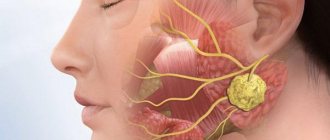
From the oral cavity, you can notice a limited area of inflammation in the form of redness and swelling of the mucous membrane. The development of the disease is accompanied by the formation of a subperiosteal abscess, which can develop into phlegmon of nearby soft tissues.
Hamatogenous osteomyelitis of the mandible
Inadequate or incomplete treatment of the hematogenous form of osteonecrosis, as a rule, ends with the disease transitioning to the chronic stage.
Signs of septic-pyemic form
The development of this form of osteonecrosis begins with a sharp jump in body temperature. Severe intoxication of the patient is accompanied by nausea and periodic vomiting.
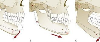
In the first few days, the patient complains of intense attacks of pain in the area of purulent inflammation of the bone. At the same time, the doctor notes a contracture, in which a person’s mobility of the lower jaw is sharply limited.
After a few days, septicopyemic osteomyelitis is manifested by pronounced swelling of the soft tissues, which indicates the formation of an abscess. The skin over the pathological area becomes inflamed and becomes bright red.
Swelling of the soft tissues of the face in the septicopyemic form of osteomyelitis
Osteonecrosis with signs of severe intoxication provokes dehydration, disturbances in water-salt metabolism and deterioration of blood clotting. Acute symptoms of osteomyelitis mainly last about 7-10 days, creating conditions for further spread of purulent infection throughout the patient’s body.
Symptoms of the toxic form of osteomyelitis
The instructions for surgical dentistry state that the toxic form of osteonecrosis is the most dangerous variant of osteomyelitis. This disease often ends in death.
In patients with toxic osteomyelitis, acute cardiovascular failure develops already on the first day, blood pressure drops, the person periodically loses consciousness and a convulsive symptom develops.
Toxic osteomyelitis
Treatment methods
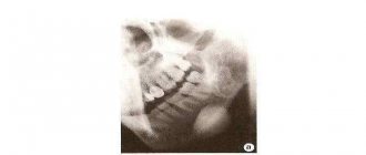
When the first symptoms appear, contact your dentist immediately. A thorough examination is carried out. An x-ray and a detailed blood test are required.
Take note:
- patients taking a course of radiation or chemotherapy for cancer in the neck should be periodically examined by an oral surgeon;
- before starting therapy, in addition to mandatory tests, a study of the microflora of the oral cavity is required;
- the reason is the presence at the site of osteonecrosis of not only pathogenic bacteria, but also fungi and viruses. When dangerous pathogens are identified, therapy adjustments and the prescription of antimycotic and antiviral drugs are required.
It is difficult to completely cure avascular necrosis. In the absence of cancer, the prognosis is more favorable. For patients registered with an oncologist, maintenance therapy improves the quality of life and reduces pain. The patient should know that treatment of osteonecrosis lasts with varying success, often dragging on for months, and relapses are possible.
Important! The success of therapy largely depends on the right attitude and strict adherence to recommendations. A change in lifestyle has a positive effect on the state of the body: giving up smoking, alcohol, feasible physical activity, walking. Healthy foods, multivitamins, mineral complexes, and medications to strengthen the immune system are recommended.
Treatment methods depend on the causes of severe bone damage:
- the focus that provoked inflammation is removed, the purulent masses spread to large areas;
- if the cause of the problem is complications after tooth extraction, antiseptic rinses and antibacterial therapy are prescribed. Drugs are selected taking into account antibiotic sensitivity tests;
- In case of chronic osteonecrosis, the affected tissue is removed. The surgeon applies splints to prevent fracture of the jaw segment;
- It is mandatory to take multivitamins and drugs that strengthen the immune system. Anti-inflammatory compounds and calcium supplements are required;
- the patient must change his diet, provide a diet with sufficient vitamins and minerals;
- physiotherapeutic procedures and drugs for detoxifying the body are required;
- From folk recipes, antiseptic rinses with decoctions of medicinal herbs are effective. Decoctions of calendula, chamomile, oak bark, and sage have not only an anti-inflammatory but also an astringent effect and strengthen the gums;
- Severe cases require surgery with the mandatory use of appropriate antibiotics.
Take note:
- if bone destruction is caused by taking bisphosphonates, doctors recommend switching to oral administration of potent medications;
- long-term refusal of specific therapy for window diseases with the help of the drug Zometa negatively affects the nature of the course of the disease;
- the break in taking bisphosphonates should not exceed 4 months;
- An experienced oncologist will determine the further regimen for taking Zometa or another drug for maintenance therapy.
In case of partial tissue damage, after stopping the destruction process, the patient can wear prostheses that he regularly used. The orthopedic dentist is obliged to warn the patient that loose gums and destruction of bone tissue are contraindications for the installation of implants.
Stock
-11%
Segment of chewing teeth with prosthesis on day 3 135,000 rub.
120,000 rub.
get -10 %
Dental restoration All-on-4 RUB 310,000.
280,000 rub.
get -13 %
Removable overdenture on 4 implants RUB 150,000.
130,000 rub.
get -40 %
Treatment of caries with a discount of 2000 rubles.
5000 rub. 3000 rub. get
Preventive recommendations
Helpful Tips:
- Attention to the oral cavity and proper hygiene procedures will help prevent dangerous diseases of the jaw bone;
- monitoring the condition of tissues after tooth extraction is mandatory;
- Watch your diet, consume enough vitamins and minerals to keep your teeth and bones strong. Vitamins A and C are especially important for gum density;
- At the first signs of infection of the mucous membrane, gingival tissue, or complicated pulpitis, contact your dentist. In advanced cases, inflammation progresses, takes on a generalized form, and affects the jaw bone. Consequences – osteomyelitis, necrotization of the affected areas, abscesses, loss of dentition;
- during the treatment of serious diseases, monitor whether inflammatory processes have appeared in the gum tissue as a side effect of taking potent drugs;
- During courses of radiation therapy in the head area, consultation with an oral surgeon is required for early detection of changes in bone tissue.
The difficulty of diagnosis and treatment lies in the asymptomatic course of the early stages of necrosis of the jaw bone. Even with slight inflammation of the gums, swelling, or problems after tooth extraction, contact your dentist. Only regular examination of the oral cavity will reveal the first signs of a dangerous disease. The most important thing is to prevent the spread of pathological changes deep into the tissues and prevent osteonecrosis.
Read more about necrosis of the lower jaw in the following video:
Features of the disease
It is also known from the biology course that bone tissue is formed from a large number of cells, which in medicine are designated by the term “osteocytes”. The progression of osteonecrosis in the human body leads to the fact that the irreversible process of death of these particular cells begins, but no change in the mineral composition of the bone is observed.
The insidiousness of osteonecrosis lies in the fact that it is very difficult to diagnose at an early stage of the development of the pathology. This is explained by the fact that most often the patient complains of discomfort, the development of which begins without injury. If a person has progressive osteonecrosis of the jaw, then any symptoms may be completely absent for a long time. Medical practice shows that osteonecrosis is most often diagnosed precisely when pathology in the form of a protruding bone in the jaw becomes noticeable.