The so-called wisdom tooth is the eighth tooth on both the lower and opposite jaws. It owes its beautiful name to the timing of eruption: in some cases, the first third molar begins to grow over the age of 20 , which, according to popular opinion, coincides with the arrival of life experience and wisdom.
Location and structure
In the dentition, the third molars occupy the most extreme position and close the dental arch. Absence during visual inspection does not mean that a person does not have them: sometimes figure eights are located in the thickness of the gums. This pathology is called retention; see what it looks like in the photographs.
The structure of a wisdom tooth is no different from the structure of other molars: neck, root system (4–5 pieces), crown. If development is disrupted, the roots can form a single conglomerate - a single-rooted tooth grows.
Why is it dangerous to pull out fangs?
Removing the upper teeth, which interfere and cause aesthetic problems, seems to be a way out only at first glance. No specialist will remove it without good reason.
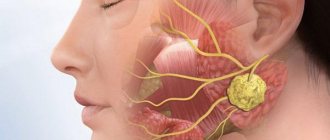
We suggest you find out which doctor treats the salivary glands
Removing these units is painful and dangerous for the following reasons:
- They play a significant role in chewing food, so their absence will complicate the process. Additionally, when removing the upper teeth, problems with diction are possible.
- If some of the eye teeth are missing, other units begin to perform their task, experiencing an unnatural load. This leads to rapid wear, weakening, disruption of the symmetry of the face, bite, and serious health problems.
- The deep position and unique shape virtually eliminate their susceptibility to caries. This means that the infection will not spread to neighboring teeth.
- These are the most stable teeth. They do not allow the incisors and molars to grind down when the jaws are compressed.
Features of wisdom teeth
The main difference between the “extreme” tooth and the rest is the fact that, in the light of recent research, it is considered not as a full-fledged organ, but as a rudiment. That is, during evolution, the function of the third molar was lost, and it gradually becomes unnecessary, so in a certain percentage of people it does not grow at all.
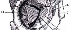
Photo: this is what the roots of a wisdom tooth look like
There are several important parameters that distinguish eighth teeth from others:
- They do not have milk precursors, which makes growth and eruption difficult: there is simply no space left on the dental arch, which leads to various pathologies.
- Larger number of roots compared to other molars: up to 5 pieces. The difficulty of its treatment or removal often depends on how many roots a wisdom tooth has.
- The roots of figure eights are significantly curved, which creates difficulties when cleaning and treating canals.
- A relatively poor blood supply leads to early aging of wisdom teeth, which is accompanied by an increase in their fragility and a greater tendency to caries. And this is despite the fact that wisdom teeth are located eighth in a row on the upper and opposite jaw, which is why the load on them is minimal.
- A large number of pathological conditions are associated with figure eights: pericoronitis, malocclusion, pathologies of the masticatory muscles and others.
All of these facts indicate that the negative consequences of having third molars are much greater than the positive effects. Many people find out what a wisdom tooth is only when it appears, and a person has to immediately contact a dentist for its treatment.
Top Eight Features
Removal of a wisdom tooth in the upper jaw - the consequences of this procedure often make the patient who is indicated for this operation panic, because no one is in a hurry to voluntarily part with parts of their body. But if the doctor insists on removal, you should listen to his words. And in order to calm panic and prepare the patient for the procedure, the specialist must explain in detail what this tooth is, what is its peculiarity, and why it is better to get rid of it in this case.
IMPORTANT!!! A good doctor always tries to save even the most damaged tooth, because... a cured and filled tooth is always better than a denture. The only exception is the upper eight - their treatment is very problematic for both the dentist and the patient.
The wisdom tooth, or, as it is correctly called in dentistry, the upper eight, is the same full-fledged component of the jaw as any other tooth. In the early stages of evolution, the eighth molars appeared simultaneously with the seventh and sixth teeth, and performed the same functions as them.
But over time and the transition from raw and hard foods to softer and thermally processed dishes, the need for these molars began to disappear. But the structure of the jaw is determined by nature, and the eighth teeth still erupt, but much later than all the others. This is what gave them their name: wisdom teeth, i.e. those that appeared with age and experience.
The appearance of teeth is always associated with pain, and in the case of eights it is a very strong and unpleasant sensation: the bones of the face and jaw are formed (and they have to change their position), and the gums have become stronger (and it is much more difficult to cut through them than in childhood). All this creates an extremely negative attitude towards the eighth molars, and the question arises - why are they needed, and isn’t it easier to get rid of them right away?
No. A wisdom tooth should be removed in two cases: when it is destroyed and can lead to the destruction of the neighboring seven, or for strict medical reasons.
IMPORTANT!!! The inaccessibility of the tooth makes it very difficult to properly care for it: “it is not cleaned” - this is the main reason why caries occurs on the molar and the destruction of its tissue begins.
A qualified and experienced doctor will never suggest removing the upper eights “just so that they don’t interfere.” The number eight is a full-fledged tooth, and as long as it does not pose a health hazard or cause discomfort, there is no need to remove it.
How wisdom teeth are cut
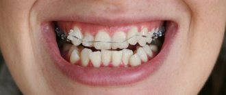
Problems with “eights” begin even when they have not had time to grow: at the stage of their eruption. Any deviation of the tooth from the vertical axis is a pathology called dystopia. Depending on the direction in which the wisdom tooth deviates, there are several types:
- Medial tilt. The molar with its apex is directed towards the front seven. Constant pressure of teeth against each other leads to damage to the enamel, which increases the likelihood of developing caries with subsequent destruction of the crowns.
- Distal tilt. With this pathology, the wise tooth is located with a backward tilt, which is often accompanied by a malocclusion - it is difficult to close the mouth, and chronic pain is noted.
- Buccal tilt. The figure eight tilts or moves completely towards the cheek.
- Linguistic tilt. Incorrect position of the tooth consists of displacement towards the tongue.
In the last two cases, the danger lies in a chronic traumatic effect on the mucous membrane of the cheek or tongue. This can lead to the development of infectious and inflammatory diseases, and with very long-term exposure – to tongue cancer.
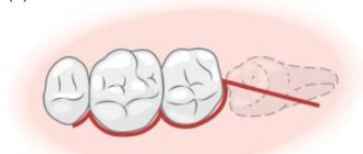
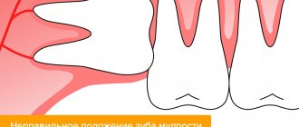
Photo: this is how wisdom teeth grow in dystopia
In addition to growth deviations, there are others. For example, a wise tooth may not erupt through the gum at all or may erupt partially, in which case it is called impacted or semi-impacted. Sometimes the molar occupies a normal position (vertical immersion) or lies on its side (horizontal position).
The eruption of wisdom teeth is very often accompanied by rather unpleasant symptoms from the jaw apparatus:
- a nagging pain appears due to the fact that the tooth has to grow through the already formed gum;
- sore throat, which occurs due to pain radiating from the eruption area;
- swelling and redness of the gums above the cutting tooth;
- local lymphadenitis - inflammation of the cervical and submandibular lymph nodes.
Photo: location of wisdom teeth in the gum pocket
Since the eruption of the eighth teeth sometimes lasts for several years , the symptoms can bother a person for quite a long time. Incomplete eruption leads to the formation of a gum hood - a fold of gum above the tooth. The peculiarity of this formation is that it is often filled with food, which is very difficult to clean with a toothbrush. When food debris becomes contaminated with microorganisms, a very unpleasant disease develops - pericoronitis.
Reasons for removing wisdom teeth
Specific indications for removing figure eights are determined by a dental surgeon. The following conditions require surgical intervention:
- incorrect position of the tooth in the gum, accompanied by injury to the cheek or tongue;
- damage to neighboring teeth - second premolars;
- the impossibility of fully treating figure eights, for example, with pulpitis;
- granulomas, cysts, pericoronitis;
- purulent diseases, which include osteomyelitis, abscess, phlegmon.
Timely removal of wisdom teeth allows you to avoid most of the problems associated with them. It is recommended to pull out third molars during the so-called cold period , that is, when a person is not bothered by toothache. During this period, you can undergo all the necessary examinations and remove the eights without much discomfort; in case of inflammation, anesthesia does not work so well.
There are no absolute contraindications to wisdom teeth removal. There are only conditions for which it is recommended to carry out specific treatment and consult a doctor later. These include:
- inflammatory diseases of the oral cavity: gingivitis and stomatitis;
- a number of systemic infectious diseases: HIV infection, viral hepatitis;
- diseases of the heart and blood vessels in the stage of decompensation;
- mental disorders.
Pregnant women are not recommended to have their teeth pulled during the first and last trimesters. Since this procedure is recommended to be carried out under anesthesia, and in the first months the fetus should not be exposed to drugs - a miscarriage can be provoked. In later stages, pain during removal can provoke premature birth.
Indications and contraindications for removal of the eighth molar
In dentistry, there are strict indications for the removal of the upper eighth molar. This:
- incorrect position of the tooth crown, which causes injury to the soft tissues of the oral cavity;
- the growth of the figure eight is oblique or horizontal, which makes it impossible to position it in the alveolus of the upper jaw;
- difficulty in treating the neighboring seven;
- caries leading to partial or complete destruction of the tooth root, pulp or enamel;
- a purulent abscess that threatens to develop into an abscess.
The first three indications also apply to intact teeth: in these situations, the doctor will insist on removing the figure eight, even if it is completely healthy.
Even such a simple procedure as tooth extraction has certain contraindications. Before performing a surgical intervention, the dentist must clarify whether the patient suffers from blood diseases (low coagulability will become an obstacle to surgical treatment), whether he has mental disorders or chronic diseases in the acute phase.
The latter include pathologies of such internal organs as the heart, liver, kidneys and digestive system. Pregnant mothers will also have to refuse the removal of the figure eight if the pregnancy is in the first or last trimester.
IMPORTANT!!! The dentist cannot refuse to remove a tooth without compelling reasons: he must justify the decision and offer a more rational option.
How to remove a wisdom tooth
Like any medical intervention, removal begins with a visit to the dentist. At the first contact with the patient, the doctor should assess his general condition and decide whether the tooth can be pulled out now or if it is worth a little preparation.
An anamnesis must be collected: the presence of chronic diseases that may complicate the course of the postoperative period is clarified. Such pathologies are diabetes mellitus, bronchial asthma, and arterial hypertension. The doctor is simply obliged to ask the patient if he has any allergic reactions.
The next step is to do an orthopantomogram. A panoramic x-ray provides the clearest picture possible, showing how the figure eights are positioned in relation to the rest of the teeth. The image allows you to more accurately determine the treatment regimen.
Photo: where is the wisdom tooth located and what does it look like on OPTG
The tooth extraction procedure itself begins with anesthesia: the doctor injects a local anesthetic into the gum with a syringe with a thin needle. The patient then waits 10–15 minutes for the medicine to take effect. Further events develop in two ways:
Easy removal
Despite the fact that a person’s wisdom tooth is located in a relatively inaccessible place, the doctor uses special forceps to grab it and remove it from the bone tissue by rocking and rotating it. An antiseptic is placed in the vacated hole. A competent dentist will definitely put a suture on the hole.
Simple category removal is often used when extracting teeth from the upper jaw.
Removing a complex category
This option is used for impacted or semi-impacted teeth, as well as for severe destruction of the molar, when there is simply nothing to grab onto with forceps, and when the figure eights are positioned horizontally. The difficulty may depend on how many roots the tooth has and how crooked they are. The frequency of use of this method is higher when removing teeth from the lower jaw.
Extraction in this case may be accompanied by an incision in the gum, drilling out the tooth along with its roots, or sawing. The operation can take up to 1.5–2 hours. After such an operation, an antiseptic is necessarily placed in the hole and stitches are applied.
What to do after deletion
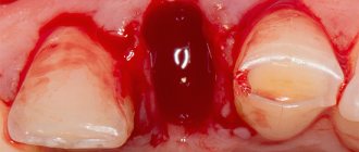
Photo: this is what gums look like after wisdom tooth removal
After simple removal, you need to follow the general recommendations:
- remove the tampon from the wound as soon as the root holes stop bleeding;
- try not to touch the hole with your tongue;
- apply cold to the cheek for 10–20 minutes;
- do not eat for 3 hours after removal;
- refrain from smoking and drinking alcohol.
In difficult cases, after extensive tooth extraction surgery, it is recommended to take the following actions:
- Take painkillers. You can do this until the local anesthesia wears off, before pain appears in the gum bordering the extracted tooth.
- Start taking antibiotics if surrounding tissues are inflamed. The specific drug and dosage are prescribed by the doctor.
After removing a wisdom tooth or any other tooth, you should not take aspirin; it can increase bleeding from the socket.
Features of tooth extraction in the upper jaw
The structure of the upper and lower jaws is significantly different. They also vary in density and location. Therefore, the method of tooth extraction will differ:
- on the upper jaw the operation is faster;
- the recovery period is shorter;
- pain relief is simpler and more effective.
Complications during tooth extraction in the upper jaw occur less frequently than in the lower jaw.
Anesthesia
The secret of high-quality pain relief is the correct delivery of the drug to the nerve. The choice of anesthesia method takes into account the patient’s age, his general condition, and the complexity of the tooth being removed.
Anesthesia of the upper jaw
The upper jaw is less dense than the lower jaw. It is covered with a cortical plate with many holes. Nerves and blood vessels pass through them. Pain relief is easier and faster than in the lower jaw
Sinus damage
Above the upper jaw is the nasal sinus, also known as the maxillary sinus. Its bottom may be close to the root of the tooth being removed. Then, during removal, sinus perforation may occur. This complication is typical for surgery on molars. This pattern is explained by the fact that the jaw in the area of the incisors is longer, and the nasal cavity is further away relative to them.
The main sign of sinus perforation is the escape of air, food and fluid through the nose. There is foam in the hole. Hospitalization in a hospital may be indicated for treatment.
Entry of part of the root into the sinus
Due to perforation of the sinus, part of the root can get inside it. This is caused by thinning of the bone plate or errors during the removal operation.
The symptoms when part of the root gets into the sinus are the same as when perforation occurs: air and contents of the oral cavity escape through the nose. But at the same time there is pain and increased body temperature. Treatment requires hospitalization.