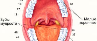
Upper jaw
The upper jaw (maxilla), paired, is located in the center of the face and connects with all its bones, as well as with the ethmoid, frontal and sphenoid bones (Fig. 1). The upper jaw takes part in the formation of the walls of the orbit, nasal and oral cavities, pterygopalatine and infratemporal fossae. It distinguishes between a body and 4 processes, of which the frontal is directed upward, the alveolar is directed downward, the palatine is directed medially, and the zygomatic is directed laterally. Despite its significant volume, the upper jaw is very light, since its body contains a cavity - the maxillary sinus (sinus maxillaris).
The body of the upper jaw (corpus maxillaris) has the shape of a truncated pyramid. It distinguishes 4 surfaces: anterior, infratemporal, orbital and nasal.
The anterior surface (fades anterior) is somewhat concave, bounded above by the infraorbital margin (margo infraorbitalis), laterally by the zygomaticalveolar crest and zygomatic process, below by the alveolar process and medially by the nasal notch (incisura nasalis). Below the infraorbital margin there is an infraorbital foramen (foramen infraorbitale), through which the vessels and nerves of the same name emerge. The infraorbital foramen with a diameter of 2-6 mm is usually semi-oval, less often oval or in the form of a slit, sometimes double. In isolated cases it is covered with a bone spike. Located at the level of the 5th or in the interval between the 5th and 6th teeth, but can shift to the level of the 4th tooth. Under this opening lies the canine fossa (fossa canina), which is the origin of the muscle that lifts the angle of the mouth.
The infratemporal surface (fades infratemporalis) is convex and participates in the formation of the walls of the infratemporal and pterygopalatine fossae. A more convex part is distinguished on it - the tuber of the upper jaw (tuber maxillae), on which there are 3-4 posterior superior alveolar openings (foramina alveolaria superiora posteriora). These holes lead into the canaliculi, which pass through the wall of the maxillary sinus and are directed to the roots of the large molars. The corresponding alveolar vessels and nerves pass through these openings and tubules (see Fig. 1).
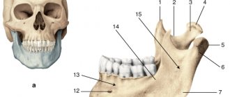
Rice. 1. Upper jaw, right:
a - topography of the upper jaw;
b — right view: 1 — frontal process; 2 - anterior lacrimal ridge; 3 - tear groove; 4 - infraorbital margin; 5 - infraorbital foramen; 6 - nasal notch; 7 - anterior nasal spine; 8 — front surface; 9 — canine fossa; 10— alveolar elevations; 11— alveolar arch; 12—body of the upper jaw; 13 - zygomaticalveolar ridge; 14 - posterior superior alveolar openings; 15—infratemporal surface; 16—tubercle of the upper jaw; 17—zygomatic process; 18—infraorbital groove; 19—infraorbital surface; 20 - lacrimal notch;
c—view from the nasal surface: 1—frontal process; 2—anterior lacrimal ridge; 3 - tear groove; 4 - cleft of the maxillary sinus; 5 - greater palatine sulcus; 6 - nasal ridge; 7 - alveolar process; 8 - alveolar arch; 9—incisive canal; 10—palatine process; 11 - nasal surface of the upper jaw; 12 - shell ridge; 13 - ethmoid ridge;
d — bottom view: 1 — incisive fossa and incisive openings; 2 - incisor bone; 3 - incisal suture; 4 - palatine process; 5 - zygomatic process; 6 - palatal grooves; 7 - palatal ridges; 8 - alveolar process; 9 - interroot septa; 10— interalveolar septa; 11 - dental alveoli;
d - alveolar canals (opened): 1 - infraorbital canal; 2 - infraorbital foramen; 3 - anterior and middle alveolar canals; 4 - posterior alveolar canals; 5 - posterior superior alveolar openings; 6 - maxillary sinus (opened)
The orbital surface (fades orbitalis) is smooth, triangular in shape, and participates in the formation of the lower wall of the orbit. Anteriorly, it ends with the infraorbital margin and laterally connects with the orbital surface of the zygomatic bone. The medial edge of the orbital surface in front connects with the lacrimal bone, for which there is a lacrimal notch (incisura lacrimalis) on the upper jaw. Posteriorly, the medial margin connects with the orbital plate of the ethmoid bone. In some cases, it bifurcates and forms cells that complement the cells of the lattice labyrinth. The orbital process of the palatine bone is adjacent to the posterior end of the medial margin. At the rear, the orbital surface, together with the edge of the greater wing of the sphenoid bone, limits the inferior orbital fissure (fissura orbitalis inferior). From the middle of the posterior edge of the orbital surface, the infraorbital groove (sulcus infraorbitalis) stretches forward, which passes into the canal of the same name, which opens with the infraorbital foramen. On the lower wall of the canal there are small anterior and middle upper alveolar openings (foramina alveolaria superiora media et anteriora), leading into small bone canals reaching the roots of the anterior and middle teeth. They carry blood vessels and nerves to the teeth.
The nasal surface (fades nasalis) forms most of the lateral wall of the nasal cavity (see Fig. 1). It connects posteriorly with the perpendicular plate of the palatine bone, and anteriorly and superiorly with the lacrimal bone. A significant part of this surface is occupied by the opening of the maxillary sinus - the maxillary cleft (hiatus maxillaris). Anterior to the cleft is a vertically directed lacrimal groove (sulcus lacrimalis), which, together with the lacrimal bone and the lacrimal process of the inferior turbinate, forms the nasolacrimal canal (canalis nasolacrimalis), which opens into the nasal cavity. Below and anterior to the lacrimal groove there is a horizontal protrusion - the conchal crest (crista conchalis) for connection with the anterior end of the inferior nasal concha. Posterior to the maxillary cleft there is a vertically directed large palatine groove (sulcus palatinus major), which is part of the walls of the greater palatine canal.
Human anatomy S.S. Mikhailov, A.V. Chukbar, A.G. Tsybulkin
Published by Konstantin Mokanov
Mandibular buttresses
1. Alveolar buttress
- goes up towards the alveolar cells of the lower jaw.
2. Rising buttress
– goes up the branch of the mandible to the neck and head of the mandible. Through this buttress, chewing pressure is transmitted to the mandibular fossa of the temporal bone.
The bones of the facial skull are involved in the formation of the orbits, nasal cavity, oral cavity, infratemporal and pterygopalatine fossae. The temporal fossa belongs to the brain skull. Due to the fact that it is closely related to the infratemporal and pterygopalatine fossa, it will be described in this section.
eye socket , orbita ,
– a paired cavity shaped like a tetrahedral pyramid.
The base of the pyramid faces anteriorly and limits the entrance to the orbit, aditus orbitae
.
The apex of the pyramid passes into the optic canal, canalis opticus
(Fig. 3.21). The orbit contains the eyeball and its supporting apparatus.
Entrance to the orbit is limited:
– nasal part, supraorbital margin and zygomatic process of the frontal bone, pars nasalis, margo supraorbitalis et processus zygomaticus ossis frontalis
;
– zygomatic bone, os zygomaticum
;
– infraorbital margin and frontal process of the maxilla, margo infraorbitalis et processus frontalis maxillae.
Rice. 3.21. The structure of the walls of the orbit.
1 - ala minor ossis sphenoidalis; 2 - canalis opticus; 3 - for. supraorbitale; 4 - for. ethmoidae posterius; 5 - for. ethmoidae anterius; 6 - os lacrimale; 7 - processus frontalis maxillae; 8 - facies orbitalis maxillae; 9 - processus orbitalis ossis palatini; 10 - sulcus infarorbitalis; 11 - for. infraorbitale; 12 - fissura orbitalis inferior; 13 - ala major ossis shenoidalis; 14 - os zygomaticum; 15 - fissura orbitalis superior; 16 - pars orbitalis ossis frontalis.
There are 4 walls in the orbit: superior, lateral, inferior and medial, each of which is formed by different bone structures. The structure of the walls of the orbit is shown in Table 3.5. The connection of the orbit with other topographic formations of the skull is described in Table 3.6.
A number of communications of the orbit are formed by the connection of individual bones: when the frontal process of the upper jaw and the lacrimal bone are connected, the nasolacrimal canal, canalis nasolacrimalis,
;
the upper jaw and the large wing of the sphenoid bone limit the lower orbital fissure, fissura orbitalis inferior
;
at the connection of the frontal and ethmoid bones, the anterior and posterior ethmoidal openings are formed, foramen ethmoidale anterius et foramen ethmoidale posterius
(Fig. 3.22).
Table 3.5
Anatomy of molars and premolars
The small and large molars are located on the sides of the jaw. Their main function is to chew and grind food, which is why they are also called chewers.
Read also: Runny nose during teething symptoms
Large molars, in comparison with other groups of teeth, have a large and massive chewing surface - a crown, on which 4 or 5 tubercles are located. The groove separating them is similar to the letter N. The tubercles, depending on which surface they continue, are divided into lingual, round in shape, and pointed buccal.
Molars are divided into three types:
- The crown is rectangular in shape ; on its surface there are three buccal cusps, including one anterior buccal, and two lingual cusps. The first of them occupy a large area, but are not so convex compared to the last two. The protrusions are crossed by an H-shaped groove. The tooth has 3 roots: the palatal one is rounded and straight, the cheek roots are flattened on the sides and tilted back.
- The crown has a square outline , on which there are two buccal and the same number of lingual cusps. The longitudinal groove lies closer to the lingual edge, and the transverse groove extends onto the vertical wall of the tooth and ends in a “blind” recess. In the cervical zone, the vestibular surface gradually decreases. The tooth has two roots: the back one is massive and straight, the front one is flattened. Longitudinal grooves run along the sides of the roots.
- Smaller than other molars. Often similar in structure to the second molar. The crown resembles a cube, on the surface of which there are four or five tubercles, in rare cases - three. The tubercles are separated by longitudinal and transverse grooves. There are 2 roots extending from the base, but they can merge into one thick and short one.
The small molars are located between the canines and molars. This situation has left its mark on the structure: they have some signs of neighboring teeth. On their wide surface there are 2 tubercles: oral and vestibular. Most often, premolars have one root, which are divided into two parts, in rare cases into three.
Classification of molars in dentistry:
- The first small indigenous one. It has a prism shape with rounded corners, reminiscent of a fang. The buccal and palatal surfaces are convex, the former being larger than the latter. The cutting edge bears the main tubercle in the middle, but unlike the fang, it is lowered lower. Between the tubercles there are grooves ending in enamel ridges. Closer to the top, the root bifurcates.
- Second small root. Smaller than the previous one and close to it in anatomical structure. There is only one root, and its branching is rare.
The design of the upper jaw is individual for each person. Elements have their place, structure and certain properties. The paired bone of the facial part of the skull is important not only in the formation of a beautiful profile, but also for the functional activity of the masticatory-speech apparatus.
The structure of the inferior nasal concha
The inferior turbinate is a paired bone. It is very thin, its side part is concave in shape. The middle part is rough due to the large number of vessels passing through it.
The upper edge of the shell is adjacent to the maxillary conchal ridges and the palatal bone plate. The shell is located directly above the maxillary cleft.
Soft Fabric Profile Components
The soft tissue nose is short, rounded and pug-shaped.
The nasal bridge is low; the nasal profile is concave and the nostrils are visible on the obverse. It protrudes very little and is quite short vertically. The human nose continues to grow in a downward and forward direction at least until early adulthood. There appears to be no noticeable reduction in the rate of nasal growth, which is typical for skeletal structures. The average annual increase of 1-3 mm in total length of the external nose is almost the same for men and women. Three processes originate from the upper edge of the shell:
- the maxillary process of the inferior nasal concha
is the largest in area, faces down and covers the border of the maxillary cleft, thereby delimiting the nasal region and the space of the maxillary sinus; - lacrimal process or anterior
- facing upward, extends to the lacrimal ossicle; - the ethmoidal process, or posterior
, is also directed upward, where it reaches the ethmoid bone and is attached to its uncinate process.