Types of equipment
In dentistry, instruments made of stainless steel and other durable alloys are used. They are additionally coated with titanium, zirconium, and Teflon. To create artificial teeth from wax, doctors choose equipment made of wood or medical plastic.
Any modeling tool consists of a handle, a rod and a working part or several such parts. The handle is made of stainless steel with notches so that the dentist’s hand does not slip during complex work. For the same purpose, rubber or plastic inserts are sometimes placed on the handle.
The rod is the connecting link between the handle of the device and the working part. Doctors choose the following dental instruments for modeling:
- Ironing iron. The doctor uses it to apply filling material or other composition to make an impression of the dentition. The tool is convenient for wax modeling and distributes the composition well over the walls of the teeth.
- Putty knife. At both ends of the handle there are small blades of different shapes (rhombus, rectangle, triangle, oval). The dentist uses them to distribute the composition and compact it. It is convenient to use a spatula to make notches on dentures.
- Stopfer. The handle has a thin pointed tip or small ball. The doctor chooses it if it is necessary to introduce filling material into a tooth cavity or to recreate the natural surface of chewing teeth.
Also, before seeing the patient, the nurse prepares other dental instruments: retractor, knife, brush, retraction thread. Manufacturers produce equipment modifications: smoothers, pluggers, spatula-knife. You can immediately purchase a set of tools for the preparatory stage of creating teeth from ceramics, metal and other materials.
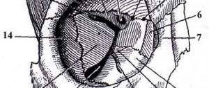
Anatomical shape of the chewing surface
| Rice. 4. Then the mesial-approximal ridge is formed (3). It begins at the buccal end of the distal accessory tubercle (2) and reaches the proximal region, where it is extended by a tongue (5). directed towards the mesial fossa. |
| Rice. 5. The main protrusion (4) of the mesial buccal cusp should be modeled. It forms the tip of the tuberosity and ends at the central fissure. It's a little wider. than the distal accessory tubercle (2). The edge of this protrusion is slightly shifted to the mesial region. |
Rice. 6. Modeling ends with a flat buccal cusp. The marginal protrusion should be formed, which together with the tubercle represents a single whole (0).
The marginal projection begins in the distal part of the mesial buccal cusp and reaches the approximal region to end there with a tongue directed towards the distal fossa. The free space is filled with a tongue (7). He reaches the hole.
Rice. 7. The image is rotated 130°, buccal: the side is now down. The construction of the lingual part of the mesial buccal cusp follows. The latter consists of a main protrusion (10) forming its top.
With this design, the protrusion should be turned obliquely towards the central fossa. It should be modeled small but relatively tall.
The approximal region is supplemented by a marginal protrusion (8), starting at the apex of the tubercle, going to the approximal region and ending with a tongue (9) towards the mesial fossa.
Rice. 8. An additional tubercle (11) is formed. It should begin in the mesial region and, in the shape of an ellipse, reach the central fossa. The additional tubercle runs parallel to the central fissure.
| Rice. 9. The mesial lingual cusp is complemented by a tubercle (12). which begins at the marginal edge of the tubercle and moves in a curved shape towards the central fossa. Both additional tubercles meet here. |
| Rice. 10. Finally, the distal lingual cusp is modeled. The main protrusion (13) forms its top. It has a uniform width, directed towards the central fossa, without reaching which it ends at an obtuse angle. |
| Rice. 11. The marginal protrusion (17) is modeled to the approximal area. From here, a wide and curved additional tubercle (15) extends to the central fossa, where it slightly overlaps the main protrusion. |
| Rice. 12. An additional tubercle (14) begins at the marginal protrusion (16). distinctly curved in the shape of the letter S and directed towards the central fossa. |
| Rice. 13. Like modeling in wax, the ceramic mass is always applied in small increments. To achieve the required consistency, you can use distilled water or modeling liquid. |
| Rice. 14. Tooth after condensation. The clear contours of the fissures have become somewhat blurred. Before firing, it is necessary to carry out correction using special instruments made from endodontic ones. After firing, correction is possible using grinding tools. |
| Rice. 15. Crown after firing. Sandblasting is carried out using aluminum oxide to remove the top, especially hard, glossy layer of ceramics. Firing is carried out at a temperature of 900 - 920-C. Due to this, the fusion of ceramic mass particles is insignificant. which makes the grinding process easier. Glazing is carried out at normal temperature. |