The term “osteoma” is used to define a disease associated with the formation of a benign tumor, usually located on the lower jaw.
Typical features of the neoplasm are: the absence of characteristic symptoms and the duration of pathological development processes. The formation of osteoma is physiologically similar to the growth of skeletal elements; the tumor can be characterized as a kind of pathological growth that can spread into or onto the surface of the affected bone.
Despite the fact that osteoma is benign, in the vast majority of clinical cases, surgical removal of the tumor is required.
What it is
As noted above, osteoma of the jaw is a tumor-like neoplasm, the basis of which is mature bone tissue. Depending on the etiological factors, odontogenic and non-odontogenic tumors are distinguished, the development of which can be a consequence of injuries, periodontal disease, jaw joints, and there is also the possibility of pathological growth after tooth extraction.
The process of formation of osteoma of the jaw is a gradual replacement of full-fledged healthy cells of bone tissue with pathological ones, located, as a rule, in a chaotic order. The development of the tumor occurs at a slow pace; often the tumor does not bother the patient for several years. However, there is a possibility that osteoma will transform into a malignant tumor.
Kinds
There are two main types of the disease.
Bizarre
The tumor is characterized by a large number of multinucleated cells. Not dangerous to humans, prognosis is favorable. The formation is identified by x-ray.
Aggressive
Occurs within 2 years after removal of the formation and degenerates into a malignant tumor. A distinctive feature of the tumor is clear contours. Education is common among the male half.
Comprehensive diagnostic measures and the patient’s medical history will help determine the type of tumor process. If there is information about a previously removed osteoblastoma, the doctor makes a diagnosis of an aggressive form of formation.
Classification
To carry out high-quality treatment, the basis of which is surgical removal of osteoma of the jaw, it is necessary to determine the type of neoplasm. There are several classification options according to the location, extent of the lesion, etiological factors, as well as the structural features of the tumor.
By origin and localization
First of all, osteoma of the upper jaw is classified into several main subgroups according to the specific etiological factors. There is a wide range of reasons that can lead to the launch of pathological tissue growth processes, these include injuries, dental diseases, and genetic mutations.
Depending on the location of pathological formations, osteomas of the jaw are classified as follows:
- Osteoma of the lower jaw. As the name suggests, this type of tumor affects the tissues of the floor of the mouth. With the rapid growth of the tumor, there is a possibility of damage to nerve fibers, as well as limited jaw mobility. Compact osteoma of the lower jaw, which has insignificant dimensions, usually remains completely static.
- Osteoma of the upper jaw can affect the orbits, palate, and maxillary sinuses, which leads to impaired vision and breathing. This form of pathology especially often requires surgical excision; such interventions include, for example, surgery on the paranasal sinus.
By structure and location
The structure of osteomas can vary significantly. The main options are:
- Compact. It is a formation that rises slightly above the surface of the bone tissue and has a wide base.
- Tubular. The structure of this jaw osteoma is a continuation of bone tissue. A neoplasm of this type has a spherical structure.
- Intraosseous. As the name suggests, this type of tumor develops in tissue structures.
There are also tumors formed exclusively from bone tissue and spongy osteomas, in which the spongy component predominates.
Diagnostics
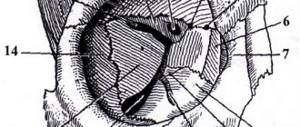
When visiting a doctor, the patient must first undergo an external examination. The doctor feels the site of the lesion and determines the external characteristics of the tumor. For a more detailed study of the pathology, an x-ray of osteoma of the lower or upper jaw is prescribed.
On the picture, the tumor appears as a darkened spot. In the presence of spongy osteoma, the dark spot is heterogeneous. If the size of the growth is large, the displacement of the surrounding tissues is clearly visible on the x-ray.
To obtain more detailed information about the condition of the jaw, the doctor prescribes a computed tomography scan. With its help, the lesion site is removed layer by layer, which allows you to examine the tumor from all sides.
Causes
Numerous factors of different nature can provoke the development of osteoma of the lower jaw. The prevailing options include, for example:
- Paget's disease, leading to deformation and enlargement of bone tissue, as well as disruption of the physiological processes of their growth and renewal.
- The presence in the oral cavity of completely destroyed teeth, devoid of a crown protruding above the surface of the jaw tissue.
- Inflammatory diseases affecting bone or soft tissue.
- Low quality, incorrect selection or incorrect wearing of dental prostheses, braces – systems, fillings.
- A large amount of dental calculus or a high density of structural formations.
Even factors such as long-term chronic rhinitis, infectious or inflammatory processes affecting facial tissues can cause the formation of a tumor.
Why does osteoma of the jaw appear?
Currently, doctors have not yet given a definitive answer regarding the reasons for the development of osteoma. Nevertheless, some patterns of its occurrence have been established. Thus, it was found that patients suffering from osteoma had previously received injuries to the jaw bone, for example, bruises. The likelihood of tumor formation also increases with constant trauma to the oral mucosa. In this case, chronic injury can be caused by:
- remains of destroyed teeth;
- tartar;
- poorly fitting dentures;
- poorly processed filling edges;
- etc.
In addition, the factors that provoke the formation of osteoma include inflammatory processes in the maxillofacial area, such as:
- chronic form of periodontitis;
- periostitis;
- osteomyelitis;
- sinusitis;
- etc.
Thus, although osteoma is not classified as an odontogenic disease, dental diseases are still among the factors that create the risk of tumor development.
Foreign bodies in the maxillary sinuses, as well as various adverse external influences, such as radioactive radiation and chemical factors, can also lead to the development of a tumor.
Symptoms
In the early stages of formation, osteoma of the lower jaw is not accompanied by the appearance of characteristic symptoms. As a rule, negative manifestations of the disease appear only when the tumor grows rapidly and is of significant size. The main features include:
- Pain syndrome, which can have a different nature and localization. It should be noted that painful sensations can occur solely against the background of damage to nerve fibers.
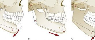
- Facial asymmetry that occurs against the background of osteoma, which is of significant size.
- Violation of the processes of chewing, swallowing, breathing, vision.
- Limited jaw movements.
- Violation of normal bite.
In addition, it is necessary to highlight additional signs of osteoma: a tumor that is significant in size and protrudes above the surface of the affected bone tissue is clearly visible visually.
Provoking factors
The development of a benign neoplasm is due to a number of provoking factors. The underlying causes of the tumor:
- mechanical damage;
- genetic predisposition;
- systematic impact on a specific area;
- congenital pathologies;
- inflammatory processes;
- infectious diseases;
- lack of timely treatment for inflammatory processes in the oral cavity;
- rheumatism;
- advanced form of syphilis;
- disorder .
On this topic
- Musculoskeletal system
Baker's cyst: when is surgery necessary?
- Natalya Gennadievna Butsyk
- December 6, 2020
People with weakened protective functions of the body are at risk. Constant colds and inflammatory diseases increase the likelihood of a benign neoplasm. Receiving the same injury is dangerous due to the development of a tumor-like process.
Osteoblastoma manifests itself at a young age and, without treatment, grows rapidly.
Treatment
Treatment of osteoma of the lower jaw is carried out exclusively through surgery. There are several main factors, the presence of which is a mandatory indication for surgery. These include dysfunction of the jaw apparatus, cosmetic defects, and limited jaw mobility.
If the tumor remains static for a long period of time, it is permissible to use only conservative therapy, which also helps to stop the growth of pathological tissues. Medication and other treatment options may be required after surgery.
Surgical therapy
The use of facial surgery, one of the tasks of which is the removal of malignant and benign tumors, is necessary to remove osteoma only if there is evidence from a specialist. During the procedure, the specialist must provide free access to the tumor tissue. As a rule, this is done through the oral cavity. Next, the osteoma is excised and the affected bone tissue is polished, after which the incision is sutured.
If the tumor is significant, after the main procedure aimed at removing osteoma of the jaw, the patient often requires plastic surgery, which consists of building up bone tissue and restoring facial symmetry. After surgical interventions, the patient must follow a number of rules for the rehabilitation period, which will help to significantly reduce the likelihood of possible postoperative complications.
Evaporation
If surgery to remove osteoma is contraindicated for a number of reasons, it is possible to use other, more gentle methods, including evaporation or exposure to laser radiation. This method is used to remove tumors of various sizes and shapes. Evaporation can significantly reduce the risk of injury to healthy tissues, as well as shorten the duration of the rehabilitation period.
Medication
Drug treatment for tumors is used exclusively as an auxiliary treatment option. The main goals of medications include pain relief. For this purpose, drugs such as Ibuprofen, Diclofenac, and a number of others are used.
Folk remedies
The use of traditional medicine recipes directly to remove tumor-like bone formation is not effective.
However, some methods of alternative treatment will help to significantly reduce the pain that may appear due to jaw pathology.
For example, the following recipe is quite effective: three large spoons of hawthorn inflorescences should be steamed with a glass of boiling water, and then let the prepared product brew for an hour. The finished infusion must be consumed at least three times throughout the day in the amount of three large spoons.
It is important to remember that the development of a neoplasm can lead to serious consequences, which include the degeneration of healthy cells into malignant ones. In order to prevent this, it is important to begin treatment immediately after making an accurate diagnosis.
If you find an error, please select a piece of text and press Ctrl+Enter. We will definitely fix it, and you will get + to karma
Diagnostic methods
Diagnosis of osteoma includes:
- Anamnesis collection.
- Objective examination.
- Laboratory and instrumental diagnostics.
First, the doctor finds out the complaints, how long ago the tumor appeared, hereditary and allergy history. Then visually and palpation determines the size, pain, and localization of the formation. The doctor looks at changes in the mucosa above the osteoma, the presence or absence of fistulas, and the functionality of the affected jaw.
The patient is given a CBC, where attention is paid to the indicators of leukocytes, segmented neutrophils, and ESR. In the presence of inflammatory changes, these indicators increase.
The main diagnostic methods are x-ray examination (or CT, MRI) and histological analysis of the formation. Based on these manipulations, the type of osteoma and the type of surgical intervention are determined.
CT is a method for diagnosing osteoma of the jaw
Differential diagnosis for osteoma is carried out with the following diseases:
- Sclerosing osteomyelitis. Its characteristic feature is pronounced sclerosis of bone tissue, the presence of serous or purulent fluid inside the tumor. The pain intensifies at night. Patients present with symptoms of general intoxication: tachycardia (pulse above 80 beats per minute), severe weakness, poor appetite, dizziness. The disease occurs with periods of remission and exacerbations.
- Osteoblastoma, osteoblastoclastoma. When the jaw is affected, the tumor is usually localized in the area of the chewing teeth. A biopsy reveals multiple giant cells with one nucleus (osteoblasts) or several nuclei (osteoclasts).
- Osteochondroma. The upper parts of this tumor have uneven, unclear contours, reminiscent of cauliflower. Most often localized on the articular heads of the lower jaw. Patients complain of crunching or clicking in the temporomandibular joint, discomfort when chewing and talking. A noticeable deformation of the face quickly develops with a displacement of the lower jaw to the healthy side. An X-ray examination reveals an increase in the size of the joint head due to the proliferation of bone tissue.
- Osteogenic sarcoma. This is a malignant disease of the jaw. With a maxillary lesion, the tumor is located on the alveolar process, with a mandibular lesion - on the entire body. The danger of this pathology is that patients seek medical help very late, when the tumor is large, has disintegrated and ulcerations appear on the oral mucosa. This is due to the long asymptomatic course of the disease. It is characterized by rapid growth, lack of clear boundaries on radiography, and homogeneous bone destruction.
Osteoma should also be distinguished from inflammatory destruction of bone tissue due to tuberculosis or syphilis. With these pathologies, the patient experiences characteristic changes in tests and accompanying specific symptoms.
Tumor removal
At the Medicine 24/7 clinic, we use both the classical method of surgical removal of osteoblastoma and minimally invasive operations. If possible, we always strive to achieve the goal with the least traumatic intervention.
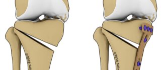
The goal of any operation is to reliably remove the tumor and minimize the risk of its recurrence. The classic operation consists of excision of osteoblastoma within healthy tissue. It is performed under general anesthesia.
Along with the tumor, part of the healthy bone is removed, which is then replaced through osteoplasty (bone material). Access is created by cutting the skin and soft tissue. In the case of a cranial bone tumor, trepanation may be required.
All operations for osteoblastoma are performed by experienced, highly qualified oncologist surgeons in the operating room of the Medicine 24/7 clinic, equipped to the highest standards with modern, high-tech equipment and instruments.
We will call you back
Leave your phone number
Indications for surgery
Osteoblastoma is removed in case of its rapid, aggressive growth, risk of malignancy, compression of neighboring organs, vascular or nervous structures due to the large size of the tumor, bone deformation, severe symptoms.
A benign tumor can occur on any bone, in any place, but in 50% of cases this place is the spine. By compressing the spinal nerve root, osteoblastoma causes pain, ranging from moderate, aching, constant to acute. Other symptoms of a spinal tumor are associated with radicular syndrome - muscle weakness in the arm or leg, sensory disturbances, numbness, paresis.
A tumor of the facial bone often leads to a pronounced aesthetic defect. If it compresses a blood vessel, blood circulation is disrupted, and if it presses on a nerve, neuralgia occurs (in particular, trigeminal neuralgia with unbearable pain).
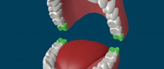
One example of facial osteoblastoma is a tumor of the lower jaw, which can cause tooth loss, inflammation, skin ulcers, purulent fistulas, and facial asymmetry.
If the tumor has affected the skull bone, it manifests itself as headache, memory loss, vision impairment, epileptic seizures, nosebleeds, and hormonal imbalance. Osteoblastoma in the sinus impairs breathing and vision.
A tumor of the femur is manifested by lameness, nagging pain in the muscle, which intensifies when walking.
If osteoblastoma occurs in the joint area, its movements are impaired. For example, a large tumor in the shoulder joint makes it impossible to move the arm. Tumors on the bones of the fingers disrupt their function and are accompanied by local inflammation, pain, burning, weakness and muscle atrophy.
These and other pronounced symptoms of osteoblastoma are indications for surgery.
Our doctors will help you
Leave your phone number