269
The mandible (from the Latin Mandibula) is the lower jaw of vertebrates. If we talk about people, the LF plays an important role not only in performing the functions of the dentofacial apparatus (chewing, swallowing, speaking), but also in ensuring the aesthetics of the lower third of the face.
The shape of the edges of the jaw body, the size and severity of the cervical-mental angle affects the beauty and expressiveness of our face, and its perception by others. And who among us is indifferent to the impression we make on other people?
General overview
Mandibuloplasty is an aesthetic operation that involves correcting the shape of the edges of the body and the size of the lower angle of the jaw.
To make it clear what we are talking about, we will provide some general information.
The human mandible consists of the following elements:
- body (the curved part on which the alveolar ridge with teeth is located);
- 2 branches directed upward and forming a cervical-mental (lower) angle with the body;
- articular processes crowning the upper parts of the branches.
Surgeries on the lower jaw are divided into two types:
- Orthognathic, aimed at eliminating dentofacial anomalies that lead to disruption of the functionality of the dentofacial apparatus (for example, lengthening or shortening of the jaw body).
- Mandibuloplasty.
If a patient with a dental anomaly requiring surgical intervention consults a doctor for mandibuloplasty, orthognathic surgery is performed first, followed by mandibuloplasty.
In cases where combined surgery is possible, orthognathic correction and mandibuloplasty are performed simultaneously.
The following types of mandibuloplasty are distinguished.
- Reducing the angles of the lower jaw.
- Increased angles of the lower jaw.
- Correction of the shape of the edge of the body of the bass.
The mandibuloplasty strategy consists of increasing or decreasing the volume of bone in certain areas of the jaw body, and involves resection (removal), reposition (movement) of bone tissue or installation of implants.
In the most difficult cases, a combination of these operations is used. As a result of mandibuloplasty, the lower part of the face acquires expressiveness and beauty, a feature that can be called “chiseling.” Beautiful people are said to have “chiselled” facial features.
In recent years, cartilage implants have been used. They are made from the patient's own cartilage using tissue engineering methods. Thanks to their “kinship” with the patient, they are absolutely compatible with his body and are completely integrated into the jaw.
Such implants do not “dissolve”. Their volume and shape are preserved for life, as is the result of the entire operation.
Reference. As an alternative to mandibuloplasty, contour plastic surgery is used, which is the injection of fillers under the skin. The use of contour plastic surgery is limited to a small area of correction and a temporary effect.
After about a year, the fillers “dissolve” and a repeat operation is required. The main advantages of this type of correction are low invasiveness and affordable price.
Why does the jaw click when chewing and how to eliminate the unpleasant symptom.
Come here if you are interested in how to correct a shift in the midline of your teeth.
At this address https://orto-info.ru/zubocheliustnye-anomalii/chelyustey/krivaya-ne-prigovor.html we will find out whether it is possible to correct a crooked upper jaw.
Indications and restrictions
The indication for mandibuloplasty is, first of all, the patient’s desire to improve, in his opinion, the aesthetics of his face, which is not high enough, which can be caused by the following reasons:
- Defects of the mandibular angles. Hypo- or hypertrophy of the cervical-mental angle, its deformation, asymmetry.
- Deformation of the edge of the LF body. Curvature, asymmetry, etc.
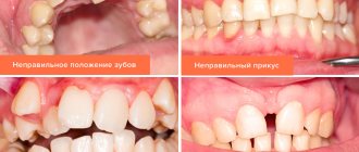
- Malocclusion – distal, mesial, cross, frontal open, deep. In addition to disrupting aesthetics, malocclusions have a negative impact on the functionality of the dentofacial apparatus - they reduce the efficiency of chewing and impair the functions of swallowing and speech.
- Chin defects. Asymmetry, too massive protruding or too small, receding with smoothing of the lower angle (“bird” profile). Chin plastic surgery has its own name – genioplasty.
Contraindications to mandibuloplasty are all cases in which any surgical intervention cannot be performed:
- Impaired hemostasis (blood formation), including due to medication.
- Oncological diseases - not specifically in the oral cavity.
- Serious chronic diseases of internal organs.
- Endocrine disorders.
- Pregnancy, lactation.
- Acute infectious diseases.
- Age up to 22-25 years (maxillofacial bones must be fully formed).
Technique
The intervention is performed under general anesthesia. A substance is injected intravenously that puts the patient to sleep; if it is necessary to prolong the effect of the drug, it is reintroduced.
To reduce the chin, the surgeon makes an incision according to the chosen tactics. In the oral cavity, this occurs at the junction of the gum and soft tissue. A peeling is performed to expose the problem area of the bone.
If a lift is needed, an incision is made in the natural crease of the lower chin. In order to remove part of the LF, an osteotomy is performed.
The bone tissue is cut transversely, and its lower section is shifted and fixed in the desired position. This allows you to remove a protruding chin. If the goal is to narrow it, then in this case the bone tissue is resected on the sides, then the fragments are removed or inserted into the central part, fixing them.
Finally, the bone is polished and the soft tissue is sutured. Excess skin, if present, is cut out and removed. Then an aseptic fixing bandage is applied. The duration of the operation is 2-3 hours.
Preparatory activities
The general goal of preparatory procedures for mandibuloplasty is to reduce surgical risk and minimize the likelihood of postoperative complications.
Activities begin with an initial consultation, during which the doctor assesses the type and severity of functional and aesthetic disorders, listens to the patient about his problem, intentions and expectations from the operation, informs him about the features and possible results of the operation.
This is followed by diagnostics - facial anthropometry, X-ray studies in the necessary projections (OPTR, teleradiography, targeted images), CT. Based on their results, the volume of bone tissue in the surgical area, the nature of occlusion, and malocclusion are assessed. A treatment plan is drawn up.
If there are concomitant malocclusions that cannot be corrected during mandibuloplasty, the patient may be indicated for orthognathic surgery to correct them.
An important stage of preparation is computer modeling , the purpose of which is to select the optimal surgical strategy, predict its results, and determine the size of the implant.
Before operations, laboratory and instrumental examinations of the patient’s health are also carried out - urine and blood tests (general and biochemical), a coagulogram (to assess blood clotting), bacteriological studies for the presence of viral infections, an electrocardiogram, etc. If required, doctors from other countries are involved in the diagnosis specialties.
15-20 days before surgery, stop taking medications that affect blood clotting. NSAIDs may also be prohibited . It is important that the doctor knows about all medications the patient is taking. If among them there are those that negatively affect hemostasis, the doctor will inform the patient that their use should be stopped for a while.
The length of the preparatory stage is about 7-10 days.
Stages of implementation
Before starting the operation, the doctor must have a ready-made implant. It is made from the patient’s autogenous bone or cartilage tissue (taken from the auricle or rib). The latter option is preferable to bone; a cartilage implant cannot be resorbed and is easier to form.
The most modern material is solid silicone, which lasts a lifetime and is absolutely safe for the patient.
The operation is performed under general anesthesia. Access to the bone is carried out through the mucous membrane of the oral cavity or from the outside, through the skin. Percutaneous access is used when neck rejuvenation is performed simultaneously with mandibuloplasty.
Moreover, the latter is ensured not only by greater tension of the soft tissues, but also by improving the contour of the lower jaw and the cervical-mental angle.
If there is no need for rejuvenation and tension of the skin, incisions are made in the oral cavity and implants are inserted through them. The operation lasts about an hour or less, and ends with stitches and a tight bandage on the jaw.
How is upper jaw teeth removed?
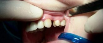
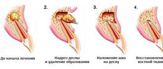
Before the operation, an examination is carried out, if necessary, radiography, consultations with other specialists. Typically, tooth extraction surgery is performed on an outpatient basis. Local anesthesia is most often used. Before the operation, the mucous membrane and teeth are cleaned to prevent infection of the socket. For tooth extraction, forceps and elevators are used, and in some cases, the tooth root is cut out with a bur. Special forceps are used to remove the wisdom tooth of the upper jaw.
Removing the upper front teeth is usually simple. Certain difficulties arise when removing the upper canine, since it has a long, massive root, the apex of which is very often curved, as well as when removing molars, since they have three roots.
When removing the upper teeth, the patient sits in a chair with a slightly reclined back and headrest. The dentist is positioned to the right and in front of the patient.
Stages of tooth extraction:
- Separation of the gums from the edge of the alveolus and the circular ligament from the neck of the tooth (with a raspatory or a trowel);
- Applying and advancing forceps under the gums, fixing them;
- Rocking or rotation of the tooth, during which the periodontal fibers that connect the tooth root to the walls of the socket are torn;
- Extracting a tooth from the socket;
- Inspection of the surgical site, extraction of small pieces of bone and tooth roots.
The success of the operation does not depend on the physical strength of the dentist, but on the correct execution of all stages of the procedure. Minor bleeding from the hole stops after 2-5 minutes, the hole is filled with a blood clot, which protects the wound from infection. To avoid damaging the clot and causing bleeding, the patient is advised not to rinse the mouth or eat food for several hours. On the day of surgery, you should not consume hot food or drinks, or take any thermal procedures; it is recommended to refrain from heavy physical activity. If all recommendations are followed, the tooth healing process will proceed quickly.
Rehabilitation period
The bandage lasts for about a week or the exact time determined by the doctor. In some cases, it is removed at the time of discharge, which usually occurs the next day after surgery.
The first days after mandibuloplasty, swelling persists, the patient experiences moderate pain, which intensifies when talking and chewing food.
The normal position is one in which swelling persists for a week. If it does not decrease, or even increases, you should consult a doctor.
To reduce pain and swelling, cold compresses are used, anti-inflammatory drugs, analgesics, and rinsing with antiseptic solutions are prescribed. Stitches are usually removed on the eighth to tenth day.
Particular attention is paid to diet. The composition of the products does not change, but the structure and temperature of the food changes. It should be predominantly liquid and at body temperature.
Eating cold and hot foods can lead to pain. Solid foods, the chewing of which requires strong tension in the masticatory muscles, should be avoided so as not to cause displacement of the implant.
The duration of the rehabilitation period depends on the type of operation, its traumatic nature (volume of surgical intervention), the age and general health of the patient. It usually takes about 2 months.
During rehabilitation, it is necessary to refrain from heavy physical activity and not expose yourself to high temperatures.
The final conclusion about the results of mandibuloplasty is made in about six months.
What methods can be used to correct a small lower jaw in men and women?
In this publication, we will consider modern methods of expanding the dentition in children.
Here https://orto-info.ru/zubocheliustnye-anomalii/chelyustey/ukorochenie.html all the most important things about shortening the upper dentition.
Fracture of the lower jaw - symptoms and treatment
Diagnosis of patients with fractures of the lower jaw is carried out in order to establish the location, number of fractures, as well as to determine injuries to nearby tissues, vessels and nerves.
Diagnostic methods are basic and additional (instrumental).
Basic diagnostic methods are carried out with a standard set of instruments in the emergency room, dental office or dressing room. The doctor finds out the patient’s complaints, learns about previous diseases and previous operations. At this stage, it is very important to establish the time of injury, since the duration of the injury plays a big role in further treatment tactics.
During the investigation, the doctor will find out whether the injury was sustained at home, at work, or during a fight, and whether it is of a criminal nature. In case of injury during violent acts, the doctor is obliged to report the incident to the police. After that, an internal affairs officer talks with the victim, finding out the details of what happened. In this regard, there is often a concealment of the real reasons for the injury, and the patient sometimes invents very ridiculous and implausible stories.
Sometimes collecting complaints and medical history is difficult or impossible due to the patient’s serious condition or alcohol/drug intoxication.
A clinical study begins with an external examination; post-traumatic swelling, changes in skin color, the presence of abrasions, hematomas, and bleeding are detected. Next, palpation is carried out, during which the nature of the swelling is revealed, inflammatory infiltrate, hematoma, and subcutaneous emphysema are excluded. Palpation of the lower jaw is carried out symmetrically, starting from the branch of the lower jaw and ending in the chin. The presence of a bony step indicates a fracture of the mandible. The degree of mobility of the fragments and the direction of their displacement are also determined.
When examining the oral cavity, special attention is paid to the mucous membrane of the alveolar part of the lower jaw, identifying violations of integrity and bleeding. The relationship of the dentition and the displacement of the central line of the lower jaw when opening and closing the mouth are determined. The landmarks are the labial frenulum, the line between the central incisors of the upper and lower jaw.
Often, the opening of the mouth with jaw fractures is limited. When rocking the lower jaw, relying on the chewing teeth and the basal edge, you can determine the degree of pathological mobility of the lower jaw and the location of the fracture.
Additional diagnostic methods:
- X-ray : an X-ray of the skull is taken in a direct projection in order to exclude fractures of other parts of the face and an X-ray of the lower jaw in lateral projections, the number of fractures, the presence or absence of displacement, diastasis (gap) and the presence of teeth in the fracture line are analyzed.
- An orthopantomogram is a more modern method, with functionality similar to an x-ray. One detailed image is taken, and it is possible to view it on the monitor screen using various programs.
- Computed tomography allows you to most accurately identify fractures of the lower jaw, measure the distance of displacement of fragments, diastasis, determine the size and number of fragments, and the presence of foreign bodies. Using certain setting modes, you can determine the violation of soft tissues (muscles, blood vessels, nerves) and the presence of hematomas.
- Assessment of the general condition of the body - examination of organs and systems of the body, identification of viral and chronic diseases. For this purpose, a physical examination, digital chest x-ray, ultrasound examination, and laboratory diagnostic methods are used. If necessary, consultations with related specialists are carried out. All this helps to select the most effective therapy for the patient, taking into account the characteristics of a particular clinical case.
Possible complications
Swelling and moderate pain are considered normal after surgery. But if they do not go away within a week, and/or some other symptoms of trouble appear (for example, bleeding), this is a sufficient reason to consult a doctor
The most common complications:
- decreased sensitivity, numbness in the lower jaw, lasting more than a week (the reason is usually pinched nerve endings due to swelling and the presence of an implant);
- bleeding;
- implant displacement – can occur in the first days after surgery;
- infection at the surgical site.
In all of the above cases, the patient must consult a doctor to identify the cause and carry out the necessary treatment measures.
How is augmentation of the angles of the lower jaw carried out, the scheme for introducing fillers
The augmentation procedure takes place in several successive stages: preparation, correction of the corners of the mandibular bone, treatment of the oval of the face. Fillers are injected with a needle or a special thin cannula.
Choosing the best filler
When choosing the best filler for injection into the lower third of the face and chin area, it is important to pay attention to the safety and density of the product. To replenish the volume, dense fillers are used. The following lines of correctional drugs are often used in clinics:
- Restylane series drugs. Hyaluronic acid in the composition ensures ideal compatibility with human tissue. The dense structure perfectly solves the problem of lack of volume. Despite the viscosity of the consistency, the filler is perfectly distributed in the subcutaneous structures, does not show through, and models the oval of the face well. Repeated administration is indicated after 5-7 months.
- Equio beauty injections. New product in cosmetology. The main feature is fast and even distribution without lumps, accumulation of substances under the skin, and no side effects.
- Surgiderm. A popular and frequently used product for correcting facial contours. Hyaluronic acid in the composition not only models the face, but also deeply moisturizes the skin, saturates it with nutrition, and stimulates the synthesis of its own collagen. The result lasts up to 6-8 months.
- Juvederm Ultra. The highlight of the filler is the presence of an anesthetic component in the injection, so the procedure is as comfortable as possible.
- Radiesse. The most effective and therefore expensive filler for filling defects in appearance with long lasting results. A repeated course of administration is indicated after 1.5-2 years.
Organic and inorganic-based drugs have a high degree of safety and rarely cause serious side effects and complications.
Rejuvenation of the lower oval of the face with HA-based fillers in 3 stages
Correction of the angles of the lower jaw with fillers is performed according to a specific algorithm. The manipulation itself includes sequential effects on the mandibular angles, followed by the chin and oval of the face. Augmentation is performed according to the following rules:
- cleansing the skin of makeup, impurities, oil and sebum with alcohol-free skin care products;
- antiseptic treatment, disinfection;
- application of a compress with an anesthetic drug or treatment with lidocaine-based gel;
- injection of filler at intervals of up to 1 mm, single injection into one point of no more than 0.3 ml;
- applying a soothing mask to reduce redness.
At the time of drug administration, the doctor manually distributes the filler under the skin using massage movements. Such manipulations allow you to create and record the desired result. Before the procedure, a profile photo of the lower third of the face is taken to evaluate the final result. The effect is observed immediately and reveals itself within a week after administration of the drug.
Price issue
As a rule, the cost of mandibuloplasty indicated by clinics in their price lists includes all costs associated with the operation:
- initial and subsequent examinations;
- expenses associated with the patient's stay in the hospital (food, provision of a robe, towel, slippers, hygiene kit);
- anesthesia and the operation itself;
- dressings;
- postoperative procedures and rehabilitation monitoring.
The cost of the operation is influenced by the type and complexity of the surgical intervention, the technology used, the general health of the patient, which may require additional procedures, the pricing policy of the clinic and its location.
In general, the entire mandibuloplasty procedure may require the patient to pay from 100 to 250 thousand rubles. In some cases, even more.
The video provides additional information on the topic of the article.
