Benign tumors of the oral cavity are neoplasms located in the oral cavity, characterized by limited slow growth and not prone to metastasis. Benign tumors of the oral cavity include papillomas, myxomas, retention cysts, Serra glands, fibromas, gingival fibromatosis, fibroids, hemangiomas, lymphangiomas. Diagnosis of oral cavity tumors is carried out on the basis of examination, palpation, X-ray examination, angiography and histological examination. Removal of oral cavity tumors is possible by surgical excision, electrocoagulation, laser valorization, cryodestruction, vascular sclerosis, or the use of radio wave method.
Causes of oral tumors
The etiology and mechanism of development of oral tumors have not yet been sufficiently studied. Risk factors include alcohol consumption, smoking, acute trauma (for example, tooth extraction), chronic trauma to the oral mucosa from the edge of a broken tooth, poorly treated filling surface, crown or denture. The risk of oral tumors is highest when smoking and drinking alcohol are combined. In the development of some neoplastic tumors of the oral cavity (papillomas, papillomatosis), the leading role is played by a viral infection (human papillomavirus, herpes virus).
Tumors of the oral cavity that occur in childhood are often associated with impaired tissue differentiation during fetal development. These include dermoid and retention cysts, Serra glands, and congenital nevi. As a rule, these neoplasms are detected during the first year of life.
Epithelial tumors of the oral cavity
Papillomas.
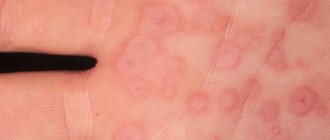
Oral cavity tumors consisting of stratified squamous epithelial cells. They are most often localized on the lips, tongue, soft and hard palate. Oral papillomas are a rounded protrusion above the surface of the mucosa. They may have a smooth surface, but are more often covered with cauliflower-type papillary growths. Usually single papillomas are observed, less often - multiple ones. Over time, these oral tumors become covered with keratinizing epithelium, due to which they acquire a whitish color and a rough surface.
Nevi.
In the oral cavity, nevi are observed in rare cases. They are often convex and have varying degrees of pigmentation from pale pink to brown. Among the tumors of the oral cavity there are blue nevus, papillomatous nevus, nevus of Ota and others. Some of them can become malignant with the development of melanoma.
Hemorrhoids kill the patient in 79% of cases
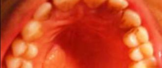
Glands of Serres.
Typically, this type of oral tumor is located in the alveolar ridge or hard palate. Serre's glands are hemispherical formations of yellowish color, up to 0.1 cm in size and of dense consistency. May be of multiple nature. Usually, by the end of the child’s first year of life, spontaneous disappearance of these formations is noted.
Removal of papilloma or neoplasm in the mouth reviews
- Irina A. Moscow. Good afternoon, not long ago I noticed strange formations in my mouth, I underwent an examination, and the doctor said that these were papillomas. I was immediately offered to excise them with a laser. To be honest, there was no talk of any self-medication. In the end, I decided to have the procedure, everything went quite well, there was no pain, but now I feel just great.
- Sonya L. G. Omsk. Hello, two months ago I was given such an unpleasant diagnosis - papillomas in the oral cavity. After reading the reviews of other patients, I decided that it was better to remove them using the radio wave method. I went to a good clinic, where they removed all the growths for me for a fairly small fee. I can say that the sooner you receive treatment, the better it will be for you.
Papillomas that appear in the oral cavity always cause a lot of pain and trouble, so you should not self-medicate, because they can only be properly removed in a clinic.
—
Connective tissue tumors of the oral cavity
Fibroids . Oral fibroids are most common in the lower lip, tongue, and palate. They look like a smooth oval or round formation, in some cases located on a stalk. The color of these oral cavity tumors does not differ from the color of the surrounding mucosa.
Fibromatosis of the gums . Not all authors classify gingival fibromatosis as a tumor of the oral cavity; some believe that it is based on inflammatory changes. Fibromatous growths are painless, dense formations. They can be local in nature within several teeth or diffuse, involving the entire alveolar process of both the lower and upper jaw. Tumor growths in fibromatosis are localized in the gum papillae and can be so pronounced that they completely cover the crowns of the teeth. This type of oral tumor requires differentiation from hyperplastic gingivitis.
Myomas . Develop from muscle tissue. Rhabdomyomas are formed from fibers of striated muscles. Most often they are observed in the form of single nodular formations in the thickness of the tongue. Leiomyomas develop from smooth muscle fibers and are usually located on the palate. Myoblastomas (Abrikosov tumor) are the result of disembryogenesis and are diagnosed in children under one year of age. They are a round tumor of the oral cavity up to 1 cm in size, covered with epithelium and having a shiny surface.
Myxomas . These oral tumors may have a round, papillary, or bumpy surface. They are located in the area of the hard palate or alveolar process.
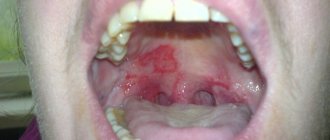
Pyogenic granuloma . Develops from the mucous or connective tissue elements of the oral cavity. Often observed after injury to the mucous membrane of the cheeks, lips or tongue. Pyogenic granuloma resembles richly supplied granulation tissue. It is characterized by a rapid increase in size up to 2 cm in diameter, dark red color and bleeding when touched.
Epulis . Benign tumors of the oral cavity located on the gums. They can grow from the deep layers of the gums, periosteum, and periodontal tissues. Epulis occurs most often in the area of the front teeth. They are classified into fibrous, giant cell and angiomatous formations.
Neuromas . They are formed as a result of the proliferation of Schwann sheath cells of nerve fibers. They reach 1 cm in diameter. They have a capsule. Neuromas are practically the only tumors of the oral cavity that may be painful on palpation.
Benign neoplasms of the oral cavity
In dentistry, benign tumors of an epithelial nature are most often encountered. Also, benign tumors can originate from fat, muscle, connective cells, nerve fibers and blood vessels. Depending on where the formations are located, benign growths are distinguished on the tongue, the inner surface of the cheek mucosa, sublingual tissues, gums, lips, soft and hard palate.
Epithelial neoplasms in the oral cavity can cause discomfort when chewing.
Epithelial
Nevus formations
In the mouth, nevi form rarely. They can be of varying degrees of color - from pale pink to dark brown, most often rise above the skin, and have a round, regular shape. Papillomatous nevi, blue nevi, and nevi of Ota form in the oral cavity. Under certain circumstances, some nevi tend to develop into a malignant neoplasm, so if you find a suspicious tumor in your mouth, it is better to consult a doctor.
Papillomas
These neoplasms consist of stratified squamous epithelial tissue. The main places of localization are the lips, tongue, palate, and mucous membranes of the cheeks. The tumors look like a round formation that rises above the surface of the skin and has a bumpy cover, similar to cauliflower inflorescences. Papillomas in the mouth often appear as single formations, rarely as multiple ones. If the formation is not removed in time, the surface becomes covered with a rough layer of epithelium, the formations become rough and acquire a white color.
Glands of Sulfur
Neoplasms are located in the area of the hard palate or alveolar process. Sera's glands look like a round formation that rises above the mucous membrane, are yellow in color, and have a dense consistency. More often they appear as multiple rashes; if the tumors appear in a newborn child, they disappear within a year without special treatment.
Vascular tumors in the oral cavity tend to become inflamed frequently and periodically cause discomfort.
Vascular
Lymphangiomas
They arise as a result of a malfunction of the lymphatic system and are detected in infancy. The formation looks like a limited or diffuse tumor that is located in the oral cavity. Tumors are prone to frequent inflammation due to interaction with saliva and food particles, as well as in chronic diseases of the ENT and digestive organs.
Hemangiomas
These formations most often form in the mouth and are also found in infants. The formation rises above the mucosal tissues, has a red tint, turns pale when pressed and decreases in size. If the integrity of the hemangioma tissue is disrupted, bleeding occurs, which then cannot be stopped for a long time.
Connective tissue
Fibroma formation
Tumors most often form on the tissues of the lower lips, tongue, and palate. The surface is smooth, oval in shape, sometimes attached to the mucosa with a stalk. The color of the neoplasm is the same as the color of healthy mucosal tissue; when injured, it bleeds and hurts. If the formation is damaged, the tissues often become inflamed and a bacterial complication occurs.
Fibromatosis on the gums develops due to a chronic inflammatory process in the oral cavity.
Fibromatosis on the gums
Fibromatous formations on the gums arise as a result of chronic inflammation of the tissues of the oral mucosa. The tumors are painless, grow on the gums between the teeth, can be small in size, but there are those that involve the entire alveolar process of the upper and lower jaws.
Myoma formations
Consist of muscle fibers. Leiomyomas consist of smooth muscle tissue, the main location is the palate. Rhabdomyomas consist of striated muscle fiber tissue and affect the surface of the tongue. Myoblastomas occur in infants, since the neoplasm is a consequence of disembryogenesis. The tumors are round in shape, up to 10 mm in size, the surface is smooth and shiny.
Pyogenic granuloma
Consists of connective and mucous tissues of the oral cavity. The main cause of occurrence is a violation of tissue integrity as a result of mechanical trauma and damage. The formation increases in size (up to 20 mm), bleeds and hurts when irritated. In these cases, you should not try to cope with the problem yourself, as there is a high risk of complications.
Epulis formations are characterized by rapid growth and bleeding.
Epulis formations
A benign neoplasm, the main location is the gums. Empulis grow from the deep layers of the gums, periodontal cells, and periosteum. Most often they are localized on the gums between the front teeth. There are fibrous, giant cell, and angiomatous empulis. Under certain circumstances, eimulis develop into a cancerous tumor, so if the growth rapidly increases and bleeds, it is worth visiting a doctor.
Neuromas
This type of oral tumor grows from the tissues of the Schwann nerve fiber sheaths. They look like a capsule, up to 10 mm in diameter, they hurt when pressed, and the patient experiences discomfort when eating. If you manage to find a tumor of unknown origin in your mouth with the above symptoms, under no circumstances should you try to squeeze out or tear off the tumor yourself. If you self-medicate, you can not only aggravate the situation, but also provoke life-threatening complications.
Vascular tumors of the oral cavity
Hemangiomas.
The most common tumors of the oral cavity. In 90% of cases, hemangiomas are diagnosed immediately or shortly after the birth of the child. There are simple (capillary), cavernous, capillary-cavernous and mixed. A distinctive feature of these oral tumors is that they turn pale or decrease in size when pressed. Trauma to hemangiomas often leads to bleeding.
Lymphangiomas.
They arise as a result of disorders of embryogenesis of the lymphatic system and are usually detected in newborns. Characterized by the formation of limited or diffuse swelling in the oral cavity. Among tumors of the oral cavity, cavernous, cystic, capillary-cavernous and cystic-cavernous lymphangiomas are distinguished. These oral tumors are prone to inflammation, which is often associated with trauma to the oral mucosa or exacerbation of any chronic inflammatory disease of the nasopharynx: pulpitis, periodotitis, chronic tonsillitis, sinusitis, etc. Some authors also point out the connection between the recurrent inflammatory process in lymphangioma and exacerbations chronic gastritis, duodenitis or colitis.
Removal of odontogenic cyst
An odontogenic cyst is a cavity neoplasm that occurs in the thickness of the jaw bones. It has a shell and liquid contents, and occurs as a result of the pathological development of teeth or their diseases.
Most often, an odontogenic cyst arises from an epithelial apical granuloma and at the first stage of development occurs without visible symptoms.
If the cyst is not removed in time, the bone bulges and swelling occurs in the oral cavity.
Removal of the cyst is carried out in our surgical method with complete desquamation of the membrane or with its opening and resection of the root apex. The teeth that are included in the cyst are subject to depulpation.
Diagnosis of oral tumors
Small oral tumors can be accidentally diagnosed by a dentist during a routine examination, during the treatment of caries and other dental procedures. An accurate determination of the type of tumor is possible only after a histological study of its structure. Such a study can be carried out on material taken during a tumor biopsy or after its removal.
Ultrasound of the tumor is used to determine the depth of tumor growth in the oral cavity, and X-ray examination is used to assess the condition of bone structures. For gingival fibromatosis, an orthopantomogram is performed, which often reveals areas of destruction of the alveolar process. Angiography is often used in the diagnosis of vascular tumors.
Removal of papilloma
Papillomas are benign tumors that are formed as a result of the proliferation of stratified epithelium of the oral mucosa under the influence of viruses, injuries, temperature irritants, and inflammatory processes.
Most often, papillomas form on the tongue, palate, membrane of the lips, alveolar process, in the area of the frenulum and pterygomaxillary folds. Treatment of papillomas in ours consists of excision of the tumor, after which the removed tissue is subject to histological examination.
Treatment of oral tumors
Difficulty speaking and chewing food in the presence of an oral tumor, constant trauma to neoplasms in this location, as well as the likelihood of their malignancy - all this is a reason for active surgical tactics. Depending on the type of tumor in the oral cavity, it is possible to use electrocoagulation, laser removal, cryodestruction, radio wave method, surgical excision, and sclerotherapy.
Removal of diffuse oral tumors is carried out in several stages. Excision of fibromatous growths is carried out together with the periosteum. Areas of destroyed bone tissue are processed with a milling cutter and coagulated. Vascular tumors of the oral cavity can be sclerosed by injecting sclerosing agents directly into the tumor vessels.