Dental restoration is a common practice in modern dentistry. People want to look good and not be embarrassed about their smile and are willing to pay money for it. Sometimes defects in teeth lead to functional disorders, which modern dentistry has effectively learned to combat. Aesthetic restoration of the dentition is a procedure necessary when teeth are damaged. For this purpose, restoration is used, which is aimed not only at restoring the structure and shape of the teeth itself, but also at completely restoring all the physiological functions that should be present in healthy teeth.
Types of artistic restoration
Restoration can be direct or indirect. In the first case, the procedure is performed directly in the patient’s mouth and lasts one day. For indirect restoration, a laboratory is used.
Direct restoration is not as reliable and copes with smaller defects. It is used when the patient needs help as soon as possible.
To perform an indirect restoration, you will have to visit the dentist several times. The advantage is an exact repetition of the natural shape of the tooth and enamel. In this way, subsequent tooth decay can be stopped and even minor bite defects can be corrected.
To make a direct restoration, composite materials or photopolymers are used. In the second case, the choice of materials and restoration options is wider. Composite materials are not destroyed by exposure to hard foods or temperature changes, and they do not cause allergic reactions.
Disadvantages of indirect artistic dental restoration
Two stages of production
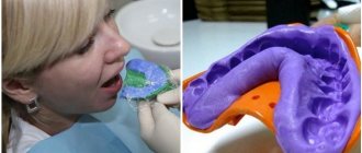
The patient has to come to the clinic twice: the first time to take an impression and make a temporary plastic structure, the second time to fix the finished work. It should be noted that the time spent in the clinic with indirect restoration is less than with direct restoration. Several days pass between these visits, during which the dental technician will make a restoration fragment that is completely suitable for the patient’s teeth (for this, impressions taken are used).
Indirect restoration method
The indirect method is called prosthetics. There are several options for restoring the appearance of a tooth:
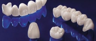
- veneers;
- bridges (when the tooth is restored from the root);
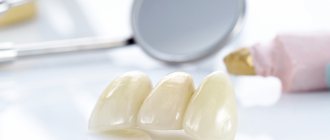
- crowns
Veneers allow you to create a Hollywood smile. Natural teeth are covered with thin dentures, giving a perfectly smooth and white surface.
At the first stage, a silicone impression is made. The rest of the work is carried out by the technologist without the patient; the prosthesis is made. This is much more convenient for the patient, because he does not have to waste time and suffer in the dental chair. There is also convenience for the doctor. The production of the prosthesis is carried out in the laboratory on a table, without interfering with the patient’s neighboring teeth or saliva.
The patient visits the clinic for the first time for consultation and impression taking. After this, a trial version is installed to identify shortcomings. Depending on the situation, you can skip this stage and put the final version right away.
The cost depends on the initial condition of the oral cavity, the chosen prosthetic method, the materials used, as well as the professionalism of the doctor. You can choose an individual treatment regimen at the first consultation after the examination.
Restoration of anterior teeth
We need the front teeth for normal chewing no less than the side teeth. If you have one or more of your front teeth destroyed, it will be difficult for you to bite off a piece of a green apple or crack a cracker.
However, the restoration of front teeth is of particular importance for a person’s social comfort. Therefore, in good clinics, aesthetic restoration of anterior teeth and their treatment are inseparable processes.
There are two restoration methods:
- Direct – involves modeling and restoring teeth directly in the patient’s mouth. In this case, composite materials are used - photopolymers and glass ionomers.
- Indirect (orthopedic) – an impression of the patient’s jaw is made. Then, structures from ceramic, zirconium or other materials are modeled and manufactured in the laboratory. In this way, it is possible to restore an almost completely lost tooth, but this already relates to the field of prosthetics.
Restoration of anterior teeth is performed using both methods. Each of them has its own advantages, disadvantages and areas of application.
The doctor decides which method will be used for you personally, based on the individual situation and the problems that the procedure should solve.
Cases in which dental restoration is needed
A dental procedure such as dental restoration is resorted to in the following situations:
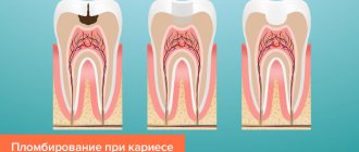
- after performing the procedure of ordinary filling of cavities with caries;
- in the presence of a pathological process of rapid tooth wear;
- for any type of tooth decay, but maintaining the integrity of the root;
- in the presence of defects in teeth such as cracks and chips, changes in the shade of enamel and the appearance of irregularities;
- if a person does not want to use orthodontic methods aimed at eliminating such a defect as large gaps between teeth;
- in cases where there is a need to change the position of a tooth in the dentition or to correct its shape
Preparatory process for performing a dental restoration procedure
If a person has a need or indication for a procedure such as dental restoration, the following steps should be performed at the first stage.
- Undergoing a detailed examination and diagnosis by a specialist such as an orthodontist, who will determine which method of dental restoration will be the most optimal solution.
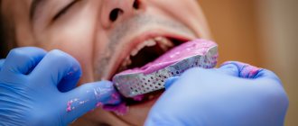
- Undergoing such a mandatory procedure as professional oral hygiene, during which all teeth will be thoroughly cleaned of plaque.
- To exclude a possible source of infection, complete sanitation of the oral cavity occurs.
- At the individual request of the client, a teeth whitening procedure can be performed before their restoration is carried out.
- Selection of restoration materials according to the color of tooth enamel. Thanks to this, the restored teeth will fit into the dentition as accurately as possible.
Before you begin the dental restoration procedure, do not forget that this method of restoration also has its own contraindications. It is strictly forbidden to perform dental restoration in cases of malocclusion, bruxism, or in the case when several chewing teeth are missing in the oral cavity.
Pros and cons of the two methods
The main advantages of the direct restoration method are:
- Maximum preservation of dental tissues, eliminating the possibility of damage to the teeth or oral cavity;
- Affordable price matches quality;
- Several teeth can be restored in one visit;
- Excellent aesthetic effect;
- Even severely damaged teeth can be restored.
Among the disadvantages of the direct method are:
- The procedure takes a long time; the patient needs to be patient in order to sit in the dentist’s chair all day;
- The dentist must have sufficient experience and professionalism to carry out restoration taking into account all the subtleties and nuances;
- After the procedure, increased oral hygiene is required, selection of a special brush and paste, regular visits to the dentist for professional cleaning and polishing;
- Over time, the color of the teeth may change and traces of restoration may become noticeable.
Advantages of using veneers
The use of ceramic veneers (as one of the most durable) with the indirect restoration method has the following advantages:
- With proper care, they will last for decades;
- Visually, they are practically no different from natural teeth;
- They are resistant to food dyes and tobacco smoke;
- The tooth is not allowed to decay further;
- Do not cause allergies;
- No discomfort for patients;
- Possibility of whitening procedures.
Disadvantages of using veneers
- Fairly high cost;
- Part of the enamel is cut down; if the veneers need to be removed, the enamel will have to be restored;
- If the preliminary installation stage is not followed, the veneers will not last long and may change color;
- The sensitivity of teeth increases, which causes pain in some patients;
- If not properly cared for, veneers can peel off and break.
It is important to understand that the main goal of dental restoration is to restore the functionality of teeth and achieve maximum aesthetic effect . That is why this procedure is very popular among patients of dental clinics who have problems with the shape and aesthetics of their teeth, who need to adjust the position of teeth in a row or eliminate gaps between teeth.
The cost of the dental restoration procedure directly depends on the chosen method and consumables; it also takes into account whether the restoration was complete or partial, and how significant the initial damage to the teeth was.
Methods of composite dental restoration
The choice of restoration method with light-curing substances depends primarily on the type of tooth affected (anterior/frontal or chewing) and the extent of damage.
Veneers
Veneers are thin plates of composite or ceramic that cover the dental surface. This is done either to correct visual dental defects or to protect against external damage. The aesthetic function of veneers is to mask widened tooth gaps or pronounced yellowness of the enamel. They are made by a dental technician or, in some cases, formed by a dentist directly in the patient’s oral cavity using a direct method.
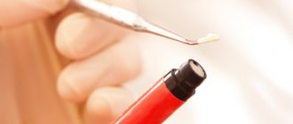
After local anesthesia, the dentist removes part of the enamel so that the veneer does not extend beyond the dentition. Then the contact surface is cleaned, degreased, and treated with an antiseptic. The composite is applied in layers, drying each with a special directional lamp. The final stage is to grind its surface, give it a physiological shape corresponding to the configuration of the tooth, and cover the surface with fluorine-containing varnish.
Tabs
The inlay is represented by a composite cast, duplicating the shape of the tooth, made by the indirect method. Inlays are necessary to close the dental cavity.
Installation of a composite inlay is carried out in two stages. On the first visit, the doctor treats the affected tooth. There is no need to grind down healthy tooth areas. Then an impression of the dentition is taken to establish the outlines of the tooth and bite, the color of the future inlay is determined and its temporary analogue is installed. An inlay is then made from a composite material based on the shape of the impression, which is installed during a follow-up visit to the dentist. Installation is carried out using special adhesives. The border of the inlay is carefully polished so that it blends with your own tooth.
Currently, this technique is rarely chosen due to its low efficiency compared to inlays made from other materials. However, the cost of the procedure remains quite high.
Seals
Before installing a composite filling, the dental cavity is first prepared: the damaged tooth area is drilled out and the cavity is given the proper shape, and the surface is cleaned for a tighter bonding of the composite. The tooth is isolated, limiting contact with liquid media, and etched for 15 seconds using phosphoric acid, after which the enamel should become dull.
Then a binding component is applied, hardening under the influence of a photopolymerizer, and the composite substance is directly installed. The elastic or flowable composite is applied in layers, the layer thickness should be about 2 mm so that it hardens properly under the influence of the light lamp. When applying layers, the dentist gives the composite the physiological shape characteristic of the tooth in order to preserve the aesthetic appearance. Before the final polishing, the bite is checked so that the protruding elements of the filling do not interfere with chewing; they are ground off. Polishing gives the material smoothness and shine.
Table. The choice of restoration method depending on the shape and stage of tooth decay
| Degree of destruction | Central (or internal) caries | Peripheral (or external) caries | Combined central + peripheral |
| Minimum | Amalgam or composite material, pin | Amalgam, composite material or polymer-modified glass ionomer | Amalgam, composite material or polymer-modified glass ionomer |
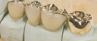
| Moderate | Amalgam on cement base | Amalgam, composite material or polymer-modified glass ionomer | On a large tooth there is an amalgam, on a small tooth there is a cast gold inlay |
| Heavy | Crown+contact rod | Crown | Crown+contact rod |
| Complete loss of a dental crown | – | – | Molars: crown + contact rod; premolars: root treatment+contact rod+crown |
Indirect aesthetic restorations
Due to improvements in the physical properties, wear resistance and esthetic potential of composites, making a choice between composite and ceramic as a material for indirect restoration has become quite difficult. There are several comparative parameters and corresponding clinical outcomes in the literature.
Resistance to wear of material and antagonist teeth
Ceramic and composite exhibit wear resistance similar to natural enamel. In particular, it was noted that the wear of the composite in the case where the antagonist is a natural tooth is influenced by two factors: the hardness of the filler particles and their size. The hardness of the filler particles should ideally be equal to the hardness of hydroxyapatite; this condition is implemented by serious global manufacturers of dental materials. In addition, composites subjected to final polymerization in an oven, through light and temperature, have significantly better mechanical (wear resistance, microhardness, etc.) and physical properties (solubility, degree of thermal expansion , elastic modulus, brittleness) compared to composites polymerized only with light. Even microinfiltration, measured by recording dye penetration, was found to be significantly lower. Due to the fact that the brightness of the composite may change, it is necessary to control the temperature for post-curing. In 1991, Rueggeberg et al. showed that the final polymerization cycle at 100 °C for 5 minutes is the most optimal. The greater wear resistance inherent in ceramics can lead to increased wear on the antagonist. In 1993, Krejci et al. We measured the wear of ceramics and enamel of the antagonist cusps. The ceramics studied in this study were cast glass ceramics (Dicor), sintered feldspathic ceramics (Biodent), pressed glass ceramics (Empress), one group of samples were polished and the rest were glazed. Pressed ceramics (and polished ones in particular) had the greatest wear resistance in in vitro locations, as previously noted by other authors. It was also found that antagonist cusp wear depends on a combination of hardness and surface texture of the ceramic, and glazed ceramic, being rougher, is more aggressive than polished ceramic.
Elastic modulus and fragility
According to many clinicians, ceramic adhesive restorations are more susceptible to fractures than composite ones, since ceramics, as a less elastic material, conducts functional stress to a greater extent to the walls of the cavity. This hypothesis is supported by the results of several studies, which indicate that chipping of the enamel edges of the cavity was observed more often in cases with ceramic inlays. It should be noted, however, that this statement is true for the first generation of reinforced ceramic compounds and has not been confirmed for later formulas.
Marginal adaptation and adherence to cavity walls
Some in vitro studies have documented enamel fractures at the interface with ceramic inlays under prolonged loads; however, it should be noted that such defects do not occur in the absence of fatigue. In a recent study by Dietschi et al. assessed cavity wall adaptation and marginal adaptation of adhesive inlays in Class P cavities. Inlays were made of composite (Tetric), low-temperature (Ducera) and sintered ceramic (In-Ceram) and were cemented to various cements and subjected to non-destructive fatigue loads. More rare cases of enamel microfractures have been reported at the border with inlays fixed to Panavia. The authors emphasize that cohesive enamel fractures are mainly associated with weak bonding to dentin, which causes stress to spread and concentrate towards the enamel margin. With regard to adaptation to the cavity walls, the same study demonstrates good adherence of both materials to the remaining enamel from the gingival margin to the occlusal surface. An uneven fit was noted at the dentin level. There is a well-established opinion that the softness of ceramic restorations negatively affects adaptation. In fact, the results of the study showed slightly worse adaptation of composite inlays. This suggests that the high elasticity inherent in the composite may have a negative impact when compared to a stiffer material that is unable to absorb stress like ceramics. The authors also note that composite inlays made in the oral cavity using a semi-direct technique have better adaptation compared to composite and ceramic indirect inlays, since the latter requires a larger cementation space (more than 30 ct); In addition, the surface layer of dentin collagen fibers is subjected to significant pressure during impression taking and cementation, this pressure can cause them to collapse and hardly effectively bond the material to dentin. Therefore, the authors suggest using the technique introduced by Paul and Sharer in 1996, which consists of applying a thin layer of dentin adhesive before taking the impression (double bonding technique). This layer, in addition to preventing collagen collapse, also protects the bonded surfaces from contamination during the manufacture of the inlay and is easily removed before the fixation procedure using a rubber buffer impregnated with pumice and hand instruments. Marginal integrity and internal adaptation of indirect composite restorations are now easily assessed by microscopic analysis of the smooth-hard tissue interface. In light of the above, it is difficult to single out a definite opinion regarding the superiority of this or that material in regional adaptation, because The thickness and properties of the luting composite are of utmost importance in the adaptation of the restoration. The choice should be made based on clinical parameters in favor of composite or “soft” polished ceramics, such as low-temperature or pressed.
The issue of choosing a restoration method is quite complex and capacious, and can only be decided by a specialist in a dental clinic. The initial consultation, which has become banal for many, nevertheless hides a lot of important things. The initial consultation is not an attempt to “extort” money from the client, but an important stage of patient-doctor communication, the stage of examination, diagnosis and the opportunity for the doctor to discuss treatment options with the patient. It is extremely reckless to refuse such a dialogue with a specialist. Sign up for a consultation at our clinic by phone, 8(495)417-41-17
Laboratory steps and costs
Composite inlays are easier to manufacture than ceramic inlays, even considering that ceramics made on a refractory model do not require additional waste of time. The great technical complexity of manufacturing and the delicate fixation procedure determine the high final cost of these artifacts, which are usually 6 times more expensive than amalgam for composite restorations and 8 times more expensive for ceramics.
Possibility of repair
Unlike ceramics, composite inlays can potentially be repaired in the mouth in situations where material needs to be added due to caries or marginal shrinkage. Inaccuracies at the marginal level can be corrected by adding a small amount of material, just as insufficient contact point density can be corrected by creating a roughness and applying a layer of material. It must be taken into account that reintervention deteriorates the quality of the final restoration.
Polishability
Surface finishing and final polishing in the oral cavity of composite inlays is definitely easier and more efficient compared to ceramics.
Stages of restoration
Cavity Preparation There are several elements that influence cavity design. These elements are: the relationship between occlusal contacts and preparation boundaries; functional load on the remaining cavity walls and type of occlusion; cavity wall thickness; the presence of defects such as erosion, abrasion or cracks. According to Fuzzi, Jackson, Dietschi, Magne, Spreafico and many other clinical experts, cavity preparation for ceramic and composite restorations differs from traditional metal restorations in large part by the presence of an extensive connection without sharp corners. There is no doubt that now there is no need to create classical retention points; the cavity should have a simple geometry with a retention form for better adaptation of the restoration. The side walls should be flat. The divergence of the side walls to the bottom of the cavity should be about 10 oC to ensure insertion of the restoration. In order to ensure good resistance of the inlay to loads (this mostly applies to ceramics), the minimum preparation depth should be 1.5-2 mm and the isthmus should be at least the same width. The edge of the cavity is prepared at an angle of almost 90o, without creating a bevel, but it must be smoothed, it is necessary to remove all enamel prisms that lack dentin support. A 100 micron grit conical cylindrical rounded bur is used for cavity preparation, and the same 30 micron grit bur is used for cavity finishing. The final preparation of the cervical border can be done with a rounded enamel knife without creating sharp corners. It is well known that with such a cavity, enamel prisms are cut at an angle and that etching does not ensure optimal adhesion; Moreover, during the period from preparation to cementation, staining or accumulation of bacterial plaque along the periphery of the cavity may occur, which subsequently impairs the effectiveness of bonding. To improve marginal sealing, some authors have suggested beveling the enamel margin immediately before cementation. However, in this case, more space is created for the cement, which leads to increased wear. When localizing the edge of the cavity below the gum level, Dietschi suggests first removing this area using direct restoration with a composite, with obligatory isolation with a rubber dam and precise fitting of the matrix. Performing a cavity wall removal procedure - “BLOCK-OUT” When restoring medium and large cavities, the advantage of performing a “pre-restoration” is not only the biological protection of the pulp, but also the creation of a geometry suitable for indirect restoration, which should have the same thickness throughout throughout. It has been proven that the fracture resistance of all-ceramic crowns depends to a large extent on the elasticity modulus of the luting cement and the material from which the core is made. Therefore, the use of materials such as glass ionomer and zinc phosphate cements, as well as pastes based on calcium hydroxide, as a base is contraindicated. Glass ionomers, in particular, which have long been used as a basis for indirect restoration, are now considered obsolete due to limited mechanical properties and poor marginal protection due to the constant resorption of the material. Composites are definitely more effective due to their strong adhesion to dentin and resistance to compressive loads. Flowable composites are a reliable alternative for base construction. Compared to traditional composites, they have a low filler content (50-70%), higher elasticity (from 3.6 to 7.6 GPa) and high flexibility. These properties, combined with high fluidity, which reduces the risk of blistering during application, significantly reduce polymerization stress. The modern direction of adhesive dentistry is based on the concept of “progressive elasticity” of restoration, due to the formation of an elastic layer, which, by partially absorbing polymerization stress, prevents damage to the hybrid layer or its separation. However, it should be noted that excessive elasticity and, as a result, significant deformation can disrupt marginal adaptation. Currently, the use of a flowable composite is recommended in the form of a substrate under the base itself, made from the same microfilled composite as the inlay. Taking an impression This stage is performed after preparation and fully corresponds to the classical procedure for taking an impression for the manufacture of fixed structures. In Class 2 cavities, where there is less than 1 mm of remaining enamel at the gingival margin or is completely absent, the gingiva is processed to expose the preparation margin. The impression is taken simultaneously using masses based on polyester or polyvinylsiloxane. Fabrication of the inlay As with all materials, esthetic or not, a collapsible plaster model is necessary, which is processed to visualize the edge of the preparation. It is necessary to block all undercuts and create space for cement. Next, the inlay is formed, occlusal contacts are adjusted, finishing and polishing are carried out. Trying on the inlay During the fitting of the inlay, its stability, marginal adaptation and tightness of the proximal contacts are assessed. During fitting, the insert should be inserted and removed without friction. You cannot check the occlusion at this stage, since a fracture of the restoration that has not yet been cemented is possible. Cementation, finishing, polishing Indirect adhesive restorations are retained by micromechanical and chemical bonding through the adhesive cement to the tooth structure. For successful fixation, the key points are reliable isolation of the working field, thorough cleaning of the cavity and decontamination. The temporary restoration is removed using hand and ultrasonic instruments, the cavity is cleaned with a brush and pumice stone: cleaning with a soda stream is not recommended due to the aggressiveness of the latter. The quality of the marginal fit of a fixed adhesive restoration has been comprehensively studied taking into account the following factors: Cement thickness It has been established that to achieve optimal marginal adaptation at the margin of the inlay should be less than 100 tg, while at the bottom and walls of the cavity a cement thickness of 300 tg is acceptable due to the use spacer and the need to fill undercuts. Secondly, composite cements have increased wear compared to restorative composites. Occlusal wear is proportional to the interocclusal distance. This wear is attributed to food bolus impact. If the gap is less than 100 um, food particles should not mechanically affect the cement layer. If this distance is greater, then in the vertical direction it will also increase with the inevitable occurrence of microinfiltration and secondary caries. Under normal conditions, cement wear is 15-20 kg in the first year with a gradual decrease and further stop of wear. Research also demonstrates higher wear resistance of microfilled composites compared to macrofills. Bondability The most reliable internal adaptation and marginal sealing is achieved when the preparation margins are in the enamel. Indeed, if adhesion to enamel is quite reliable, then the strength of adhesion to dentin can vary. It has been proven that adhesion failure is mainly observed between the hybrid layer and the composite. This may be due to the fact that the hybrid layer was not completely impregnated with the adhesive. At the same time, it has been found that overly aggressive etching, excessive drying and compression of the collagen fiber network during impression taking and cementation cause their denaturation and/or collapse, forming a partially impregnated layer in which, as Sano points out, micro-spaces arise that allow penetration enzymes and hydrogen ions (H*), which in turn cause hydrolysis of resin and collagen - a phenomenon called nanoleakage. Paul and Sharer showed that applying an adhesive after preparation can stabilize adhesion, since, on the one hand, it prevents contamination during impression taking and wearing of a temporary restoration, and on the other hand, it protects the dentin from the stresses caused by the above maneuvers. This technique (double bonding technique) will also reduce postoperative sensitivity, which is believed to be a result of bacterial contamination and hydrodynamic phenomenon. This theory is supported by the fact that temporary filling material quickly leaves the edges of the cavity, losing its protective properties. Application and polymerization of adhesive cement The internal surfaces of the restoration and cavities must be prepared for bonding. Cement is introduced into the cavity and onto the inner walls of the inlay. When working with dual-cure cements, it is necessary to insert the inlay quickly enough, because polymerization occurs faster at oral temperature. If the same restorative composite is used for fixation, then the use of ultrasonic instruments is recommended for easier adaptation of the inlay into the cavity, due to its higher viscosity. Excess cement is removed using a probe and a brush soaked in adhesive resin. Adequate light activation is necessary to ensure maximum material conversion, and this is true for light-curing composites. It is recommended to expose each wall of the inlay for 1 minute. In terms of finishing and polishing, there are currently several polishing systems on the market designed for finishing restorations in the oral cavity. The quality of the surface of an aesthetic restoration depends on the structure of the material, the environment in the oral cavity and the scrupulousness of the doctor at this stage of its manufacture. The goals of finishing and polishing are to: achieve integrity between the inlay and the cavity walls; smoothing rough surfaces; correction of any surface defects. It is known that the presence of oxygen affects the polymerization of the surface, therefore, to avoid the formation of an inhibited layer, the surface can be coated with a glycerol-based gel. Finishing of flat and accessible proximal surfaces is carried out with flexible discs, in the gingival area - with abrasive strips (strips), and fine and ultra-fine grain diamond burs are used for processing the occlusal surface. Silicone heads, brushes impregnated with diamond paste and an aluminum oxide paste for final polishing ensure a smooth and shiny surface of the restoration. Finally, the margins of the restoration are sealed with a low-viscosity resin. This is necessary not only to fill edge defects, but also to reduce wear of the material, due to the penetration of the sealer into micropores. Strict adherence to the sequence of the above steps leads to a controlled and predictable result. The described technique in relation to the restoration of a tooth with endodontic treatment is a very conservative approach compared to other restoration methods (for example, covering with an artificial crown), often used in such cases. From this point of view, we propose a simple protocol based on a clinical case. Conclusion The likelihood of successful treatment and longevity of an indirect adhesive composite or ceramic restoration is inextricably linked to the correct assessment of the clinical case and strict adherence to the operating protocol. Repeated examinations of teeth restored with indirect inlays allowed us to achieve an absolutely satisfactory result. In particular, Fuzzi and Rappelli, in a study of long-term results of dental restoration with ceramic inlays, reported an unacceptably high failure rate. Schulte et al., evaluating the marginal fit of Ips-Empress inlays after 3 and 5 years, concluded that the prognosis of such restorations is good, even if the problem of cement wear remains unresolved. However, it should be emphasized that this study used dual-cure cement, which has lower wear resistance compared to light-cured composites. Similar conclusions were obtained by Thordrup et al. when examining the condition of direct and indirect composite and indirect ceramic restorations after 5 years. Giving both materials their due, it should be noted that composite inlays have objective advantages. Firstly, the cementation procedure is simpler; In addition, the finishing and polishing capabilities of ceramic restorations are limited because ceramics are more fragile and require careful handling. In addition, in our opinion, the possibility of correction during preventive examinations before and after fixation represents a decisive advantage of the composite over ceramics. It should also be noted that more complex laboratory procedures are the reason for the higher cost of ceramic restorations. Therefore, in light of all of the above, it is clear that in conservative indirect aesthetic restoration, ceramics are gradually losing their position in favor of composites.
| Name of service | Price, rub.) |
General manipulations | |
| initial visit - examination and consultation | for free |
| re-examination and consultation | 500 |
| certificate of reorganization | 500 |
Treatment of caries and its complications (pulpitis, periodontitis) | |
| * the price includes everything you need: | |
| application anesthesia, injection anesthesia (1 carpule), removal of an old filling, trephination of the tooth crown, cavity opening, cavity formation, medicinal treatment, insulating pad, medical-isolating pad, polishing and of course the work of a qualified specialist | |
| Anesthesia | 500 |
| Filling made of glass ionomer cement or chemically cured composite (1 surface) | 2000 |
| additional surface | 500 |
| Treatment of cervical caries | 5000 |
| filling made of light-cured composite from the world's leading luxury manufacturers (3M; Keer; GS) 1 surface | 4500 |
| additional surface | 500 |
| production of veneer using the direct method (cost depends on the clinical situation) | 5000-12000 |
| first degree of difficulty | 5000 |
| second degree of difficulty | 6000 |
| third degree of difficulty | 7000 |
| fourth degree of difficulty | 10000 |
Non-surgical treatment of radicular cysts and cystogranulomas (root canal preparation included) | |
| temporary canal filling for 4-6 months | 2000 |
Preparation of root canals for filling (opening of the tooth cavity, removal of dental pulp or its decay, medicinal treatment, expansion, formation of root canals) | |
| 1 (one) channel | 1000 |
| 2 (two) channels | 1500 |
| 3 (three) channels | 2000 |
| 4 (four) channels | 2500 |
Temporary filling of root canals with medicinal paste | |
| 1-channel tooth | 1000 |
| 2-channel tooth | 2000 |
| 3-channel tooth | 3000 |
| 4-channel tooth | 4000 |
Permanent filling of root canals with paste or gutta-percha pins (medication included) | |
| 1-channel tooth | 3000 |
| 2-channel tooth | 4000 |
| 3-channel tooth | 5000 |
| 4-channel tooth | 6000 |
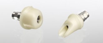
Installing an anchor pin in the root filament | |
| metal | 500 |
| fiberglass | 1000 |
| removing the anchor pin | 1000 |
Unsealing the root canal | |
| eugenol paste, gutta-percha | 500 |
| Resorcinol-formalin paste, cement | 1000 |
| Installing a temporary filling | 500 |