Features of dental treatment for pregnant women
Pregnancy is not an absolute contraindication to any dental procedures. However, the patient must warn the doctor about her situation, and also indicate the exact duration of pregnancy.
Main nuances of therapy:
- while carrying a child, caries, pulpitis, periodontitis and inflammatory gum diseases (gingivitis, periodontitis, stomatitis) can be treated;
- To fill a tooth, you can use both chemically curing materials and light-curing composites; photopolymer lamps are safe for the fetus;
- enamel bleaching is prohibited;
- Dental treatment is carried out under local anesthesia (injection of Ultracaine, Articaine), the expectant mother must not be allowed to endure terrible pain in the dentist’s office;
- General anesthesia is strictly contraindicated.
Early and late dental treatment
The entire period of pregnancy is conventionally divided into 3 periods (trimesters).
First trimester (up to 12 weeks)
In the 1st trimester (the earliest period), all the vital organs of the child are formed. The placenta is just beginning to form; it cannot yet protect the fetus from negative influences. Therefore, it is undesirable to carry out any medical intervention during this period. However, the dentist can prescribe local drugs to relieve inflammation (Chlorhexidine, Miramistin, Cholisal).
Second trimester (from approximately 13 to 24 weeks)
In the second trimester, the risk of dangers decreases significantly. The placenta serves as a reliable protective barrier for the baby. This is the optimal period for dental treatment and other dental procedures.
Third trimester (from 25 weeks to delivery)
In the 3rd trimester, increased sensitivity of the uterus to drug effects occurs. In addition, during this period the woman’s body is quite weakened. Therefore, “extra” stress in the dentist’s office is extremely undesirable. If possible, it is better to postpone dental treatment during lactation. However, this does not apply to emergency cases, such as acute toothache.
Research results
At the time of examination, pregnant women were diagnosed with dental and periodontal diseases in 100% of cases. The KPU index was on average 8.75+0.9. At the same time, the number of carious cavities was 48%, extracted teeth – 8%, and filled teeth – 44%. There were no significant differences between the groups in terms of caries prevalence.
The enamel acid resistance test averaged 4.93+0.29 points, which indicates a decrease in caries resistance compared to non-pregnant women of the same age group (3.18+0.19 points). During the physiological course of pregnancy, this test was 4.18 + 0.39 points; with mild vomiting – 4.34+0.41; for congenital and rheumatic heart defects – 5.58+0.31; for late gestosis – 6.15+0.39 points.
It is characteristic that in pregnant women of all examined groups the highest rate of TER, that is, the lowest level of acid resistance, was observed at 8-10 and 32-34 weeks of pregnancy (Table 2). Thus, during normal pregnancy, the level of TER at 8-10 weeks reached 5.63 + 0.23 points, at 32-34 weeks – 5.12 + 0.24 points; for mild vomiting – 5.94+0.23 and 4.83+0.23 points, respectively. The lowest level of caries resistance during these periods was in groups with late gestosis and heart defects (reached 7.89+0.46 points).
Table 2. Characteristics of the TER test at different stages of pregnancy
| Gestation period (weeks) | TER in women with normal pregnancy | TER in pregnant women with mild vomiting |
| 5-7 | 3,70±0,25 | 4,03±0,24 |
| 8-10 | 5,63±0,23 | 5,94±0,28 |
| 11-12 | 3,96±0,26 | 4,58±0,25 |
| 13-20 | 3,56±0,22 | 3,61±0,25 |
| 21-31 | 3,67±0,24 | 3,53±0,24 |
| 32-34 | 5,12±0,24 | 4,83±0,23 |
| 35-40 | 3,58±0,23 | 3,88±0,25 |
According to the literature, the most critical moments with increased uterine tone occur at approximately 8-12 weeks of pregnancy and 28-32. Data obtained from the study of acid resistance of tooth enamel show that a decrease in resistance coincides with critical periods of gestation.
The condition of the hard tissues of the tooth was assessed at the initial examination, after 7 and 12 months using the KPU indices and the TER test. Women were divided into subgroups to evaluate the effectiveness of the use of adaptogens in dentistry when prescribed drugs at different stages of pregnancy for two weeks.
In women who took vitamin E from the 8th and 32nd weeks of pregnancy, the increase in the intensity of caries over 7 months was 0.55+0.10, over a year – 0.75+0.08; when prescribing Eleutherococcus extract – 0.62+0.09 and 0.81+0.12 for the corresponding periods of time. At the same time, in pregnant women in the control subgroup, the increase in the intensity of caries was 1.37 + 0.16. Consequently, the reduction in caries growth under the influence of vitamin E for 7 months was 59.85% and for a year – 55.88%. The administration of Eleutherococcus extract led to a decrease in the increase in caries intensity by 54.71% over 7 months of pregnancy and by 52.35% over one year. Thus, the use of the studied drugs in two courses during critical periods according to the TER test turned out to be the most effective.
Anamnesis collection for periodontal diseases revealed that 46.7+2.8% of examined women did not complain about bleeding and discomfort from the gum tissue, 49.3+2.2% complained about bleeding from the gums when brushing their teeth. Eight pregnant women had periodic bleeding when eating solid food, three of the women complained of spontaneous bleeding from the gums.
During the examination of periodontal tissues, it was found that the simplified Green-Vermilion index was on average 2.28 + 0.06, which indicates unsatisfactory oral hygiene, with 55% of this indicator being due to dental plaque (DI-S). According to the RMA index, the inflammatory process covers the papillae and marginal gums; on average, this indicator was 54.2+3.8%. The intensity of the inflammatory process in the gums according to the GI index was at the lower limit of moderate severity (1.1+0.02).
As a result, 20.3+3.6% of women were diagnosed with mild gingivitis, 52.6+3.8% with moderate gingivitis, and 11.8+2.7% with severe gingivitis. In 15.3+2.2% of pregnant women, a clinical diagnosis of mild marginal periodontitis was made.
Pregnant women undergoing hospital treatment for late gestosis were divided into two subgroups. Women of the first subgroup were taught oral hygiene using standard methods, and pregnant women of the second subgroup, in addition to training in standard methods of oral care, had their gums covered with SDAP.
When examining women in the first subgroup, the Green-Vermilion oral hygiene indicator in individual analysis was distributed as follows: 29.0% of women had satisfactory oral hygiene, 32.3% - unsatisfactory and 38.7% - poor. After training in individual hygiene and motivation, after two days the OHI-S index became, on average, 26% lower for the subgroup (the difference is significant). 19.4% of pregnant women had good hygiene, 25.8% had satisfactory hygiene, 29.0% had unsatisfactory hygiene and 25.8% had poor hygiene. By the third visit after 7 days, this indicator was good and satisfactory in 47.0% of women, unsatisfactory in 17.6% and poor in 35.3%. On average, for the subgroup OHI-S was 2.09+0.1. This is 13% less than during the initial examination, and 13% more than immediately after oral hygiene training. OHI-S parameters changed due to its DI-S component, since dental plaque removal was not performed at this stage (Table 3).
Table 3. Changes in dental indicators by visit in pregnant women with gestosis of the first subgroup.
| Indexes | First visit | Second visit | Third visit |
| OHI-S | 2,4±0,06 | 1,78±0,07 | 2,09±0,1 |
| DI-S | 1,16±0,07 | 0,54±0,08 | 0,71±0,11 |
| CI-S | 1,24±0,07 | 1,24±0,07 | 1,38±0,12 |
| G.I. | 1,11±0,05 | 0,99±0,06 | 0,9±0,09 |
| P.M.A. | 56,7±8,9% | 51,1±9,0% | 47,3±12,9% |
At the first visit, in 19.4% of pregnant women the inflammation was localized within the papillae, in 48.4% it involved the marginal edge and in 32.3% there were areas with inflamed alveolar gum. On average, the PMA index reached 56.7%. After training in oral hygiene, on average, the PMA index for the subgroup decreased by 10%, and after a week by another 7% and amounted to 47.3 + 12.9%. The intensity of the inflammatory process according to the GI index at the first visit was mild in 48.4% of women, and moderate in 51.6%. On the third day after oral hygiene training, the GI index decreased by 11%, and a week later by another 8%. As a result, 64.5% of pregnant women had mild inflammation in the gums and 35.5% had moderate inflammation.
Thus, in women, after training in oral hygiene and motivation, the plaque index decreased by 2 times. And the intensity and prevalence of the inflammatory process in the gums is 1.2 times.
In pregnant women in the second subgroup, the Green-Vermilion index at the initial examination was 2.1 + 0.07, plaque accounted for 57%. After three times treatment of the gum mucosa with SDAP, the OHI-S index decreased by 27%, and the DI-S index component changed by 2 times. After training in individual oral hygiene, the plaque index decreased by another 2 times and amounted to 23% of the OHI-S index. At the last visit, 69% of women in this subgroup had good and satisfactory oral hygiene, which is 2 times more than at the initial examination (Table 4).
Table 4. Changes in dental indicators by visit in pregnant women with gestosis of the second subgroup.
| Indexes | First visit | Second visit | Third visit |
| OHI-S | 2,22±0,08 | 1,67±0,13 | 1,22±0,11 |
| DI-S | 1,19±0,09 | 0,61±0,12 | 0,28±0,07 |
| CI-S | 1,03±0,09 | 0,61±0,12 | 0,94±0,13 |
| G.I. | 1,08±0,07 | 0,59±0,05 | 0,51±0,05 |
| P.M.A. | 53,4±9,98% | 32,9±12,6% | 24,3±10,7% |
An analysis of the condition of the marginal periodontium showed that 11.4% of women had inflamed gingival papillae, 63.6% had inflamed marginal margins, and 25% had areas of inflammation of the attached gums. The prevalence of the inflammatory process in periodontal tissues after triple treatment with SDAP decreased by 1.5 times. After oral hygiene training, the PMA index decreased by another 22%. The treatment made it possible to change the picture of the prevalence of inflammation: 81.3% of women had inflamed gingival papillae and 18.7% had damage to the marginal gums. On average for the subgroup, the PMA indicator was 24.3 + 10.7% (with an initial indicator of 52.6%). The intensity of the inflammatory process according to the GI index was 1.08+0.06. At the same time, 59.1% of women had mild inflammation and 40.9% had moderate inflammation. After treating the gums with SDAP, the GI index decreased by 1.8 times. And after training in individual oral hygiene, it decreased by another 10%. After treatment, all women had mild inflammation in their gums. The average for the subgroup was 0.51+0.05 points.
Studies have revealed that applications of anti-inflammatory drugs reduce the plaque index by 2 times and reduce the intensity of inflammation by 1.7 times. Individual oral hygiene improves by 2.1 times, and inflammation in the gums decreases by 1.2 times after one-time training and motivation. The plaque index decreases by 4.3 times, and the intensity of inflammation in the marginal periodontium decreases by 2.2 times with the combined effect of anti-inflammatory drugs and training in individual oral hygiene. The data obtained made it possible to propose in the case of severe bleeding from the gums the following stages in carrying out professional and training in individual oral hygiene: on the first visit, it is better to use anti-inflammatory drugs topically on the gum mucosa and recommend their use at home 1-3 times a day for 2-3 days. At the second visit, reapply local anti-inflammatory treatment and provide instruction and training in individual oral hygiene. Professional oral hygiene and personal control are best carried out from the second or third visit, when swelling and bleeding from the gums are significantly less.
Dental diagnostics during pregnancy
Treatment of pulpitis and tooth extraction during pregnancy cannot be done without diagnosis. Traditional radiography (sighted x-ray) is not the best option for pregnant women. Fetal cells are in the process of dividing, so they are especially sensitive to radiation.
But if there is a need for such diagnostics, it is better to carry it out in the second trimester. Be sure to cover your stomach and pelvic area with a protective lead apron.
The safest option for women during pregnancy is digital radiovisiography. This method is characterized by minimal radiation exposure - 90% less compared to film X-rays.
Is it possible to do x-rays?
It is not advisable for pregnant women to undergo X-rays, and it is strictly contraindicated in the first trimester. But at a later date, if there is evidence, such a procedure is possible. Fortunately, modern dentistry uses equipment with highly sensitive sensors and minimal radiation. During the examination, the patient’s chest and abdomen are covered with a lead apron.
Attention!
To avoid the risk of complications in the fetus, it is worth taking care of prevention at the stage of pregnancy planning. Before deciding to expand the family, a woman needs to consult a dentist, cure all carious lesions, chronic diseases of the gums and periodontal tissues, and, if necessary, undergo procedures to strengthen tooth enamel. Those who are preparing to become a mother need to pay special attention to hygiene and proper oral care, because during pregnancy the risk of developing caries increases.
Safe anesthesia for pregnant women
Local anesthetics are used that do not cross the placental barrier. Another requirement for painkillers is a low degree of impact on blood vessels.
Lidocaine is not suitable for expectant mothers, as this drug can cause muscle weakness, cramps and a sharp decrease in blood pressure.
The best option is anesthetics based on anticaine:
- Ubistezin;
- Ultracaine;
- Alfacaine;
- Artifrin.
These drugs do not harm the baby because they act locally. They also have a reduced concentration of vasoconstrictor components (adrenaline, etc.), which is safe for the mother.
Tooth extraction during pregnancy
Tooth extraction is a surgical operation that is always accompanied by psycho-emotional stress. Of course, it is undesirable for women while carrying a child.
Therefore, tooth extraction is carried out only in extreme cases:
- crown or root fracture;
- deep carious lesion, which causes purulent inflammation;
- formation of a cyst whose diameter exceeds 1 cm;
- persistent acute pain that cannot be eliminated with conservative therapy.
Wisdom teeth removal is generally not performed during pregnancy. This operation often ends with alveolitis (inflammation of the socket) and other complications requiring antibiotics.
Is there an impact on the baby?
Maintaining dental health during pregnancy is important not only for the expectant mother, but also for the child. Any infectious foci in the body of a pregnant woman pose a potential danger to the fetus. Microbes and the toxic substances they release can be absorbed into the bloodstream and enter the placenta along with the blood, causing infection of the child.
The risks are especially high in the first trimester of pregnancy due to the formation of internal organs and systems. If infection occurs at this stage, there is a risk of fetal malformations. With later infection, premature birth, hypoxia and fetal malnutrition are possible. In addition, some microorganisms can cause an increase in uterine tone, opening of the cervical canal and damage to the membranes of the fetus, which greatly increases the likelihood of miscarriage.
Implantation and dental prosthetics during pregnancy
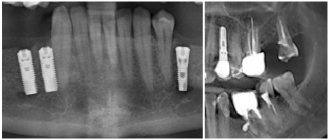
During pregnancy, you can have any type of prosthesis, including crowns and bridges. The exception is dental implants.
Implanting a dental implant often requires a lot of vital energy. But during pregnancy, all resources are aimed at developing a healthy baby.
In addition, after implantation, anti-inflammatory and painkillers are required, which are contraindicated for the expectant mother.
Dental treatment during pregnancy can be done absolutely free if you use the compulsory medical insurance policy. A list of all government institutions, as well as private dentistry, can be found on our website.


