Anomalies of teeth
The causes of dental anomalies are very diverse and can be divided into endogenous and exogenous.
Endogenous causes, in turn, include genetic and endocrine factors. Genetic characteristics can be determined by the characteristics of the dental system inherited from parents - the size and shape of teeth, jaws, the structure and function of soft tissues, etc. Dental anomalies are often included in the structure of congenital defects and hereditary diseases - clefts of the upper lip, soft and hard palate, alveolar process; Shershevsky-Turner syndrome, Seckel syndrome, Waardenburg syndrome, Down syndrome, osteogenesis imperfecta and many others.
Dental anomalies may be associated with endocrine factors that have a significant impact on the formation of the dental system. Thus, with hypothyroidism, there is a delay in the eruption and replacement of teeth, enamel hypoplasia, deformation and osteoporosis of the jaws, adentia, changes in the shape and size of the crowns of the teeth. Peculiarities of jaw development and dental anomalies are observed in hyperparathyroidism, hypocortisolism, pituitary dwarfism, etc.
Exogenous causes of the formation of dental anomalies are divided into prenatal, intranatal and postnatal, general and local. General prenatal factors include adverse environmental influences on the developing fetus, multiple pregnancy, the presence of amniotic cords, fetal hypoxia, intrauterine growth retardation, toxicosis of pregnancy, intrauterine infections, stressful situations during pregnancy, etc. Intranatal factors contributing to the development of dental anomalies, include complicated childbirth - umbilical cord entanglement, asphyxia, intracranial birth injuries, prolonged anhydrous interval, respiratory distress syndrome. In the postnatal period, dental anomalies can be associated with childhood diseases: rickets, hypovitaminosis, insufficient exposure of the child to fresh air, deviated nasal septum and impaired nasal breathing, hypertrophy of the palatine tonsils, etc.
Among local factors, the most significant etiological role in the development of dental anomalies belongs to artificial feeding, dividing the use of a pacifier, thumb sucking, feeding preschool children with soft food, etc. Dental anomalies can be a consequence of dental trauma, short frenulum of the upper lip or tongue, complicated caries, leading to to early removal of teeth, osteomyelitis of the jaw, leading to the death of tooth germs, etc. The presence of impacted or supernumerary teeth can lead to abnormalities in the position of the teeth.
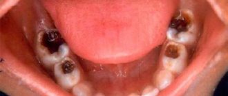
Abnormalities in tooth color are observed in the presence of plaque, penetration of food dyes into tooth cracks, exposure to nicotine, demineralization of enamel, pulp necrosis, dental granuloma, fluorosis, tooth trauma accompanied by internal hemorrhage, filling of canals with a resorcinol-formalin mixture, etc.
Etiology and pathogenesis of acquired and congenital dental anomalies
Many authors distinguish between the concepts of “anomaly” and “deformation”. Anomalies in the development of teeth and jaws are understood as congenital persistent deviations from the generally accepted norm (anatomical), increasing with the age of children, accompanied by functional and cosmetic disorders.
Deformation as acquired persistent changes in the shape and function of teeth and jaws that occur in the postnatal (after birth) period as a result of impaired growth and development. They progress over the years, disrupt the functions of the chewing system, and change the appearance of the child.
Anomalies can be hereditary, congenital or acquired. Hereditary (family) anomalies are anomalies in which not a “ready-made” anomaly is inherited, but a predisposition to deformations of the masticatory system. Such anomalies include true diastema, edentia, supernumerary teeth, true progeny (mesial bite) and prognathia (distal bite), deep overbite.
Congenital anomalies develop in the embryonic period. The reasons for their occurrence are pathological processes associated with the development of the fetus, its incorrect position, changes in the pressure of the amniotic fluid, mechanical compression of the fetus from the outside, illness of the parents, especially the mother (toxoplasmosis, syphilis, alcoholism, dysfunction of the endocrine glands, radiation injury, drug addiction, toxic effect of certain medications during pregnancy). Congenital anomalies occur more often as a result of impaired fusion of the maxillofacial processes, which results in clefts of the upper lip, alveolar process, hard and soft palate. This group of anomalies also includes unilateral microgenia.
Classification of prostheses
Acquired anomalies occur after the birth of a child due to unfavorable conditions for its development under the influence of various factors.
Artificial feeding of a child . During the act of sucking, the child covers the nipple with his lips, holds it, squeezes out milk with the alveolar processes and swallows. Sucking movements promote the normal development of the lower jaw with its surrounding tissues and lead to the elimination of the usual distal position of the lower jaw in the newborn. When sucking from a bottle, the mixture usually pours out rather than being squeezed out. This may be the cause of dental anomalies (for example, the development of a distal bite).
Child's diseases . One of the reasons for the development of dental anomalies can be diseases in which the function of the endocrine glands is disrupted. Insufficient function of the thyroid gland leads to delayed growth of jaws, teeth and delayed teething. Dysfunction of the pituitary gland leads to micro- and macrognathia, dysfunction of the parathyroid glands negatively affects calcium metabolism, which reduces the resistance of the jaws to deformation.
The disease rickets leads to a disturbance in the metabolism of calcium and phosphorus, calcification of bone tissue as a result of a lack of ergocalciferol in the body, which leads to the pliability of bone tissue and the occurrence of dental anomalies under the influence of ordinary factors (muscle traction, tongue pressure, chewing pressure) or bad habits. Rickets is associated with the development of a deep or open bite, narrowing of the dental arches and jaws (Fig. 6), delayed teething, and enamel hypoplasia.
Other diseases of early childhood also play a role in the development of deformations of the masticatory system: dyspepsia, infectious diseases, tuberculosis, toxoplasmosis and others that weaken the child’s body’s defenses.
Adenoid growths (polyps in the upper respiratory tract) cause difficulty breathing through the nose and the child switches to breathing through the mouth. With this type of breathing, the mouth is constantly open. At the same time, the muscles of the cheeks tense, squeezing the lateral areas of the dental arches. When you inhale, positive pressure is generated in the mouth. The stream of inhaled air presses on the palatine vault, causing deformation of the hard palate - the formation of a high vault (Gothic palate) and narrowing of the dental arches. When breathing through the mouth, the tongue lowers, touching the lateral areas only with the lower dentition, and does not counteract the pressure of the cheek muscles on the upper dentition and alveolar processes. Such a violation of muscle balance leads to compression (narrowing) of the upper dentition and jaw (Fig. 7).
When the lingual tonsil enlarges, to facilitate breathing, the child moves the tongue forward, and at night also moves the lower jaw, which leads to the development of mesial occlusion (progenia). When the pharyngeal tonsil enlarges, the child raises (throws back) his head, freeing the epiglottis from the pressure of the tongue, moves the lower jaw posteriorly, separating the root of the tongue from the soft palate, thereby clearing the way for the passage of an air stream. All this leads to the development of distal occlusion (prognathia).
Bad habits are common among children, but dental anomalies and deformations occur only in the presence of predisposing conditions, in particular inferiority of bone structures after rickets and other diseases.
Acquired anomalies also occur when the child is in an incorrect position during sleep. With a high headboard, the child's head is tilted anteriorly, the neck muscles are weakened, the lower jaw moves anteriorly, which contributes to the development of mesial bite (progenia) (Fig. 5).
If a child sleeps with his head thrown back, the neck muscles become tense and the lower jaw moves posteriorly (distally). This makes it difficult to grow, and distal overbite (prognathia) may occur. If you constantly place your hand under your cheek while sleeping, a cross (oblique) bite may develop (Fig. 8).
Playing the violin sometimes has the same effect. Incorrect body position at a desk, desk, or while walking plays a role in the occurrence of deformities.
Sucking fingers, tongue, cheeks, pacifiers, various objects, biting lips, tongue sometimes causes various types of deformations and anomalies of teeth, dentition, bite (distal, mesial, open, cross) depending on the manner of sucking or biting. For example, if a child, when sucking, places his finger (fingers) obliquely from top to bottom, a distal bite develops; if they are positioned horizontally, an open bite often develops (Fig. 9).
When biting the lower lip, the lower jaw moves back or the anterior portion of the lower dental arch flattens - a distal bite occurs.
Prosthetics for increased gag reflex
Diseases of the teeth and jaws . Early removal of baby teeth retards the growth of alveolar processes and jaws; erupting permanent teeth erupt outside the dental arch due to lack of space. The destruction of the first permanent molars by caries and the associated decrease in bite height can lead to the formation of a deep overjet or deep bite (Fig. 10).
Osteomyelitis of the jaws in children, disrupting bone growth zones, leads to deformities. Thus, osteomyelitis of the angle of the lower jaw causes the appearance of unilateral or bilateral microgenia. Scars after noma and burns lead to a delay in the development of certain areas of the jaws (especially the lower) and the occurrence of deformation.
Improper swallowing, improper articulation of the tongue, lips, and cheeks also play a role in the development of deformities of the masticatory system.
Anomaly in tooth size
Excessively enlarged teeth have a huge base compared to the base of healthy teeth. Often, such a pathological deviation is diagnosed in older patients, when a permanent dentition has already formed. An exception to clinical cases is the diagnosis of overly large teeth in older children and teenagers, when they still have a temporary bite. Most often, the anomaly of increased tooth size affects the incisors, sometimes other teeth in the row.
The reasons for the development of the pathological process in modern medicine are known and well studied. The main one is dysfunction of the body's endocrine system, as a result of which several tooth buds merge into one, which leads to gigantism. The increased size negatively affects the process of teething located in close proximity, since it can slow it down.
In rare cases, the location of giant teeth may be outside the dentition. In addition to functional disorders, this pathology can cause mental distress in the patient due to the unaesthetic appearance of the teeth.
As a therapeutic therapy, dentists perform the removal of a pathological tooth; it is possible to remove it along with adjacent teeth. Removal is performed in conjunction with prosthetics, which helps eliminate large gaps between teeth. If it is possible to remove an abnormal tooth without damaging nearby growing teeth and correct their position, then after removal there is no need for prosthetics and filling voids.
Teeth that are abnormally small are often an abnormality of the permanent bite, affecting the incisors of the upper, lower, or both jaws. Unlike giant teeth, small teeth have small crowns.
Experts note the fact that today the true cause of the development of anomalies of small teeth is unknown. One of the etiological factors in the development of pathology is the genetic predisposition of the patient. It is also known that most often in patients with an anomaly of small teeth, parents suffered from such anomalies as small teeth in one, and large jaws in the other.
A characteristic feature of the anomaly is the location of the teeth at a great distance from each other, which leads to a violation of the aesthetic appearance and psychological disorders of the patient. Treatment includes prosthetics and the installation of crowns on pathological teeth.
Anomalies in the number of teeth
One of the most common pathologies is a deviation from the standard number of teeth in one row. The main symptom of deviation is single or multiple absence of teeth due to their failure to erupt. Also, a smaller number may be due to the combination of several teeth into one during the period of their development and eruption. Most often, such an anomaly occurs on the front teeth and incisors located in the lateral part. This localization of the process causes microdentia - a disease characterized by tooth reduction or absence in the pathological zone on one or the other side of the upper jaw. In dentistry, the anomaly of missing teeth is called adentia. It can be partial or complete. Additionally, a tooth may develop in the jaw but not erupt, which is called impaction. When teeth are retained, their formation is almost complete, but the roots of the tooth are absent. The absence of teeth is due to a violation of the formation process, when the tooth germ has not formed. Most often, the cause of an abnormal number of teeth is a person’s genetic predisposition. The above anomalies are treated with prosthetics, sometimes in combination with orthodontic treatment.
Along with an incomplete set of teeth, supernumerary teeth also occur - when the number of teeth in a row significantly exceeds their normal number. The localization of such an anomaly most often occurs in the area of the front teeth. The shape of teeth located in an unnatural place is spike-shaped, but in some cases they can be similar to nearby teeth. The main reason for the increased number of teeth is dysfunction of the development of the dental epithelial plate. The treatment technique for such dental anomalies is selected individually for each case and depends on factors such as the location of the superset of teeth, the influence of the teeth on the permanent teeth. If the permanent teeth on the jaw are displaced, then treatment consists of removing the abnormal teeth and subsequent orthodontics. In cases where the grown abnormal tooth does not disturb the position of permanent teeth, its removal is not performed - the shape of the tooth crown is changed using prosthetics.
Anomalies in the position of individual teeth
Anomalies in the position of individual teeth can be observed in the absence of other disorders in the dental system. However, as independent forms, these irregularities are quite rare. Most often, anomalies in the position of individual teeth are combined with bite deformities. Based on this, a treatment plan is drawn up.
In relation to three mutually perpendicular directions, the following anomalies in the position of individual teeth are distinguished. With labiobuccal (labial, vestibular) eruption, the tooth is located on the vestibular side of the dentition. The entire tooth or only its crown can be located vestibularly. Most often, the incisors and upper canines find themselves in this position. The latter can also erupt in a high vestibular position. The reasons for such an anomaly are the deep position of the rudiment, the pathology of its development, lack of space, and retained milk teeth. The labiobuccal position of the tooth usually causes a noticeable disturbance in the patient's appearance.
During palatal (lingual) eruption, the tooth is located medially from the dentition (Fig. 279). In this case, there may also be a palatal (lingual) inclination of the crowns of the teeth or a palatal (lingual) position of the entire tooth. Most often, incisors, canines and premolars of both the upper and lower jaws are in this position. The described abnormal position of the teeth is observed during the period of primary, mixed and permanent dentition. These anomalies can disrupt the movements of the lower jaw, speech, and when the tooth is tilted lingually, the tongue is injured. The reason for palatal (lingual) teething is lack of space (narrowing of the dentition, retained milk teeth, presence of supernumerary teeth).
Orthodontic treatment for vestibular or palatal (lingual) teething is reduced to freeing up space (if there is none) by expanding the dentition or removing any teeth (usually the first premolars). Moving teeth into the dentition is carried out using removable or non-removable orthodontic appliances (Angle, Mershon appliances, crowns with levers and hooks for rubber traction, Katz guide crowns, plates with vestibular arches, screws, springs). Massaging the area of an abnormally located tooth is useful. It is also necessary to eliminate bad habits.
With mesial eruption (displacement), the tooth is located closer to the midline. This happens with early removal of milk or permanent teeth, with partial edentia, abnormal position of tooth buds or abnormal position of other teeth.
With distal eruption (displacement), the tooth is located further than its place. This is due to the abnormal position of other teeth or the abnormal position of the tooth buds. Mesially or distally erupted teeth can be simultaneously inclined towards the vestibular or oral side.
Moving mesially or distally erupted teeth into their place should be carried out when this is dictated by functional and aesthetic considerations or the need to create space for a prosthesis. Movement is carried out using non-removable devices with rubber traction or plates with springs (Fig. 279, b).
The position of the teeth in the vertical direction is determined according to the occlusal plane. If the tooth on the upper jaw does not reach this plane, then it is said to be in a high position (supraocclusion); if it falls below it, it is said to be in a low position (infraocclusion) (see Fig. 242). If a tooth on the lower jaw does not reach the occlusal plane, its position is said to be low; if it rises above it, it is said to be high. Permanent teeth can erupt in a supra- or infraocclusion position due to different curvatures of the dental and alveolar arches.
If there is space, treatment of low eruption in the lower jaw and high eruption in the upper jaw is carried out by stretching these teeth using fixed devices (Angle apparatus, rings with hooks, rubber traction). If there is no space, the dental arch is first expanded.
For high tooth eruption in the lower jaw and low eruption in the upper jaw, therapeutic bite plates are used. Under their influence, the bone tissue of the alveolar process is rebuilt and the tooth is placed in its normal position.
Rotation of a tooth around a vertical axis is called tortoanomaly. Rotations from several degrees to 90° and even 180° are observed. In the latter case, the palatal surface of the tooth crown is on the vestibular side. There are tooth rotations: along the frontal axis, when the crown is tilted labially or palatally, and along the sagittal axis, when the crown is tilted mesially or distally. An axis-rotated tooth can simultaneously be in a palatal, lingual, or other position. Rotations along the axis of the permanent incisors, canines and premolars are noted. Patients with this anomaly usually complain of an aesthetic defect. The causes of tortoanomaly are lack of space in the dentition, incorrect position of the tooth germ, supernumerary and retained milk teeth.
Treatment consists of creating space by expanding the dental arches and removing permanent supernumerary or retained primary teeth. Subsequently, the tooth is set in the correct position by creating two counteracting forces using the Angle apparatus, crowns with levers, hooks, rubber rods, removable plates with arches, and springs (Fig. 280, a, b, c). Elimination of rotations along the axis is a difficult orthodontic intervention. Treatment of this anomaly is accompanied by tension in the interdental ligaments and periodontal fibers, which are not adapted to reconstruction. The tense state persists for a long time, and therefore a longer period of using removable or non-removable retention devices is necessary. If this rule is not followed, a relapse occurs.
Transposition of teeth is a position where teeth change places (Fig. 281). The reason is the atypical position of the primordia.
Treatment is carried out depending on the functional and aesthetic disorders.
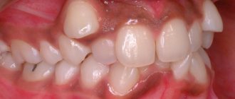
If the teeth cannot fit in the dentition (there is not enough space) due to a narrowing of the jaw or dentition, a discrepancy between the size of the jaw and the size of the teeth or the overall width of the dentition, they speak of a close position of the teeth, or their crowding. Crowded teeth (most often the front ones, sometimes the lateral ones) are in various abnormal positions and interfere with the patient’s appearance and speech.
Treatment consists of freeing up space by removing any teeth, widening the dentition and moving the teeth into the correct position. For this purpose, plates with screws, springs, arcs, Angle and Mershon devices are used.