Main symptoms and photos
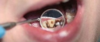
Such enamel damage can be recognized by the appearance of the teeth. Sometimes it is not even necessary to contact a specialist for this. The most characteristic clinical manifestation of the pathology is a change in the color of tooth enamel and the appearance of stains on its surface .
Sometimes changes affect the shape of the teeth. Grooves or simply small depressions may appear on the protective layer. Other symptoms depend on the form of hypoplasia.
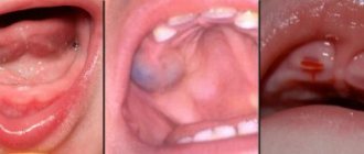
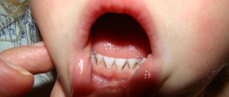
Photo: enamel hypoplasia of permanent teeth in a child
Photo: enamel hypoplasia of primary teeth
Causes of dental enamel hypoplasia in newborns
The disease can affect teeth even before they begin to erupt. This happens due to maternal health problems during pregnancy. The cause of the development of hypoplasia of the baby tooth buds of a child in the womb can be:
- severe toxicosis;
- toxoplasmosis;
- rubella;
- incompatibility of the blood of mother and baby according to the Rh factor.
Certain forms of hypoplasia occur for reasons that are not related to the gestation period:
- prematurity;
- congenital allergy;
- birth injuries;
- asphyxia during childbirth.
Typically, the disease affects those dental units that developed during the influence of a negative factor. Therefore, a specialist can determine at what week of gestation hypoplasia of the enamel of baby teeth appeared in a child.
What is hypoplasia?
- Enamel hypoplasia is a pathology, the main symptom of which is the partial absence of tooth enamel. In the early stages, it manifests itself in the form of pigment spots on the surface of the tooth, grooves, pits, and chips. The last stage of this disease is considered to be aplasia or the complete absence of enamel. In all cases, this pathology is congenital in nature and is a consequence of metabolic disorders in the embryo. Dental hypoplasia is a frequently diagnosed abnormality. Currently, about 40% of clinically healthy children suffer from this disease. The reverse process to this disease is enamel hyperplasia - the appearance of excess tooth tissue.
- The disease can be detected both in a preschooler who has only baby teeth, and in older children who have already developed molars. The disease can be identified during the first examination by a specialist. Based on the location of the spots, the dentist can determine at what point in the development of the fetus this disease began and what led to it.
- When hypoplasia of the enamel of baby teeth is diagnosed, the child is registered with a dentist, and in the future he will have to visit the doctor’s office several times a year and undergo prophylaxis. If the disease is detected in the early stages, it is possible to quickly cope with aesthetic defects, as well as prevent further development of the disease.
- Clinical manifestations of the disease depend on its stage and type. If metabolic processes have mild disturbances, there is mainly a change in the color of the enamel. They usually appear as single yellowish spots. Unlike carious formations, they do not cause discomfort and are not stained with food coloring. With deeper processes, the formation of grooves and depressions in the tissues is usually observed, and in the last stages of the disease the enamel layer may be absent altogether.
Causes of dental enamel hypoplasia in children and adults
The disease affects baby teeth, but the first signs of the disease can be noticed when the bite changes. Sometimes symptoms of the disease can only be detected on the molars in adulthood. This usually means that hypoplasia has affected the tooth enamel after birth. Most often, the development of the disease occurs in the first months of life. At this age, many factors can affect the body:
- poor metabolism;
- disturbances in the functioning of the endocrine system;
- problems with the stomach and intestines;
- insufficient bone mineralization;
- infectious diseases and their improper treatment;
- lack of protein in the diet;
- congenital syphilis.
If hypoplasia of tooth enamel appeared in a child older than one year, then the reasons for its development could be the following:
- disturbance of phosphorus-calcium metabolism;
- excess fluoride in water;
- Iron-deficiency anemia;
- severe form of diathesis;
- severe allergies;
- taking certain medications;
- injuries to teeth while they are still growing;
- heredity.
Many of these factors arise due to poor nutrition of the child. Therefore, parents who carefully monitor their baby’s diet are less likely to encounter enamel diseases.
Causes of the disease
Almost always, hypoplasia is a congenital disease, although it manifests itself after birth.
The rudiments of baby teeth are formed in the first three months of the prenatal period. At the same time they are mineralized. The presence of any provoking factors during this period practically guarantees the occurrence of enamel pathology in the baby.
Main intrauterine causes:
- gestosis during pregnancy, especially in the early stages;
- infectious diseases of the mother during pregnancy, such as measles, rubella, toxoplasmosis, severe forms of influenza;
- poor maternal nutrition during pregnancy, especially a lack of meat products in the diet;
- premature birth;
- traumatic injuries to the child during the birth process;
- hemolytic disease;
- Rhesus conflict;
- endocrine and renal pathologies in the mother.
Causes of the disease after birth:
- acute pathologies of the stomach and intestines;
- errors in the child’s nutrition in the first year of life, in particular, the use of low-quality formulas that contain insufficient amounts of vitamins and microelements;
- neurological abnormalities in the child;
- influence of background radiation or other toxic substances;
- improper calcium metabolism.
Types of tooth enamel hypoplasia
There are two main types of the disease: local and systemic. It is very easy to differentiate them. Local enamel hypoplasia is characterized by damage to only a few teeth, most often premolars. It manifests itself when negative factors affect the child already in adulthood. Such a disease cannot appear during fetal development. Therefore, the disease found on permanent teeth is most often of the local type.
Most often, the local form of the disease manifests itself in the form of pink spots. The shape of the teeth also suffers: depressions and grooves appear on their surface. All this negatively affects the overall sensitivity of the dentition and can lead to a number of negative consequences.
Systemic damage is different in that it spreads to all of the child’s teeth at once. It is the most common type of disease, since it forms either at a very young age or during fetal development. At this time, the enamel coating is more susceptible to negative effects.
Causes of development of enamel hypoplasia
Hypoplasia develops during the embryonic period, when the intrauterine development of the fetus is disrupted due to the impact of internal and external factors on the maternal body. Internal causes of the development of enamel hypoplasia include disturbances in the primary formation of germ layers.
External factors for the development of hypoplasia are:
- disturbances of metabolic processes in the fetal body during the period of tooth formation;
- infectious diseases suffered by the mother during pregnancy (toxoplasmosis, rubella, ARVI, etc.);
- toxicosis of pregnancy (early and late);
- congenital allergies and pathologies of the cardiovascular system;
- incorrect intrauterine position of the fetus;
- oligohydramnios;
- exposure to radiation, high temperatures and toxic substances;
- trauma (including birth trauma);
- mother's bad habits;
- illnesses of the mother during pregnancy, transmitted to the unborn child through the placenta (rubella, toxoplasmosis, influenza);
- prematurity;
- hemolytic disease;
- artificial feeding of a child;
- encephalopathy.
Hypoplasia of the enamel of primary teeth occurs when exposed to unfavorable factors both during pregnancy and in the early postpartum period. Hypoplasia of permanent teeth begins to develop if the child’s body is exposed to adverse causes in the first few years of his life. Also, predisposing factors for the development of enamel hypoplasia may be:
- severe forms of childhood infectious diseases;
- rickets;
- purulent diseases;
- pneumonia;
- allergies;
- severe forms of diathesis;
- diseases of the gastrointestinal tract, kidneys, parathyroid and thyroid glands;
- iron deficiency anemia.
In addition, hypoplasia of tooth enamel can be detected in children of preschool and primary school age. In this case, the disease is systemic in nature and damage to several teeth is more often observed. The development of dental hypoplasia largely depends on the age at which the child suffered the disease. Thus, enamel hypoplasia can manifest itself on milk and permanent teeth, which leads to an increased risk of dental diseases in older age.
Hypoplasia of the enamel coating of baby teeth is associated with metabolic processes in the child’s body, which develop in the sixth month of life. At the same time, the localization of tooth enamel hypoplasia depends on the age at which the child suffered the disease, which became the main pathogenetic link. So, if a child suffers from diseases in the first months of life, then they lead to hypoplasia of the enamel of the cutting edge of the central incisors and the cusps of the sixth teeth. During the formation of the second incisors and canines in the ninth month, diseases lead to hypoplasia of the enamel of the lateral incisors and the cutting edge of the canines. If the diseases cause profound changes in the child’s metabolism, then areas of enamel hypoplasia are observed along the entire length of the crown and on the surface of the tooth. The uneven structure of the enamel indicates the duration of diseases suffered during the period of tooth formation. If a child has suffered severe forms of illness, this also affects the depth of changes in the enamel.
Inflammatory processes in the area of the apex of the roots of primary teeth contribute to the occurrence of local enamel hypoplasia. This type of hypoplasia is more common on small molars, since their rudiments are located between the roots of primary molars. Hypoplasia of hard tissues of teeth, which are formed in the same period of time, is systemic, hypoplasia of a single tooth is positioned as local.
Forms of dental enamel hypoplasia
Systemic hypoplasia is divided into three forms, each of which is distinguished by a number of symptoms:
- superficial lesion;
- moderate enamel underdevelopment;
- severe systemic enamel hypoplasia (aplasia).
Each degree of pathology is characterized by certain symptoms. The superficial form of the disease manifests itself as a change in the color of the upper tooth tissues. Small, sometimes completely invisible spots of yellow and white shades appear on it. They have a clear boundary that will not change throughout their lives. Affected areas form not only on the visible part of the tooth, but also on the back.
Hypoplasia of moderate severity is sometimes called spotty, and this is a more serious violation of the enamel structure. With this pathology, the relief of the tooth changes noticeably. Small depressions in the form of dots appear across the entire surface of the enamel. Their color can be very dark, which is why the patchy form of enamel hypoplasia is often confused with caries.
Less commonly, small compactions called ridges appear on the teeth. The middle stage of development of the disease can also be grooved, then depressions in the form of clear stripes appear on the enamel. One gets the impression that the teeth in this place are worn away.
Aplasia is an extremely severe stage of the disease. With it, areas appear on the teeth where there is no enamel at all. The degree of sensitivity increases significantly. A person experiences discomfort and pain under the influence of almost every irritant.
Aplasia occurs only in the complete absence of treatment. In this case, the patient may encounter other complications.
Based on the shape of the spots on the enamel, hypoplasia is classified into the following types:
- spotted;
- erosive;
- grooved;
- wavy;
- aplastic.
Types of disease
Hypoplasia of dental enamel has a broad classification depending on the extent of damage, genetic predisposition, involvement of hard tissues, clinical picture, and period of development.
Based on the clinical picture of the lesion, the following types of disease are distinguished:
- Erosive – the damage is deep and cup-shaped.
- Spotted - the enamel is covered with flat spots of a characteristic size and contour.
- Grooved - there are linear indentations located horizontally relative to the upper edge.
Based on the involvement of hard tissues, they are divided into:
- hypoplasia of the entire tooth;
- damage to the enamel (this pathology accounts for about 50% of cases of defects detected in adolescents and mature people).
Based on the presence of a genetic predisposition to the occurrence of the disease, the following are distinguished:
- hereditary;
- acquired (obtained during fetal development, during childbirth or in the first months of life).
Regarding the period of development, hypoplasia of the enamel of milk and molar teeth differs.
There are several types of non-carious lesions (hypoplasia) according to the prevalence of the lesion, especially highlighted by doctors:
- systemic – in which almost the entire row is damaged;
- localized – 1-2 teeth are affected, the disease does not spread to others;
- aplasia – with it there is a complete absence of enamel on several teeth.
Systemic and localized (local) dental hypoplasia occurs more often in people, so we need to talk about these types in more detail.
Systemic hypoplasia
Systemic enamel hypoplasia is a lesion of hard and soft tissues of teeth that form at the same time. Has three stages:
- Change in enamel color.
- Underdevelopment of enamel.
- Complete absence of enamel.
A type of systemic enamel hypoplasia is:
- Pflueger teeth: have not fully developed cusps, which is why the tooth can take on a cone shape. Also, an indicator of this particular form of hypoplasia is the larger size of the crown at the cheek than at the chewing surface.
- Hutchinson's teeth: the main feature of this disease is the barrel-shaped shape of the front incisors, which have a thicker neck than the cutting surface. Another important characteristic of this type of pathology is the presence of a crescent-shaped depression near the cutting edge.
- Fournier's teeth are similar in shape to Hutchinson's pathology, however, in this case, hypoplasia does not provide for the presence of a crescent-shaped notch.
Another form of systemic hypoplasia is tetracycline teeth in children. This pathology can be triggered by taking drugs containing tetracycline during pregnancy, as well as in the first months of the child’s life. It gives the teeth a yellow, sometimes brown color, most often appears on the incisors and can have a heterogeneous color and structure, most often lying down in stripes. Such pigmented teeth cannot be whitened in the future. The intensity of the color, its color, as well as the type directly depend on the doses and when exactly the pregnant woman or already born child was prescribed to take such a drug. Knowing this feature, it is advisable to avoid taking substances containing tetracycline during pregnancy.
Local hypoplasia
This type of disease is often acquired and often manifests itself in the form of small spots on the teeth or shallow grooves.
Local dental hypoplasia occurs more often as a result of injuries to the primordia of molars and rarely has a genetic predisposition. Most often the premolars (4th teeth) of children are affected. As in the case of systemic hypoplasia, with local hypoplasia, not only partial damage to the enamel can be observed, but also its complete absence. This form, however, is rare.
Consequences of the disease
The first stage of hypoplasia of primary teeth seems completely harmless, but if the disease is not treated, it leads to terrifying consequences:
- exposure of dentin – the next layer of the tooth after the enamel;
- serious carious lesions;
- malocclusion;
- problems with the gastrointestinal tract due to malnutrition.
All this sounds daunting, but often they forget to include the main consequence in this list. We are talking about embarrassment and complexes. Smiling for a photo or laughing in company becomes a real challenge. Problems with self-esteem in childhood will continue to manifest themselves in adulthood, so diagnostic and treatment measures must be taken seriously.
results
Patients with left-sided hemihypoplasia accounted for 61% (41 people), with right-sided hemihypoplasia - 39% (26).
Examination of the general neurological status did not reveal any serious abnormalities.
Based on the results of the analysis of the questionnaire and study design to identify signs of autonomic disorders, the presence of autonomic dystonia syndrome was found in 63 (94%) patients in the main group and 11 (31.4%) in the control group ( p
<0.001). The syndrome of vegetative dystonia in the main group was observed mainly of the hypotonic type, less often of the mixed type. Moreover, according to the anamnesis, 49 (73%) patients with hemihypoplasia had already been previously observed with this diagnosis by a neurologist.
The most common complaints in patients with hemihypoplasia identified when using the questionnaire were: tendency to faint (94%), numbness (coldness) of the fingers and toes (60%), fatigue (60%), sleep disturbances (60%), gastrointestinal tract functions (52%), paroxysmal headaches (52%). According to the study design, the following symptoms were most often found: deterioration of well-being when the weather changes (94%), hyperventilation syndrome (94%), the presence of vegetative-vascular crises, migraines, tendency to faint (94%), astheno-neurotic syndrome (75%).
According to the results of a study of the Kerdo index, parasympathicotonic regulation predominated in patients with hemihypoplasia (70.1%). An increase in the tone of the parasympathetic nervous system was confirmed by the presence of persistent diffuse dermographism for more than 10 minutes in 65.7% of patients. When performing a cold test, normal autonomic reactivity was noted in 12 (17.91%) patients, decreased (weak sympathetic reaction) - in 23 (34.33%), perverted (parasympathetic reaction) - in 32 (47.76%). An assessment of the oculocardiac reflex revealed normal autonomic reactivity in 32 (47.8%) patients, decreased in 20 (30%), perverted in 15 (22.2%). The results of the study of the solar reflex showed normal autonomic reactivity in 28 (41.8%) subjects and decreased (pulse is not slow enough) in 29 (58.2%). These data indicated a tendency towards a decrease in autonomic reactivity and a predominance of asympathicotonic reactions, which indicated unsatisfactory adaptation, manifested by the fact that in stressful situations, as a rule, syncope, dizziness, and poor transport tolerance were noted.
According to the results of the orthoclinostatic test, in patients with hemihypoplasia, the asympathicotonic variant of insufficient activity support was significantly more likely to prevail (55%). This indicated insufficient activity of the sympathoadrenal system. However, in 45% of patients, autonomic support of activity was within normal limits, which could be considered as a fact of preservation of adaptive mechanisms and reversibility of the autonomic dystonia syndrome under the influence of corrective measures.
According to the results of a study of the autonomic nervous system using electropuncture diagnostics using the NPO “Eurasia” method, a predominance of yin potentials was revealed in 91.04% of the subjects of the main group, which confirms the data of autonomic tests on the dominance of the parasympathetic nervous system. According to the data of electropuncture diagnostics in patients with hemihypoplasia, the average value of the asymmetry of electrical conductivity on the sides was 15.6 ± 0.88 a.u., which significantly exceeded the similar indicator in the control group (7.4 ± 0.4 a.u. p
<0.001). In 52 (77.61%) patients with hemihypoplasia, there was a predominance of electrical conductivity of biologically active points on the side opposite to hemihypoplasia, which indirectly indicates a decrease in the tone of the sympathetic nervous system on the side of hemihypoplasia and the presence of asymmetry between the right and left parts of the autonomic nervous system.
A difference in the amount of electrical conductivity of biologically active points on the arms and legs was established, with a predominance of electrical conductivity of points below the diaphragm in 95.52% of those examined in the main group and 45.71% in the control group ( p
<0.001). Also, in patients of the main group, there was a predominance of electrical conductivity indicators of yin meridians in 91.04% of cases compared to 34.28% in the control group. At the same time, the electrical conductivity indicators of the Yang meridians prevailed in 65.72% of those examined in the control group and only in 8.96% of patients in the main group.
Unequal length of the upper limbs with hemihypoplasia is not accompanied by functional disorders, while shortening of the lower limb leads to the development of an asymmetrical defect in posture. This can explain the detected difference in the amounts of electrical conductivity.
Among the most interested meridians in the main group, the BL (bladder) and SI (small intestine) meridians were most often noted. It is known that the acupuncture points of these meridians are used to influence the so-called miraculous meridian dumai, which is a circuit of biologically active action in the presence of a chronic process associated with pathology primarily of the spine and back muscles [10]. The interest in these meridians can be explained by the presence of an asymmetrical defect in posture against the background of uneven leg heights. In the main group, the LR (liver) meridian and its paired GB (gallbladder) meridian were most often affected. These meridians, according to the provisions of oriental medicine, are interested in the disruption of functional processes associated with a certain periodicity. These include dysregulation of the autonomic nervous system, dyskinesia of the gallbladder, and disturbances in the rhythm of sleep and wakefulness.
Based on the difference in temperature on the right and left on thermograms in patients with hemihypoplasia, the presence of asymmetry in the microcirculation of the upper and lower extremities was assessed. According to thermography results, the temperature gradient between the right and left extremities was at least 0.5 °C, and was most pronounced in the distal parts of the extremities. So, on the hands it was 0.94±0.17 °C, on the feet - 1.5±0.29 °C. Thermal asymmetry had no correlation with the side of shortening ( p
>0,05).
Using ultrasound, we compared the lumen diameter of the paired arteries on the left and right, and also studied the asymmetry of the resistance index. Normally, asymmetry of the lumen of paired arteries is allowed up to 20%, pathological asymmetry is more than 30% [11]. All arterial lumen diameter sizes were within the age norm. The lumen diameter of the vertebral and anterior tibial arteries did not differ in patients with left-sided hemihypoplasia. In other cases, the diameter on the hemihypoplasic side was significantly smaller than on the intact side ( p
<0.05). However, the asymmetry of the lumen of the vessels did not exceed acceptable limits. The most significant difference in the diameters of paired arteries was observed in patients with right-sided hemihypoplasia when comparing the diameters of the anterior tibial, ulnar and radial arteries, but even in this case the magnitude of the asymmetry did not reach pathological.
The RI values for the right and left arteries were also compared, which made it possible to judge the value of peripheral vascular resistance and was calculated using the formula: RI=(Vmax–Vmin)/Vmax, where Vmax is the maximum and Vmin is the minimum linear blood flow velocity. According to the results of the study, the asymmetry of RI resistance between paired arteries on the right and left did not exceed 3%.
Differential diagnosis of dental enamel hypoplasia
The main method for identifying enamel hypoplasia is differential diagnosis. It consists in the fact that a specialist, upon seeing the first manifestations of dental damage, first excludes all other possible causes. This method is effective because the early symptoms of the disease are similar to those of other oral diseases. Such spots can occur due to caries or due to taking certain medications.
To diagnose hypoplasia directly, a specialist needs to do the following:
- Conduct a complete and thorough oral examination.
- Determine at what age the first symptoms appeared.
- Get acquainted with all the information about the course of pregnancy.
- Study the lifestyle of the child and parents.
All this requires effort and time. Therefore, it is easier to carry out a differential diagnosis and first exclude other diseases with similar symptoms.
Treatment of dental enamel hypoplasia
Treatment of enamel hypoplasia is simply impossible. You cannot take any pills or perform a procedure that would get rid of this disease. Instead, attention is paid to preventing further development of the disease.
If the differential diagnosis revealed hypoplasia at the initial stage of development, the specialist gives the patient recommendations on dental care. To prevent the enamel from deteriorating further, it is very important to clean it from plaque in a timely manner, regularly strengthen it and have it professionally cleaned. For this, in-office procedures are used, as well as special pastes and gels.
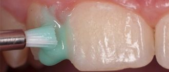
If the enamel has already become spotted, aesthetic dentistry is included in the therapeutic course. If there are depressions, composite compounds are used, which a specialist uses to level the enamel surface. The same material is used when there are rollers. Only first, the dentist removes them, and composites are used to restore the protective function of the enamel.
With aplasia, more serious measures are required. Without a protective layer, the tooth cannot perform its functions, so external correction will not solve the problem - crowns are installed on such teeth. Since after prosthetics the same dental procedure will have to be carried out throughout your life, this method is used only in the most advanced cases.
Prevention
Since complete treatment and recovery are impossible for dental enamel hypoplasia in children, more attention should be paid to the prevention of the disease. It can be divided into two stages: during pregnancy and after birth.
While carrying a baby, the mother must follow these rules:
- Healthy food;
- be careful when taking any medications (this can only be done after consultation with a specialist);
- Always complete treatment for any diseases that arise during pregnancy.
All these measures are necessary to prevent systemic enamel hypoplasia. To prevent local damage, the following is required:
- proper balanced nutrition for the child;
- teaching oral hygiene from a very early age;
- regular visits to the dentist starting from 2 years of age;
- avoiding dental injuries and timely treatment when they occur;
- complete treatment of any disease, especially acute forms of chronic ailments.
In 40% of cases, completely healthy people suffer from hypoplasia. Therefore, you should not think that in the absence of risk factors you will not encounter such a diagnosis. Dental health should always be treated with special attention.
Clinical manifestations of enamel hypoplasia
In dental practice, there are three forms of systemic enamel hypoplasia:
- mild degree (color change);
- severe form of enamel hypoplasia (underdevelopment);
- aplasia (absence).
A weak form of enamel hypoplasia most often manifests itself in the form of white, less often yellowish spots of the same size, with clear boundaries on the teeth of the same name. The spots form on the vestibular surface and do not cause any discomfort. When a stain appears, the outer layer of enamel is not painted with dyes. During a certain period of child development, the size, shape and color of the spot practically do not change. The thickness of the enamel in the area of the stain is the same as in the area of intact enamel next to it.
A severe form of enamel hypoplasia is expressed by its underdevelopment, which manifests itself in the form of wavy, dotted and grooved enamel.
Wavy enamel is revealed when the surface of the crown dries. In this case, small ridges are observed on the tooth enamel, between which there are depressions covered with unchanged enamel.
Pinpoint depressions in the enamel with hypoplasia are located on the vestibular and lingual surfaces at different levels in teeth of different groups. In this case, the enamel in the places of the depressions gradually becomes pigmented, but remains dense and smooth. The number of cellular elements in the pulp decreases.
With grooved enamel, several grooves are observed on the teeth, which alternate with unchanged tooth tissues. Rarely, grooves are present along the entire height of the crown of the teeth of some groups. This form is called scalene hypoplasia. It is characteristic that even with the grooved and scalariform forms of hypoplasia, the integrity of the enamel is not compromised.
Aplasia manifests itself in a specific area. With this form of hypoplasia, pain may occur when exposed to irritants. It is manifested by the absence of enamel on part of the crown, but more often appears at the bottom in the form of a cup-shaped depression or in a groove covering the crown of the tooth.
In addition to these varieties, enamel hypoplasia has a number of changes in the shape of teeth, namely:
- Hutchinson's teeth. With this pathology, the upper central incisors have a screwdriver and barrel-shaped crown. Their size at the neck is larger than at the cutting edge. In this case, the semilunar recess may be covered with enamel, but sometimes enamel is observed only at the corners of the tooth, and in the middle part the dentin is not covered with enamel;
- Fournier's teeth. They have central incisors with a screwdriver-shaped crown, the same as Hutchinson’s teeth, but there is no semilunar notch along the cutting edge;
- Pflueger's teeth. With this form of hypoplasia, the first large molars (molars) are affected. The size of the crown near the neck is larger than that of the chewing surface, and the tubercles are underdeveloped, which gives the tooth the appearance of a cone.
Hypoplasia of permanent teeth can be local (affects one tooth) and systemic (the pathological process affects the hard tissues of several teeth at the same time). All these types and forms of dental enamel hypoplasia lead to the fact that microbes act more aggressively on dentin, penetrating into it and causing deep caries. Since enamel hypoplasia rarely occurs in isolation, the subsequent serious complication is damage to other tooth tissues (cementum, dentin and pulp). Many children subsequently develop malocclusion.