Article:
The tongue is a small muscular organ of the human body.
The tongue is located at the bottom (lower wall) of the oral cavity and, with the teeth closed, almost completely fills it, while in contact with the hard palate, gums, and teeth. Anomalies of the tongue can be either congenital or acquired; they are often the result of certain pathological processes occurring in the body. Currently, malformations of the main speaking organ are quite rare. Many of them are serious problems that often require surgery.
- Genetic diseases . May be inherited or develop as a result of new mutations, diseases such as: Beckwith-Wiedemann syndrome, Down syndrome, Sotos syndrome, hereditary mucopolysaccharidosis
- Exogenous factors . This group of reasons includes exposure of the fetus to ionizing radiation, carcinogens, chemicals, radioactive radiation, and pesticides in food products consumed by a pregnant woman. The development of maxillofacial anomalies is facilitated by bad habits of parents (drinking alcohol, drugs, smoking), and the expectant mother taking medications with teratogenic effects (this is a drug composition that is prohibited during pregnancy).
- Endogenous factors . Include infectious and metabolic diseases of the pregnant woman: trichomoniasis, toxoplasmosis, viral infections, hypovitaminosis and hypervitaminosis A, hypothyroidism, etc. The number of congenital craniofacial dysmorphias increases in proportion to the mother’s age at the time of conception.
A generally accepted classification of tongue defects has not been developed.
Based on the main criteria (size, structure, method of attachment), anomalies can be divided into the following groups:
- Anomalies of size - macroglossia (hypertrophied tongue), microglossia (reduced tongue).
- Anomalies of structure/shape - aplasia and aglossia (absence of tongue), cleft tongue (double. Cleft tongue can be complete or incomplete.
- Anomalies of attachment - ankyloglossia. This anomaly limits the mobility of the tongue in the oral cavity, and, consequently, its functions.
- Combined anomalies – folded (furrowed, scrotal) tongue. Characterized by the presence of folds and grooves on the surface of the organ
Macroglossia - massive tongue. Such a tongue does not fit in the oral cavity and literally falls out of the mouth. The child develops an incorrect bite due to constant pressure on the dentition. Teeth imprints are visible on the lateral surfaces of the tongue. Due to the constantly open mouth, the mucous membrane dries out, which causes discomfort. This is also a big disadvantage for correct sound pronunciation and food intake. A dental surgeon comes to the rescue again; he performs an operation to reduce the muscle mass of the tongue, that is, plastic surgery.
Microglossia is characterized by underdevelopment and reduction in the size of the tongue. Depending on the severity of the defect, a disturbance in sucking occurs, and subsequently in speech. There are cases when the anomaly is asymptomatic. The small tongue is not able to take the desired articulatory position: the amplitude of its movements is too small. In the case of microglosia, a pediatric dental surgeon performs tongue plastic surgery. At what age? Only the doctor can decide this. After the operation there is a rehabilitation period and sessions with a speech therapist.
Aglossia is a rare anomaly in which the tongue is completely absent. With aglossia - the complete absence of an organ - the baby cannot suck, so this rare anomaly requires tube feeding
Ankyloglossia is a congenital anomaly that is discovered shortly after birth. It is characterized by partial or complete shortening of the lingual frenulum. The average length of the frenulum is 1.5 cm. Deviations from the norm (shortened or very short): 1. The frenulum is thin, almost transparent, but prevents the rise of the tongue. 2. The bridle is thin . Its anterior edge is attached close to the tip of the tongue. Because of this, when the tongue is raised upward, the tip bifurcates into a “heart”. 3. The bridle is a dense short cord. It is attached close to the tip of the tongue. The upward movement of the tongue is limited. When you try to stick your tongue out of your mouth, its tip curls up and the back of the tongue “bulges out.” 4. A dense short cord of the frenulum is tightly fused with the muscles of the tongue: attached almost to the tip of the tongue. All movements of the tongue are sharply limited. 5 . It is difficult to understand where the frenulum is and where the tongue is, they are a single whole . It seems that the tongue has fused with the bottom of the oral cavity. With such a language, it’s not only impossible to speak, it’s impossible to eat!
All these deviations from the norm in the structure of the hyoid frenulum will at first complicate the process of feeding the child, and subsequently will prevent the timely appearance of not only the sounds [P] - [P'], but also the whistling sounds [S], [Z], [C]; hissing [Sh], [F], [H], [Sh]; sonors [Y], [L], [L'], back-lingual [K], [G], [X].
Parents for the most part do not like to listen to arguments in favor of surgery on the hyoid frenulum. They rely on exercises that supposedly stretch the frenulum. A similar misconception is noted among speech therapists. We would like to disappoint you: the frenulum does not stretch during speech therapy work, although with the help of special articulation exercises you can significantly increase the mobility of the tongue, due to which the impression is created that something has “stretched” somewhere. In fact, the time for producing sounds is extended, wasting the energy of both the child and the speech therapist.
What else can a child’s tongue “tell” about?
Recently, children with “geographical language” have become increasingly common. The surface of such a tongue is covered with streaks of white, brown and pink colors, with outlines reminiscent of a map of the continents, hence the name. At the same time, the mucous membrane of the tongue is shiny, as if polished, and taste buds are faintly visible on it. This is the state of the tongue to talk about the need to visit an allergist and gastroenterologist . A visit to a speech therapist is also required. As a rule, the sensitivity of such a tongue and mobility are significantly limited, so it is necessary to carry out articulatory gymnastics and the production of certain sounds.
Sometimes the surface of the tongue is dotted with deep grooves “Folded tongue”, like wrinkles. A sort of old tongue. He is painfully dry and inactive, which negatively affects sound pronunciation. A similar feature occurs when there is a disruption in the endocrine system, so do not put off a visit to the endocrinologist.
Attention should also be drawn to the child’s too pale, loose and flaccid tongue. In such a language, muscle tone is reduced (hypotonicity), its movements are slow and unclear, and it is sometimes impossible for him to take the necessary articulatory posture to pronounce a particular sound. Hence the errors in sound pronunciation: “porridge in the mouth.”
If the tongue is too tense, which is why its back is upturned, like a “hump,” and in the protruded position the tongue turns blue, the tip of the tongue deviates to the side, there is hypertonicity. In both the first and second cases, the child should be shown to a pediatric neurologist; perhaps such phenomena are a consequence of birth trauma, encephalopathy or some other diseases.
Conclusion.
Language is a map of internal problems.
The noted anomalies in the structure of the articulatory organs have different effects on sound pronunciation.
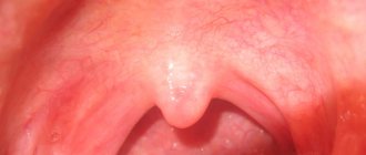
Congenital anomalies of the pharynx
With congenital choanal atresia, it is also possible to close the mouth of the auditory tube. This developmental anomaly is usually accompanied by a high palate and a short uvula, often fused to the posterior wall of the pharynx. The most common anomalies include congenital deformity of the upper lip (“cleft lip”). This is the result of non-closure of the nasal sulcus (the space between the middle nasal and maxillary processes in the embryo. Unilateral clefts are more often observed on the left and are more common than bilateral ones. Usually, simultaneously with the “cleft lip”, a cleft is formed between the lateral incisor and the canine, which can be limited to the edge of the alveolar process or spread on the hard and soft palate. This pathology has the following clinical manifestations. Dysphagia syndrome causes choking and reflux of food into the nasal cavity when swallowing, a pronounced nasal sound subsequently leads to impaired speech formation. Difficulties arise when the child sucks the breast. Usually, when sucking, the soft palate drops and closes the oral cavity from behind, and from the front the oral cavity is closed by the action of the so-called orbicularis oris, which lengthens the child’s lips covering the nipple. With a “cleft lip,” the integrity of the so-called orbicularis oris is disrupted, sucking becomes difficult or impossible. Children are fed with a spoon or with the help of a zoid . Aspiration syndrome leads to the development of recurrent pneumonia. During puberty, the ectopic part of the thyroid gland in the area of the tongue root can cause impaired swallowing (dysphagia) and breathing (stenosis). The child may develop articulation abnormalities due to protrusion of the upper (prognathism) or lower (progenial) jaw. Reasons affecting developmental anomalies of the dental system include impaired nasal breathing. Congenital cysts and fistulas of the neck are one of the most common congenital anomalies. A painless round swelling of the appropriate localization gradually develops. The cyst often becomes inflamed. Cysts are more noticeable when examined by swelling on the neck. When the cyst suppurates, median fistulas of the neck are formed, which are also closely connected with the periosteum of the hyoid bone, sometimes ending blindly in it or directed to the blind opening of the tongue. Lateral cysts are localized at the anterior edge of the sternocleidomastoid muscle, have a round shape, and a soft-elastic consistency. Large cysts can make swallowing difficult and limit the mobility of the larynx. The external mouth of the lateral neck fistula is located at the base of the sternocleidomastoid muscle, the internal one is in the pharynx, at the base of the tonsil or in the tonsil fossa. The walls of swish consist of dense fibrous tissue and are lined with stratified squamous epithelium.
Diagnosis of common diseases
The tongue is an organ capable of displaying any deviations in the functioning of internal organs, so it was often used for diagnosis hundreds of years ago.
In case of general pathologies, first of all, the shade of the tongue changes and a plaque appears.
Each organ has its own characteristics of the manifestation of these symptoms:
- Gastrointestinal diseases are characterized by the appearance of white plaque. By its shade one can judge the degree of progression of the pathology. The darker it is, the more advanced the disease. With oncological damage to the gastrointestinal tract, the tongue becomes covered with a dense gray coating;
- disruption of the gallbladder is indicated by a yellowish plaque. If the problem is accompanied by a violation of the patency of the bile ducts, then it acquires a greenish tint;
- With pathology of the pancreas, the plaque becomes black. The same effect is observed with dehydration;
- purulent processes in the abdominal cavity can be diagnosed by the cyanosis of the deposits.
What changes in the shape, surface and color of the tongue may indicate the presence of general diseases of the body, watch the video:
Simultaneously with the appearance of plaque, there is an unpleasant odor from the mouth, which is not relieved by freshening agents and frequent brushing of teeth.
If you swallow your tongue, what happens?
What happens if you swallow your tongue? Yes, in principle, it’s okay, since it’s impossible to swallow your own tongue. It is possible that if a person is unconscious, their airway may be blocked for a short time as the tongue muscle weakens and retracts into the back of the larynx. It only takes him a few seconds to return to his original position. Helping the tongue stay in place is a small “piece” of tissue located directly underneath it, called the frenulum. It is she who does not allow us to swallow our own tongue.
A little history - is it possible to swallow a tongue?
People have been afraid of swallowing their tongue since the late 19th century, when during first aid training there were specific instructions that when a person loses consciousness or has a seizure, it is necessary to grab the tongue with forceps and pull it forward, or However, if there are no tongs, then do it directly with your fingers. Many people even now stick everything they can get their hands on into the mouth of the person having seizures. Not the best idea, by the way, since this makes the victim unable to breathe. But the one who saves has only the best, albeit erroneous, motives. If you see someone faint, you shouldn’t immediately put everything you can find nearby into his mouth. You just need to lay the victim on his side and provide him with freedom of breathing. And to do this you need to raise your chin.
Throughout the day, a person swallows about 2 thousand times. Those cases when he does this deliberately do not count. Swallowing is an automatic process and consists of 12 separate muscle movements. During paralysis and Alzheimer's disease, a person may sometimes lose the ability to swallow. In this case, speech therapists teach him this again. Why speech therapists? The fact is that swallowing uses the same muscles and in the same combination as speech.
When near death, a person’s swallowing reflex often turns off. As a result, saliva and mucus accumulate in the throat, which causes the so-called. "death rattle". But before you write him off, you need to check whether his airways are clear.
What is tongue retraction?
Retraction of the tongue should be understood as a displacement of the tongue root to the back of the pharynx, which prevents the penetration of air into the larynx. This can happen during anesthesia, coma, shock. To restore airway patency, the patient's head should be tilted back as far as possible. In this case, one palm should be on the patient’s forehead, and the other under his neck. If there is no effect from ventilation of the lungs, then the lower jaw should be moved forward so as to open the mouth. In this position, moving the root of the tongue and epiglottis forward allows you to open the larynx and ensure free access of air into the trachea.
When the tongue sinks, you should turn the victim’s head to the side and hang it slightly off the bed, pushing the lower jaw forward. The tongue must be removed using a tongue holder, and air ducts are used to hold it (sometimes oral and nasal). To hold the lower jaw, you should use the following techniques:
- standing behind the victim, place your thumbs on the lower jaw on both sides of the midline and pull it down until its incisors are in front of the incisors of the upper jaw;
- using the index and middle fingers, push the lower jaw forward beyond the corners and hold it in this position;
- rest your fingers on the mastoid processes of the temporal bone. It should not be soft tissue of the neck, so as not to compress the jugular vein or facial nerve.
Causes of mechanical asphyxia
It can occur due to foreign bodies entering the respiratory tract. And this could be: vomit, tampons, mucus, blood, dentures. It also occurs when the tongue retracts.
During mechanical asphyxia, the patient must be:
- laid on your back on a hard surface;
- His head should be turned to the side;
- The first and second fingers of the left hand are crossed;
- The mouth is open;
- The oral cavity is cleaned with a gauze napkin, which must be wrapped around the second or third finger of the right hand.
- Next, the head should be turned straight and thrown back as much as possible.
In this case, one hand should be placed under the neck, the other should be placed on the forehead and the head should be fixed back, which helps restore patency of the airways. The lower jaw must be moved forward while performing these actions.