The uterus is a hollow organ consisting of a smooth muscle elastic layer that can stretch. The shape of the organ resembles a pear, with the wide part upside down.
Normal weight does not exceed 80 g (during the childbearing period).
The uterine fundus is located on top, and the lower part ends with the cervix.
The uterus is the main organ of the female reproductive system. The main function is the mechanical protection of the fetus and the maintenance of its vital functions from the 5th day from conception until the day of birth.
Abnormal development of the genital organs has a congenital etiology. Formed during embryogenesis.
Diseases account for about 65% of uterine congenital pathologies.
The most common pathology in the structure of the organ is a bicornuate uterus.
Classification of bicornuate uterus
- Saddle-shaped - from the name it is clear that the inner part resembles a saddle due to the depression in the bottom and the intercourse of the uterine horns. This pathology accounts for about 1% of reproductive anomalies. In the cross section there is a significant expansion of the cavity. The uterine fundus is not completely separated.
- Incomplete bicornuate uterus - the formation of “horns” is observed in the upper part of the organ, covering the area above the middle.
- A complete bicornuate uterus - the uterine cavity is divided by a septum from the sacrouterine fold to its bottom. The horns are directed in opposite directions at an angle.
Saddle uterus is a pathological development of the organ. It causes miscarriage and inability to conceive properly.
Causes of saddle uterus
The formation of the organ begins from 12-14 weeks of embryogenesis.
The paramesonephric ducts join to form two uterovaginal cavities separated by a sagittal septum.
As the fetus grows, the organ undergoes changes. It stretches and an isthmus forms, which subsequently separates the vagina from the uterus.
The sagittal septum resolves and one muscular cavity is formed.
If a malfunction occurs during the process of intrauterine formation, the result is abnormal development of the organ. The uterine septum does not completely dissolve or remains unchanged.
The reason for the formation of the saddle-shaped uterus is intratubal pathogenesis at the end of the gestation period.
Factors causing failure in the process of formation of the female organ:
- unhealthy lifestyle (alcohol abuse, smoking, substance abuse, drug addiction);
- hypovitaminosis (for example, lack of vitamin B9);
- infectious diseases: rubella, toxoplasmosis, herpes, influenza, cytomegalovirus (all diseases are identified during pregnancy and childbirth registration);
- cardiac pathologies causing a hypoxic state of the fetus;
- diseases associated with endocrine dysfunction (thyrotoxicosis, diabetes mellitus);
- late toxicosis;
- gestosis;
- hereditary factor.
It is possible to suspect a saddle uterus in two cases: by ultrasound in the first weeks of pregnancy and at the planning stage.
The disease has no clinical symptoms.
Fixation Features
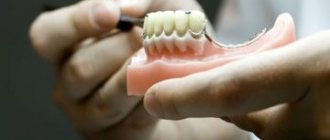
Orthopedic structures made of plastic are most often installed as a temporary product while the main prosthesis is being manufactured.
This is due to the fact that the service life of a plastic prosthesis, even with proper care, does not exceed 2 years. In addition, they are fragile and often break.
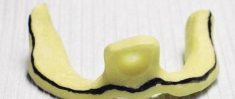
Test fixation of bridges can be done without a centering compound (a mixture of petroleum jelly and zinc oxide is used). In this case, it is recommended not to remove the bridge for several hours.
If the bridge does not fit, silicone cement is used. It does not completely harden within 24 hours. During this time, the quality of the bridge fit can improve significantly. Otherwise, the prosthesis is placed on weak cement, which is diluted with a modifying paste.
In this case, the structure remains in the oral cavity for several days (and sometimes up to several weeks). In addition to improving the fit of the prosthesis, such actions help the patient get used to the appearance of the dentition , and allow him to get used to new sensations in the oral cavity.
Also at this stage, all the problems that arise when the teeth are closed appear, which are easily eliminated before complete fixation is performed.
Before installation on a permanent adhesive, the orthopedic structure is thoroughly cleaned, and the supporting elements are treated with an antiseptic.
It is important to know! Temporary crowns are removed using special means that eliminate the adhesive effect of the composition.
Reasons for the appearance of odor from under the crown and ways to solve the problem.
In this publication, we will look at the best methods of prosthetics for periodontal disease.
Here https://www.vash-dentist.ru/protezirovanie/nesemnyie-p/problem-s-zubami-v-heyhe.html we will tell you how much it costs to insert teeth in Heihe.
Diagnostics
To clarify the diagnosis, the following diagnostic methods are used:
- Hysteroscopy. This method is intended for examining the uterine cavity using a hysteroscope - an optical device that transmits an image to a monitor. Hysteroscopy is performed for a number of indications, as well as to assess the possibility of conception.
- Ultrasound examination to determine the thickness of the myometrium. During an ultrasound examination, the doctor can suggest a pathological structure of the organ. This can be unequivocally stated in case of severe deformation of the uterine fundus and thickening of the myometrium.
- Hysterography is a form of ultrasound examination. A special contrast agent is injected into the uterine cavity, which changes color as it passes through, which is recorded on the monitors. This method allows you to identify the structure of the organ and is carried out under the control of an ultrasound machine.
- MRI is an innovation throughout medicine. The magnetic resonance imaging method makes it possible to evaluate not only the structure of the organ, but also to identify concomitant diseases and neoplasms (if any).
During a routine gynecological examination, it is impossible to suspect the disease.
Care and service life
Restored dentition using a plastic bridge requires hygienic care, in which the following rules and recommendations must be observed:
- To avoid damaging the integrity of the product, you should avoid eating food at different temperatures. Exclude foods with viscous consistency from your diet.
- You should not eat food that has a coloring effect, as in this case the denture will change color and will be noticeable in the oral cavity.
- Movements when cleaning the dentition should occur from the gums to the chewing surface.
- Hygienic brushing of teeth should be carried out after every meal. If this is not possible, use floss or dental rinse.
- A month after the installation of the bridge, undergo an examination by a dentist.
The warranty on plastic products does not exceed 2 years. With proper care and timely preventive inspection (every six months), the structure can last longer.
But at the same time, it will lose its aesthetic appearance due to the ability of plastic to darken.
It should also be borne in mind that reducing the chewing load will help increase the service life of the bridge.
Is pregnancy possible and how does this affect the fetus?
When a diagnosis of “saddle uterus configuration” is made, the likelihood of conception decreases. However, this type of organ structure is not always the cause of infertility.
According to statistics, the physical structure of the organ (with a saddle shape) does not affect the ability of a fertilized egg to implant in the uterine cavity.
This process may be disrupted due to other urogenital diseases.
There is a high probability of conceiving and giving birth to a healthy child.
The fetus, protected by the uterus and its placental barrier, does not experience discomfort due to the pathological structure of the organ.
The shape of the organ does not affect physical and mental development. The danger lies in the possibility of developing uterine hypertonicity and termination of pregnancy caused by a number of reproductive pathologies.
In this case, the sciatic shape of the uterus is an aggravating circumstance.
Splinting clasp prosthesis with clasps.
Clasp fixation is used for well-defined teeth and fairly high clinical crowns, as well as when it is necessary to splint teeth with poor mobility (a multi-link clasp is introduced into the design of the prosthesis).
Advantages: no need to prepare your own teeth and less load on the supporting teeth compared to locking fasteners.
Disadvantages: aesthetics, not very reliable fixation.
Poses for conception with a saddle uterus
The altered shape does not prevent male reproductive cells from entering the uterine cavity, so there are no specific positions for conception with this configuration.
Recommended positions for conception, regardless of the structure of the uterus:
- “Partner from below” pose,
- doggy style pose.
During sexual intercourse in these positions, sperm rush into the uterine cavity, and from there into the fallopian tubes to fertilize the egg.
The sciatic shape does not prevent their penetration, but can cause improper attachment of the fertilized egg in the uterus. Subsequently, this may lead to abnormal placenta previa.
Treatment during pregnancy
The pathological structure of the uterus can be eliminated surgically. Reconstruction is carried out during hysteroscopy of the organ.
Surgical intervention is indicated in case of unsuccessful attempts to conceive a child (the chance increases significantly after reconstruction of the uterus) and in cases of habitual termination of pregnancies.
If the cause of infertility or recurrent miscarriage lies in other pathologies of the reproductive system, then surgery to change the shape of a hollow organ will not lead to success.
There is no special specific treatment for the sciatic uterus.
The therapy used is necessary when complications develop:
- threat of early labor;
- bleeding in the postpartum period;
- placenta previa;
- incorrect position of the fetus in the womb.
Treatment applied:
- Drugs that improve blood supply to the placenta (Curantil, Actovegin).
- Antispasmodics.
- Hormone therapy (Utrozhestan and Duphaston - locally).
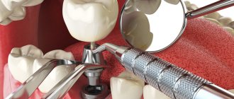
Types of dentures
Rice.
4. Schematic representation of the most common types of dentures: 1—dental inlay (indicated by an arrow); 2 - hollow metal crown (a - crown, b - cement, c - dentin, d - gum); 3 — pin tooth with plastic lining (indicated by arrow); 4 — bridge prosthesis (indicated by arrows) on a plaster model of the jaw; 5 - small saddle-shaped prosthesis, fixed on the supporting teeth by means of telescopic crowns (a - internal crowns are put on the supporting teeth, b - external crowns - in the prosthesis); 6 — clasp (arched) prosthesis for the upper jaw on a plaster model; 7 - clasp splinting prosthesis with flip-over hooks (indicated by arrows) for fixing the anterior movable teeth; 8 - removable partial laminar denture for the upper jaw, fixed on the supporting teeth with metal clasps (indicated by arrows) on a plaster model; 9 - complete removable denture for an edentulous upper jaw. Rice. 5. Dento-maxillofacial prosthesis, replacing defects of the nose, upper jaw and lip, as well as teeth of the upper and lower jaws; fixed with glasses. In accordance with the types of defects of the teeth and dentition, the following prostheses are distinguished (Fig. 4). 1. Dentures for replacing defects in the crowns of teeth; these include inlays made of porcelain (Fig. 4, 1) and various alloys (see Dental inlays). 2. Dentures for restoring damaged dental crowns - artificial crowns and pin teeth (Fig. 4, 2 and 3). 3. Dentures for replacing individual teeth or groups of teeth; these include fixed bridges and removable ones - small saddle, clasp and partial plate (Fig. 4, 4-8). 4. Dentures for replacing the dentition - complete plate removable dentures (Fig. 4, 9). 5. Dentures for replacing dentition defects, equipped with devices for splinting mobile teeth. 6. Dento-maxillofacial prostheses used to replace defects in the dentition and areas of the jaws and face. This group also includes a post-resection hollow prosthesis for the upper jaw, proposed by H. Pichler in 1923. Along with this, facial ectoprostheses are also manufactured to replace the ear, nose, and defects of hard and soft tissues of the maxillofacial area (Fig. 5) .
The choice of prosthesis design depends on the size of the dentition defect, the condition of the periodontal tissues and the prosthetic bed. To restore individual damaged teeth and replace small defects in the dentition, fixed prostheses (inlays, crowns, pin teeth, bridges) are usually used. When a dentition defect is combined with periodontal disease (see Periodontal disease), clasp (arched) dentures are indicated, the use of which makes it possible, along with replacing defects, to splint mobile teeth. In the absence of a significant number of teeth, when fixation of clasp dentures can cause overload of the periodontium of the supporting teeth, partial plate dentures are indicated. After the loss of all teeth, dentures for toothless jaws (so-called complete removable dentures) are recommended.
Artificial crowns are used to restore the shape of tooth crowns, as well as to fix fixed dentures and orthodontic appliances. The materials for making crowns are gold and silver-palladium alloys, chromium-nickel stainless steel, plastic, porcelain and a combination of metal with plastic or porcelain. Crowns are indicated in the absence of an inflammatory process at the apex of the tooth root.
Pin teeth are used to restore completely destroyed tooth crowns with a well-preserved and stable root with a canal accessible for treatment. Despite the wide variety of designs, pin teeth always consist of two parts: a root metal pin and the tooth itself (crown). The pin is inserted into the tooth root canal (after filling the apical part of the canal) and strengthened with cement. The crown of a pin tooth can be metal, plastic, porcelain or veneered, i.e. metal with plastic or porcelain lining. Pinned teeth include Logan crowns, which are standard pinned teeth with a porcelain crown.
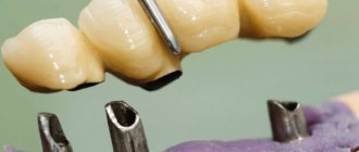
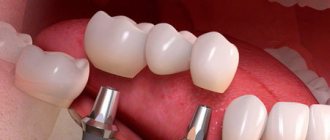
Bridges consist of supporting elements (crowns, teeth, inlays) and an intermediate part (body). They can be solid metal or lined with plastic or porcelain; They are used when 3 teeth are lost, if the periodontium of the supporting teeth is not changed. Bridge support should usually be bilateral, especially on molars. A single-sided bridge is used to replace a defect in one front tooth, usually for cosmetic purposes. Prosthetics of one molar is not indicated.
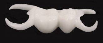
Removable dentures for replacing dentition defects have a base, retaining elements (clasps) and artificial teeth. The basis is a plate of plastic or metal that exactly repeats the relief of the prosthetic bed, i.e., the mucous membrane, alveolar process, hard palate, on which the prosthesis rests, and the necks of the teeth from the palatal or lingual surface, to which it touches. The base is reinforced with artificial teeth and retaining devices - clasps. Chewing pressure is transmitted to the base, and from it to the prosthetic bed. In the same way, chewing pressure is transmitted on dentures in the absence of teeth.
Small removable saddle dentures are located within the dentition defect and the corresponding section of the alveolar ridge. Clasp dentures are the most rational design of partial removable dentures. A clasp (arch) prosthesis has, in addition to artificial teeth and a base, an arch and its branches; the base covers only the toothless areas of the alveolar process, leaving the palate or that part of the alveolar ridge where there are teeth free. The parts of the base, called saddles, are connected to each other by an arc - a clasp. The basis of the clasp prosthesis is a metal frame (archwire, clasps, occlusal pads, fastenings for plastic, etc.).
Plate partial dentures are usually used to replace large defects in the dentition. Complete dentures, in which the base plate covers the entire prosthetic bed and which do not have supporting teeth, are manufactured for prosthetics of toothless jaws.
Rice. 6. Schematic representation of dental clasps: 1 - retaining single-arm wire clasp; 2— Retaining single-arm wire loop clasp; 3 - support-retaining wire clasp; 4 — Cast Bonville support-retaining clasp (top view). Rice. 7. Schematic representation of the locking fastening of a denture: on the model, in the supporting teeth there is a matrix with a bed for the inlay part (matrix); the inlay part is fixed to the prosthesis (the prosthesis is above the model), at the top left is the connection of the inlay part with the matrix.
Fixation of removable dentures during chewing and talking is achieved in various ways. The most common means of fixing partial removable 3. items are clasps (Fig. 6). According to their purpose, they are divided into retaining and support-retaining. Retaining clasps counteract the displacement of the prosthesis in the vertical and horizontal directions, and the pressure from the prosthesis is transferred to the prosthetic bed. The design of support-retaining clasps is more advanced; they consist of a rigid supporting element - an occlusal pad and two shoulders. The occlusal pad is located between the chewing tubercles of the tooth or in a bed specially created for it; By means of occlusal pads, vertical pressure is distributed to the supporting teeth.
For fixation, locking fasteners are also used (Fig. 7), which consist of a matrix having a bed for the inlay part (matrix); the matrix is placed in the crown of a natural tooth or soldered to an artificial crown, the inlay part is connected to the prosthesis.
Teeth that serve as support for the fixing elements of the prosthesis (crowns, inlays, clasps) are called abutment teeth. When choosing abutment teeth, their location, the height and shape of the crowns, the condition of the periodontium, etc. are taken into account. Errors in the choice of abutment teeth lead to their functional overload and, as a consequence, to pathol, mobility of these teeth.
Fixation of complete removable dentures is based on the use of anatomical points of retention, adhesion and vacuum formed under the base of the denture. Anatomical formations that prevent the prosthesis from shifting during chewing serve as points of anatomical retention: the arch of the hard palate, the alveolar ridge of the lower jaw, the alveolar process, the cusps of the upper jaw, etc.
When making prosthetics for toothless jaws, great importance is attached to the flexibility of the tissues of the prosthetic bed; compliance is the vertical or horizontal displacement of the mucous membrane under the pressure of the prosthesis base. When making prosthetics, it is important to determine vertical compliance, since the uniform distribution of chewing pressure throughout the entire prosthetic bed depends on it. The mechanism of vertical compliance is associated with the emptying of the vessels of the mucous membrane under the pressure of the prosthesis. The areas of the mucous membrane of the hard palate, where there is a rich network of vessels, are the so-called. buffer zones, as if shock-absorbing the prosthesis. The buffering properties of the mucous membrane of the hard palate vary from person to person and change throughout life due to changes in blood vessels under the influence of age-related factors, as well as various diseases. Changes in the secretion of the mucous and minor salivary glands, leading to dryness of the mucous membrane, worsen the fixation of the prosthesis.
Adhesion (sticking) occurs between the prosthetic bed and the base of the prosthesis in the presence of a thin layer of saliva. A vacuum under the prosthesis is created when it shifts, when the space between it and the mucous membrane increases, and the penetration of air is prevented by the mucous membrane adjacent to the edges of the prosthesis - the so-called. edge valve.
The manufacturing technology depends on the design of the 3rd piece. Metal crowns are stamped or cast. Bridges can be made in parts (first the crowns, then the body of the prosthesis), which are then soldered, or they can be cast simultaneously on a fire-resistant model (solid-cast bridges). The frame of clasp dentures is usually cast entirely or in parts, which are then soldered.
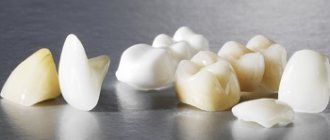
Artificial teeth for fixed dentures can be solid metal, but they meet little cosmetic requirements. More often, porcelain or plastic facets are reinforced in a metal stock. The porcelain veneer can be placed on the vestibular surface of the cast crown or cover the entire crown (metal-ceramic crowns).
Rice. 8. Set of artificial teeth for the upper and lower jaws (made of plastic).
Teeth made of plastic (see Polymer materials, in dentistry) or porcelain (see) are manufactured industrially (Fig. in a large number of sets. They have different sizes, shapes, shades of color, which allows you to select teeth that match the individual characteristics of a person.
During the polymerization of the plastic of the prosthesis base, plastic teeth are connected to it monolithically; Porcelain teeth are strengthened in the base by means of crampons or channels into which plastic penetrates. Crampons are wire pins made of gold alloy, platinum or stainless steel; At one end they are fixed in the porcelain of the tooth, at the other - in the plastic of the base.
Precise impressions of the jaws (negatively depicting the prosthetic bed) for the subsequent manufacture of a prosthesis (of any shape) are obtained using special impression materials (see) or plaster (see); In this case, a main impression (from the prosthetic jaw) and an auxiliary impression (from the opposite jaw) are required. Impressions are divided into anatomical (approximate, without taking into account the functional mobility of the mucous membrane) and functional (final), which reflect the mobility of the tissues of the prosthetic bed, which is especially necessary for the manufacture of a complete removable denture. Based on the impression, a plaster model is cast, the base of the prosthesis is modeled in wax, and artificial teeth are installed; The correct placement of teeth, bite height, etc. are checked by the doctor directly in the oral cavity of the prosthetic patient. The subsequent stages - plaster casting of the prepared prosthesis in a mold, melting wax, preparing plastic dough, molding the base, pressing and polymerization of plastic, finishing the prosthesis, etc. - are carried out in a denture laboratory (see) by a dental technician (see Dental technician).
Features of childbirth
Problems arise during labor.
Due to abnormal development of the uterus, the baby cannot get out of the womb on its own. Weak labor is observed, threatening the development of severe hypoxia.
Natural childbirth with a sciatic uterus is contraindicated.
As an alternative, preference is given to cesarean section under epidural anesthesia.
Author: Elena Yuryevna, obstetrician-gynecologist of the highest category Specially for the site kakrodit.ru