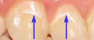
Dental demineralization is a widespread problem among both adults and children. It not only worsens the appearance of the dentition, but is also a prerequisite for the development of caries, its initial stage. Demineralization of tooth enamel is a pathological process of loss of mineral components by dental tissue, resulting in dullness, hypersensitivity, fragility of teeth, roughness, porosity of their surface, and often the presence of cracks (photo below). The main sign of pathology is the presence on the surface of the teeth of matte white, slightly grayish, or brownish spots of varying sizes, which are located in most cases in the cervical areas, but can cover the entire crown (photo below). Due to the presence of such spots, the pathology is also called focal demineralization of the enamel.
In this article we will tell you the reasons for focal demineralization of enamel, what it looks like in the photo, what features it differs in children, and we will also introduce you to treatment methods and what needs to be done for this disease.
Causes of tooth demineralization. What to do?
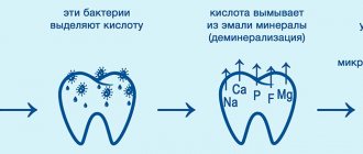
The causes of tooth demineralization are associated with the activity of cariogenic bacteria. An acidic environment (pH below 4.5) is excellent for their reproduction. It is provided by the remnants of carbohydrate foods rich in sugar. In addition to the acidic pH, cariogenic bacteria additionally produce acids as a result of their vital activity. All this damages the enamel. Therefore, one of the main reasons for tooth demineralization is excessive, and most importantly, regular consumption of sweets.
There are also other reasons for tooth demineralization:
- Poor oral hygiene is one of the main reasons.
- Presence of dental pathology.
- The predominance of soft foods high in sugar in the diet.
- Presence of dentures or orthodontic structures.
- Malocclusion or other dental anomalies.
- Changes in hormonal levels.
- The presence of metabolic diseases (rickets, diabetes, Addison's disease).
- Lack of fluorine, phosphorus, magnesium, bad habits.
Due to these and some other reasons, focal demineralization of tooth enamel occurs. What to do with this pathology and is it possible to get rid of it?
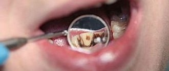
Treatment must be timely, since otherwise further development of caries with all its consequences cannot be avoided (photo below). Eliminating risk factors is the first thing to do if you or your child begins to experience demineralization of tooth enamel. What to do in the future, what medications and products to use, can only be advised by a qualified dentist.
You should not self-medicate or expect the enamel to recover on its own. Focal demineralization is a reversible process, but only if any therapeutic measures are taken at an early stage.
Types of enamel demineralization
Using microradiography in combination with two-dimensional microdenentometry of carious enamel, Crabb (1968) established the processes of remineralization along with demineralization.
Using an electron microscope, at the early stage of enamel caries, large apatite crystals were often identified between adjacent prisms in narrow channels of active demineralization. Based on this, the author suggested that the process of recrystallization is an integral part of the carious process. This assumption was developed in the works of Johnson (1967), Shiota (1970), Silverstone (1971), etc. Thus, according to morphological studies, progressive demineralization of enamel in the area of the carious lesion under favorable circumstances can be combined with remineralization. Apparently, if the latter predominates over demineralization, the carious process stops.
On ultrathin sections of decalcified and non-calcified enamel in areas of suspended caries, bacteria and isodiametric crystals were found, which were identified by crystallographic methods as vislocites. In the zone of enamel hypomineralization, apatite crystals along the periphery of the prisms are slightly disoriented and microspaces are found. Isodiametric crystals predominate in neighboring zones. The organic matter resembles embryonic enamel. According to Plackova and Vahl (1967), recrystallization is associated only with the intensity of the pathological process and depends on the physicochemical properties of the enamel.
Vahl and Plackova (1967), during electron-optical studies of a brown enamel spot (suspended caries), also found isodiametric crystals in widened microcracks, which, according to their data, are more resistant to demineralization than rod-shaped crystals of oxyapatite. Isodiametric crystals are identified as tricalcium phosphate. Recrystallization crystals in the brown spot of suspended caries are identified as calcium phosphate wyslockite and are also more resistant to demineralization than hydrokepapatites.
Plackova and Stepanek (1965), studying the submicroscopic structure of a brown enamel spot, determined that the substrate of the organic component of the brown spot (suspended caries) after demineralization consists of an acid-resistant dense spongy network. In addition, bacteria and an unidentified, electron-beam-dense, homogeneous mass are identified. It is assumed that this is a substrate of exogenous origin (disintegrated components of saliva), which penetrated into carious enamel and mineralized in it.
The results of electron microscopy carried out by A.V. Galyukova (1972) confirm early data that the pigmented spot does not always have a clear crystalline structure due to the presence of structureless organic matter. At the same time, changes in shape, fragmentation, and the appearance of new atypical crystals were found in the enamel crystals. Suspended caries is characterized by the presence of round rhombohedral crystals next to unchanged enamel crystals. The author did not find any areas of destruction. In the area of the enamel-dentin border with a pigmented spot, the crystalline structure is veiled by structureless organic matter.
The intermittent type of demineralization is a transition phase between the progressive and stabilizing form of demineralization. Morphological changes in the enamel indicate the simultaneous occurrence of two opposite processes - de- and remineralization. As we have already indicated, light brown carious spots should be classified as an intermittent type of lesion. With the intermittent type of demineralization, the enamel surface in the area of the pigmented spot is completely covered with bacteria.
According to G.P. Pakhomov (1974), brown spots occupying less than a quarter of the proximal surface are characterized by partial damage to the enamel in the form of a triangle with a rounded apex, under which an intact enamel-dentin border is visually determined. The crystal lattice constants are reduced compared to intact enamel, which indicates its compaction. In the area of brown carious spots, occupying 1/3 of the proximal surface of the tooth, complete damage to the enamel was detected. However, the enamel-dentin junction is broken only in 50% of cases. A.I. Rybakov and A.V. Granin (1970) indicate that in the area of the pigmented spot, the enamel-dentin boundary is destroyed over some extent.
During a microscopic examination of teeth with brown spots, occupying 1/3 or more of the proximal surface of the tooth crown, G. N. Pakhomov discovered a cone of completely affected enamel with a destroyed enamel-dentin border and an underlying sclerotic and pigmented cone in the dentin. The author established a significant difference in the parameters of the elementary cell of the crystals of the affected enamel, compared with the intact one, as well as the differences between the internal and external zones of the carious lesion.
Data on a decrease in constants in the surface layer of enamel compared to the internal one, according to the author, indicate a significant compaction of the crystal lattice in this zone, which is consistent with studies of zones of carious lesions in polarized light, data obtained by microradiography and other methods of instrumental research. The constants “a” of the crystal lattice of the inner layer of the affected enamel are much higher in value in comparison with the same parameters of intact enamel and the outer layer of carious enamel, which indicates an expansion along the axis of the unit cell of the crystals of the inner layer of enamel at the affected area.
In the area of black spots, the enamel is affected with rupture of the enamel-dentin junction and the formation of a sclerotic cone in the dentin. In transmitted light, such spots are black, and the area of carious demineralization can take the shape of a cone or rectangle. The results of a study of the crystal structure of enamel apatites with a pigmented carious spot quite clearly illustrate phase changes in tissue mineralization of different directions, the general tendency of which is expressed in the fact that the larger the carious spot, the deeper and more significant the pathological changes in the crystals. In cases of pigmented spots, the carious process proceeds slowly, which indicates the intermittent nature of demineralization in this type of caries with a tendency to stop.
Demineralization of tooth enamel in children
Since the main reason for the development of this pathology is the regular consumption of sweets, it is not surprising that demineralization of tooth enamel is most common in children under ten years of age (photo below). The development of this pathology is also facilitated by the lack of thorough oral hygiene, which is also typical for children of this age.
Demineralization of tooth enamel in children appears as irregular spots of varying sizes, or lines of dull white or slightly brownish color. At the initial stages, they are not very noticeable and are not located on the entire crown, but mainly near the necks of the teeth, or on the side parts of the enamel. As the disease progresses, the spots become darker in color (photo below) - this indicates damage not only to the enamel, but also to the dentin. The gates to carious infection open.
It should be remembered that demineralization of tooth enamel in children also requires treatment, even if these are baby teeth!
Prevention
The effectiveness of caries and demineralization prevention has long been proven by leading dentists. Preventive measures include simple and easy to follow rules:
- High-quality oral care. It is recommended to brush your teeth or at least rinse them with a special solution or water after each meal. You can take ready-made antiseptic rinses with you to work. Additionally, such products may contain anti-inflammatory components that are beneficial for the gums.
- The use of hygiene products with a high-quality composition, which also contains fluoride. Before purchasing a paste, you can check its availability in the composition description.
- Regular preventive examinations of the oral cavity, visiting the dentist 1-2 times a year.
- Timely treatment of dental diseases. Contact a specialist when the first complaints and violations appear. It is also important not to delay treatment of ENT diseases.
- Compliance with the principles of a healthy diet, restrictions in the diet of simple carbohydrates. After eating sweets, it is recommended to brush your teeth to remove residual sugars, which are a breeding ground for cariogenic bacteria.
- The use of multivitamin complexes, especially in cases of decreased immunity and recovery from severe infectious diseases and operations.
- Timely correction of the bite, removal of old fillings, which increase the risk of infectious processes and gum inflammation.
Pay special attention to the quality of personal hygiene. If there are provoking factors and chronic diseases, visit the dentist more often. During the examination, the professional uses special mirrors and instruments that make it easy to identify areas of calcium leaching and whitish spots, as well as other characteristic signs.
Some diseases contribute to a decrease in the protective properties of the mucous membrane. To strengthen the immune system, you can use drugs with immunostimulating properties. They prevent frequent relapses of dental pathologies.
When using removable appliances to correct your bite, do not forget to wash them regularly using antiseptic solutions. To clean braces, you need to purchase a round brush.
Focal demineralization of tooth enamel, photo
Above we described what focal demineralization of teeth looks like. Photos of patients with this pathology will be presented below. At the initial stage, focal demineralization can affect one or a small number of teeth; it is weakly expressed and often does not attract the patient’s attention at all.
Focal demineralization in adults is characterized by the extensiveness of the process - it is not uncommon that at the initial stage an entire row of teeth can be affected.
Demineralization of teeth, the photo of which is on the left, becomes more and more pronounced in the absence of treatment - the spots change their color from white to light, and then dark brown. Subsequently, focal demineralization ends with the formation of carious cavities.
Preventive measures
A preventive complex to prevent the leaching of minerals from tooth enamel requires compliance with a number of rules:
- Regular and proper oral hygiene. Dentists recommend brushing your teeth after every meal, alternating between calcium products and fluoride toothpastes.
- Annual visit to the dental office. Many experts advise increasing inspections to twice a year.
- Compliance with healthy eating rules. Including food enriched with vitamins and minerals in the diet, reducing the consumption of fast carbohydrates.
- Normalization of water-salt balance.
- Use of complex multivitamins.
Compliance with these recommendations for prevention is a guarantee of reducing the risk of dental diseases.
Demineralization is a common pathology that affects the condition of tooth enamel. As a rule, it is a symptom of the development of carious disease.
Since caries in the later stages can cause tooth loss, it is necessary to contact specialists at the first stage of the destructive process, since in this case it is possible not only to avoid removal, but also filling procedures.