In some people, the enamel has an uneven color and uneven structure, covered with stains, chips and other defects. This disease is called fluorosis, it has a chronic course. The pathology is typical only for certain areas, so it can be prevented in a timely manner.
Fluorosis - classification
There are 5 types of the disease in question, which differ in symptoms and severity. The first 3 forms of fluorosis are mild and do not provoke loss of dental tissue; the remaining types lead to partial loss. The classification includes the following variants of the disease:
- dashed;
- spotted;
- chalky mottled;
- erosive;
- destructive.
Streaked form of fluorosis
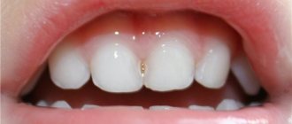
This type of pathology has the mildest course; it is rarely noticed in the early stages. The streak form of fluorosis is characterized by the appearance of small chalky stripes on the teeth, mainly the upper incisors. At first they are almost indistinguishable, but over time they merge into a spot. If you look closely at it, you can find obvious longitudinal strokes. This kind of dental fluorosis can be easily cured with timely and correct therapy. This type of disease does not cause complications or destruction of enamel.
Spotted form of fluorosis
This type of disease is accompanied by the appearance of pronounced light spots on the surface of several teeth, especially the upper and lower incisors. Gradually, the described enamel fluorosis progresses, small spots merge with each other, forming large formations. Sometimes they change color from white to yellow or light brown. The peculiarity of this type of disease is that the enamel in the spot area is very smooth and shiny.
Chalky-mottled form of fluorosis
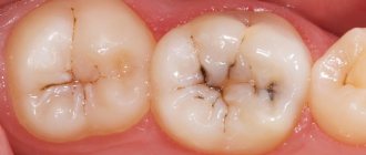
This type of pathology is characterized by various manifestations that are highly noticeable visually and cause psychological discomfort. It is important to immediately begin to treat such fluorosis - the chalky-mottled form is accompanied by the formation of well-defined white, yellow or brown spots with a matte surface on all teeth. Without timely treatment, the enamel is subject to destruction. This dental fluorosis in some cases is combined with thinning in certain areas. It looks like shallow depressions about 0.1-0.2 mm deep with a diameter of 1-1.5 mm.
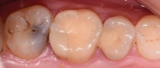
Erosive form of fluorosis
With this type of disease, the enamel has a pronounced yellowish or brown tint. Against the background of pigmentation, areas with large defects are clearly visible. The disease fluorosis of the described form leads to extensive and deep erosions. If no therapeutic measures are taken, the enamel quickly wears off and the dentin is destroyed. Sometimes the progression of the pathology ends in the loss of large sections of teeth.
Destructive form of fluorosis
The presented type of disease is considered the most severe option. Chronic destructive fluorosis causes severe pigmentation of the enamel, which becomes dirty yellow or dark brown. Multiple chips, erosions, stains and other defects are immediately visible on its surface. Destructive dental fluorosis simultaneously provokes the destruction of their tissue. The body tries to compensate for this process by secreting replacement dentin, but this is not enough. Teeth become brittle and often break under slight pressure.
Forms and symptoms of the disease
The forms of dental fluorosis due to the occurrence of the disease are as follows: endemic and professional. As is clear from the reasons, in the first case the disease occurs as a result of the characteristics of drinking water in the area where a person lives. The disease begins with the appearance of white spots or stripes on the vestibular (that is, external) side, most often starting from the incisors of the upper jaw. In the second, the cause is work in production, and the disease is accompanied by other typical complications: osteoporosis, osteosclerosis.
Based on symptoms and severity, the disease is divided into 5 forms:
- streaked: chalky streaks or stripes appear on the outer surface of the incisors. In some cases, they are clearly visible, but they are often weakly expressed and invisible in the photo, and during normal inspection they can be seen on a dry surface. The stripes can also merge into spots, but even in this case the stripes in the structure of the common spot will be clearly visible. This form is light in nature,
- spotted: a characteristic symptom is the presence of a large number of whitish spots that merge with each other, forming large spots. Their surface is shiny and smooth, has undefined boundaries, the transition to healthy enamel is smooth and gradual. This form is also considered mild,
- chalky-mottled: the enamel of the affected teeth has a non-smooth structure, with dots and spots with clear boundaries located on it. There are also such signs as yellowish enamel, on which there are small deepening specks with a pigmented bottom. This form is also characterized by the fact that the enamel is subject to rapid abrasion, and dentin of a dark brown shade quickly becomes noticeable underneath it. Experts classify this form as a condition of moderate severity,
- erosive: areas of destruction in this case do not look like specks, but large erosions. There is no enamel at the site of the lesion at all, the chewing surface of the teeth is subject to severe abrasion. Dentists call this form a severe form of the disease,
- destructive: erosive lesions affect not only the enamel, but hard tissues as a whole. Fragility leads to fractures, chips, and the shape of dental crowns changes. The deposition of replacement dentin prevents the opening of dental cavities. In order to “acquire” this form of the disease, you need to drink water with fluoride exceeding 10 mg/l. Like the previous form, this one is characterized by severe severity.
Important! The pronounced degree of the disease can be characterized by different forms of lesions in different groups of teeth. As experts note, the manifestations remain for life and do not go to another stage even if the concentration of the microelement in the water decreases.
Cause of fluorosis
The described pathology begins to develop even before the eruption of permanent teeth. Endemic fluorosis affects people living in areas with high levels of fluoride in water. The optimal concentration of this substance is considered to be 1 mg/l, the maximum is 1.5 mg/l. If a child drinks water with a higher amount of fluoride for more than 3 years in a row, the disease fluorosis is already damaging his permanent teeth, even if they have not yet grown. It is extremely rare that the disease also affects milk enamel.
An adult who moves to an area with excess fluoride in the water is not susceptible to fluorosis. There is a risk of damage to permanent enamel only if the concentration of the specified chemical is above 6 mg/l. The chance of getting fluorosis depends on the level of fluoride in the water. Number of affected population in endemic areas according to element concentration:
- less than 1 mg/l – 10-12%;
- 1-1.5 mg/l – 20-30%;
- 1.5-2 mg/l – 30-50%
- more than 2 mg/l – over 50%.
Fluorosis - symptoms
It is easy to notice the beginning of the progression of pathology even in the early stages. Dental fluorosis and the severity of its clinical picture depend on the severity of the disease. The more damaged the surface of bone structures is, the easier it is to diagnose the disease. External signs of fluorosis include enamel defects on symmetrical, predominantly front teeth:
- stripes;
- specks;
- strokes;
- spots;
- erosion;
- depressions;
- chips and others.
Stages of fluorosis
In dentistry, the disease in question is classified into 5 degrees of severity:
- Very light.
The manifestation of fluorosis is insignificant and almost imperceptible. The color of the enamel practically does not change. - Easy.
There are rare milky white spots or short stripes on a few teeth. The total surface of the lesion accounts for up to 25% of the total enamel. - Moderate.
Strokes, spots and chalky inclusions are clearly visible visually. The area of the tooth crown is damaged by approximately 50%. - Average.
In addition to white matte defects (chalky), there are yellow and brown spots, sometimes large. Due to the thinning of the enamel and exposure of dentin, there is susceptibility to tooth decay and other oral infections. - Heavy.
More than 70% of tooth crowns are damaged. The enamel pigmentation is very pronounced and has an uncharacteristic yellow or brown color. Surface defects quickly deepen and turn into extensive erosions. The enamel wears off and chips, the teeth are severely deformed and often destroyed. If fluorosis progresses at this stage, complications arise. A common problem is tooth loss and damage to skeletal bone tissue.
Forms
Dentists distinguish the following forms of fluorosis:
- dashed;
- spotted;
- chalky speckled;
- erosive;
- destructive.
Let's take a closer look at each of the forms:
- Stroke is the mildest form of the disease. It is manifested by the formation of small chalky stripes on the enamel of the front teeth, which are almost invisible and can only be examined by an experienced dentist.
- Spotted is heavier than streaked. It is manifested by the formation of chalky stains on the surface of the enamel of the front teeth, which can be located on any part of the tooth enamel. The spots may be whitish or light yellowish.
- With chalky mottled disease, not only the front teeth, but almost all of the patient’s teeth are affected. The disease is manifested by the formation of pigmented spots ranging from whitish to dark brown in color.
- Erosive is one of the most difficult. With this form of the disease, small defects - erosions - form on the surface of the tooth enamel. If the patient has at least one erosion, it means that the disease is progressing and developing quite rapidly.
- Destructive is the most difficult. In this form of the disease, tooth enamel is gradually destroyed and worn away.
Fluorosis - diagnosis
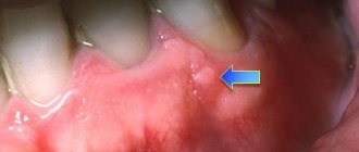
Not only a dentist, but also a pediatrician and a therapist can detect damage to enamel due to excess fluoride in the body. Symptoms of the disease and taking an anamnesis help to identify it accurately. Differential diagnosis of fluorosis is necessary when caries is suspected in the spot stage. It is very easy to distinguish these pathologies based on specific characteristics.
Fluorosis affects the vestibular and lingual surfaces of the teeth and is characterized by multiple damage to the enamel. Caries is localized in the contact and cervical areas and looks like small, single whitish spots. Another feature of fluorosis is its appearance immediately after the eruption of permanent teeth. Caries also occurs on milk bone structures.
Fluorosis: etiology, clinical picture, diagnosis, differential diagnosis, treatment.
Dental fluorosis is a disease that develops as a result of an increased amount of fluoride entering the body. An early sign of fluorosis is tooth damage.
Etiology. The cause of fluorosis is considered to be excessive long-term intake of fluoride compounds into the child’s body, coinciding with the period of mineralization of the teeth.
fluoride concentration
Sensitivity or tolerance of the body
· individual metabolic characteristics
· nutrition
· sanitary, social and climatic living conditions.
Species: endemic (drinking water); production (workers of cryolite and Al production), iatrogenic (additional intake of fluorine into the body).
Classification (Patrikeyev):
· dashed stripes-stripes of white color, weakly or strongly expressed on the vestibular surface of the incisors. Most noticeable when the tooth dries out.
· Spotted - spots are located in different parts of the tooth crown. The intensity of the white color disappears from the center to the periphery. M.b. light yellow, dark brown pigmentation.
· chalky-mottled
· erosive
· destructive
Pathogenesis.
1. Secretory phase.
— Cytotoxic effect on ameloblasts, the quantity and quality of synthesized enamel proteins.
— Direct influence on crystal growth.
2. Maturation phase - cytotoxic effect on the enzymatic system and enamel organ.
3.Direct influence on crystal growth.
4.Direct effect on mineral metabolism.
Fluoride—vessels of the dental sac—intercellular space—ameloblasts—calcium binding protein—hydroxyfluoroapatite
Clinic.
Fluorosis: -limited - short-term use by the child of water with a high fluoride content.
-generalized – long-term residence of a child in an endemic area.
In temporary dentition, molars and canines (small white spots on the vestibular surface) are most often affected. Enamel smooth and shiny. Risk factors
: artificial feeding, early complementary feeding.
More often on permanent teeth. The first permanent molars and central incisors of the upper jaw are affected.
· Mild degree: small single chalky spots on the vestibular surface of the teeth.
Dysmenialization↔reminarization.
· Moderate to severe: pigmented, confluent patches ranging from light yellow to dark brown of varying sizes and shapes. On the maxillary incisors, pigmentation is located horizontally along the equator or cutting edge. Irreversible processes of demineralization.
Diagnostics. 1) History
2)Complaints
3)Physical examination.
· Inspection of the dentition: teeth cleaning, professional hygiene; drying teeth.
Determine the intensity of brown pigmentation of enamel with a 2% aqueous solution of methylene blue
(L.K. Nikolishin).
o 0 points—no staining.
o 1 point – weak staining
o 2 points – medium intensity staining
o 3 points - intense brown coloring
4) Laboratory: analysis of the patient’s drinking water and urine for fluoride content.
5) Instrumental: determination of electrical resistance indicators of hard dental tissues. Healthy enamel does not conduct electricity.
Differential diagnostics. At the spot stage: spotted form of hypoplasia, caries at the spot stage. More severe forms: superficial and medium caries, erosion, wedge-shaped defects.
Treatment. Goal: normalization of dental aesthetics.
General: -water defluoridation
-replacement of sources with high fluorine content with optimal ones
-removal of young children for the summer months outside the endemic focus
-diet (protein, dairy products, vitamins B, C, D) + exclude fluoride preparations
-exclusion of artificial feeding and complementary feeding of children
The treatment is complex.
Medication : 1) remineralizing treatment (phosphorus-calcium preparations (prescribed by a pediatrician), vitamin-mineral complexes). Course 1-3 times a year for 1 month.
2) Retherapy:
· solutions (calcium glycerophosphate 2.5-5%, calcium gluconate 10%) for 15-20 minutes with three replacements of the solution (5-7 minutes each). The course of treatment is 15-20 procedures 2-3 times a year (depending on the severity of fluorosis).
Treatment of dental fluorosis is more effective using electrophoresis of remineralizing solutions, which can be used in children over 10 years of age. Treatment is carried out in courses of 10 sessions of 15 minutes every other day in a clinic according to standard methods 2-3 per year.
· gels (ROKS, Belagel, Remogel)
· calcium phosphate containing pastes (10-15 minutes)
Non-drug treatment:
· Bleaching - solutions based on hydrogen peroxide or carbamide peroxide of various concentrations (9-38%)
Indications: mild forms of fluorosis (damage depth no more than 150 nm).
Contraindications: intolerance to the components of the whitening system; pregnancy; age under 18 years.
Complications: increased sensitivity of teeth during or after whitening; complications from the pulp.
· Enamel abrasion
Megaabrasion—removal of deep colored layers of enamel. Perform coarse-grained
diamond heads at low speeds to prevent excessive enamel removal and
the occurrence of cracks. The prepared surface is ground with rough discs to preserve
enamel roughness.
Macroabrasion is the removal of the outer layer of painted enamel. A fine-grained turbine diamond head with water supply is used. After abrasion, the enamel is ground and polished with a 30-grid tungsten carbide bur, decreasing abrasiveness discs and composite polishing paste.
Microabrasion - a microscopic layer of enamel is simultaneously eroded and ground with a special mixture, after which a completely intact enamel surface remains.
· Production of veneers.
· Covering affected teeth with artificial crowns.
· Combination of methods.
Dental fluorosis - treatment
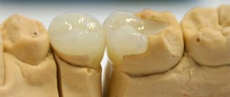
Therapy for enamel damage is developed taking into account the form and stage of the pathological process. If erosive or destructive fluorosis is diagnosed, treatment involves restoring the shape and color of the crown of the teeth using:
- orthopedic structures (veneers);
- composite filling materials;
- fixing pins with artificial crowns.
Streaked, chalky-speckled and spotted fluorosis of the surface of the teeth is accompanied by changes only in the shade of the enamel, less often there are minor damage to its thin upper coating. To treat such forms of pathology, local bleaching and remineralization, which are carried out in several successive courses in the dentist’s office, are sufficient.
Pastes for fluorosis
Personal hygiene is extremely important in the treatment of this disease. One of the auxiliary ways to treat fluorosis is the constant use of special toothpastes. Such products should not contain fluoride; it is desirable to contain easily digestible calcium, whitening components and substances that prevent the development of caries. Recommended toothpastes for teeth affected by fluorosis:
- President Unique.
Contains pantothenate, lactate and calcium glycerophosphate - compounds that strengthen and restore enamel. The composition also contains papain, which dissolves plaque, and xylitol, which slows down its formation and neutralizes the acidic environment in the oral cavity. - SPLAT Maximum, Biocalcium.
Both pastes contain papain and polydon, which promote gentle but effective plaque removal. Calcium is presented in the form of hydroxyapatite and lactate - easily digestible forms. SPLAT Maximum additionally contains an enzyme complex, zinc citrate and licorice extract, which provide comprehensive oral care. - Asepta.
The paste contains nothing extra - calcium hydroxyapatite, papain and potassium citrate. The product helps improve the condition of enamel, prevents plaque formation and reduces tooth sensitivity. - ROCS
The active component is calcium glycerophosphate, which is easily absorbed and integrated into damaged areas of enamel. To prevent plaque formation and caries, the paste contains bromelain and xylitol. - New Pearl.
The most affordable option for fluorosis. This product does not contain enzymes, whitening or plaque-removing substances, but contains the most “friendly” calcium compound – citrate.
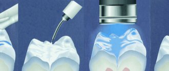
Teeth whitening for fluorosis
Visible defects can be removed from enamel only in the dentist’s office. Professional whitening for fluorosis is performed in one of 3 ways:
- Laser.
First, the teeth are coated with a hydrogen peroxide-based gel. To reveal the normal color of the enamel, they are illuminated with a laser beam. - Chemical means.
Solutions of carbamide and hydrogen peroxide and inorganic acids (hydrochloric, phosphoric) are used as bleaching agents. Immediately after the procedure, teeth are remineralized with calcium gluconate or Remodent and coated with fluoride varnish. - By grinding.
The top layer of enamel is removed using a special abrasive paste based on carborundum, hydrochloric acid and silicon gel. After grinding, the treated crown is coated with perhydrol and irradiated with a quartz lamp. At the end of the manipulation, remineralization is performed.
It is not possible to completely whiten your teeth in one session of any of the listed procedures. At subsequent visits to the dentist, active drugs will be used only on the darkest areas of the enamel, but to achieve the desired results, 3-20 manipulations will be required (the number depends on the degree of fluorosis and the color of the defects). Repeated treatment must be carried out after 6-8 months, during which dentists advise taking glycerophosphates and calcium preparations orally, and strictly observing the rules of personal oral hygiene.
Treatment methods
The choice of treatment method for fluorosis depends directly on the form of the disease. Dentists resort to the following procedures:
- bleaching
– carried out every six months and helps hide the appearance of streaked, spotted and chalky-mottled forms; - remineralization
- strengthening tooth enamel by saturating it with calcium and fluoride (effective only for the first three forms of fluorosis); - artistic restoration with composite materials
– if necessary, restore the shape and color of crowns in erosive and destructive forms; - prosthetics with veneers or crowns
are an alternative to artistic restoration.
Restoration with composites will last on average for two to five years. Ceramic veneers and crowns will last ten to twenty years.
In the treatment of fluorosis, it is very important to maintain impeccable oral hygiene, use remineralizing toothpastes and control over the intake of fluoride into the body.
Teeth whitening to prevent fluorosis
Prevention of dental fluorosis
Preventive measures can be individual and collective. In the first case, prevention of fluorosis includes:
- limiting the consumption of foods with fluoride (spinach, sea fish, animal oil);
- control of the quality and composition of drinking water;
- enrichment of the diet with vitamins C, D, calcium gluconate;
- use of toothpastes without fluoride;
- regular consumption of dairy products.
It is especially important to prevent the disease in newborns in endemic areas. Parents should not introduce complementary foods early; it is advisable to avoid artificial milk formulas. It is recommended to give water either from bottles, or replace it with milk or juices. As the child grows up, doctors advise taking him to a safe area (in terms of fluoride concentration) every summer for 2-3 months.
The local government is taking collective action to prevent dental fluorosis among the population. Prevention involves:
- replacement of the water source for consumers;
- reducing the amount of fluoride through special filtration;
- mixing water from different sources (for example, wells and springs).