Facial neuritis is an inflammatory disease. A very common form of facial nerve damage. A separate variant of this pathology is called Bell's palsy.
The term “neuritis” itself refers to damage to the peripheral nerve. In this case, both the myelin sheath (an electrically insulating sheath to exclude external influences from other nerve signals) and the axial cylinder (folds containing the processes of nerve cells) are damaged.
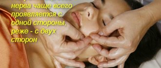
Damage leads to paralysis of all facial muscles. Typically, the nerves on one side of the face are damaged (unilateral neuritis). But this is enough for a person to be unable to close his eye (this is lagophthalmos, or otherwise hare’s eye), wrinkle his forehead, or bare his teeth.
But not only external inconvenience arises - food begins to get stuck between the cheek and gum, troubles arise with the eye of the damaged side of the face (they are called Bell's symptom).
But that's not all. Sensory (gustatory) and parasympathetic (secretory) fibers pass through the trunk of the facial nerve. Inflammation will affect them too. As a result, taste sensitivity on the affected side may be severely affected.
What is facial neuritis
Facial neuritis is an inflammatory disease that affects the 7th pair of cranial nerves (cranial nerves).
For reference. The facial nerve is responsible for the innervation of the facial muscles, therefore, when it is inflamed, facial expressions are disrupted (the patient cannot smile or frown), facial asymmetry develops, and the process of chewing food becomes significantly more difficult or impossible.
Damage to the facial nerve is most often unilateral, but two percent of patients may have a bilateral form of the disease.
Facial neuritis is characterized by a long course. Treatment in a hospital takes on average 20 to 30 days. However, complete recovery may take from 3 to 6 months.
In approximately five percent of patients, the work of the facial muscles is not fully restored (as a rule, the risk group includes patients with head injuries and tumors affecting the brain).
In 10% of patients who have suffered facial neuritis, relapses of the disease are observed.
What is the facial nerve
The facial nerve is the 7th nerve of 12 pairs of the cranial nerve. The facial nerve (FN) exits the brain in the area between the pons and the medulla oblongata. This is a motor nerve, its main function is to innervate the facial muscles.
However, due to the fact that it includes PN fibers (the intermediate nerve responsible for the innervation of the lacrimal gland and providing taste sensitivity to the anterior 2/3 of the tongue), the facial nerve acquires a mixed (motor-sensitive) character.
For reference. Quite frequent neuritis of the facial nerve is facilitated by the fact that along its length the facial nerve passes through many narrow bone canals.
Therefore, even with slight inflammation or poor circulation in the surrounding tissues, when the nerve fibers swell, they are compressed and pinched in narrow bone canals.
In the absence of timely medical care, irreversible damage to the cells and fibers of the FN occurs.
Causes of inflammation of the facial nerve
Neuritis of the facial nerve is a fairly common pathology.
For reference. The disease occurs with equal frequency in both men and women. More often, FN lesions are recorded in the cold season. The maximum number of patients occurs in regions with a cool and damp climate.
The main causes of the disease include:
- hypothermia (exposure to a draft or air conditioner on the neck and ear area leads to spasm of muscles and blood vessels, blood supply is disrupted, nerve inflammation occurs, and general hypothermia is observed, which contributes to a decrease in immunity);
- alcohol abuse (alcohol intoxication can cause damage to nerve fibers and lead to the development of neuritis);
- uncontrolled forms of arterial hypertension (against the background of increased intracranial pressure, damage to the nucleus of the facial nerve is possible);
- herpes viruses (especially with herpes zoster, which affects the trigeminal nerve, in which case inflammation may spread to the fibers of the facial nerve);
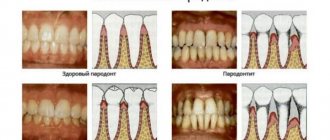
- mumps viruses, enteroviral and adenoviral infections, polio pathogens, past sinusitis, otitis, odontogenic infections;
- poor quality dental treatment;
- pregnancy (against the background of changes in hormonal levels, when combined with other risk factors in the first trimester, inflammation of the FN fibers may develop);
- diabetes mellitus, accompanied by damage to blood vessels and nerve fibers;
- pronounced forms of atherosclerosis of cerebral vessels and neck vessels, as well as multiple sclerosis , which is accompanied by autoimmune damage to the myelin sheaths in nerve fibers.
Basic treatment approaches
Treatment of neuritis of the facial nerve is a set of prescriptions, the implementation of which is possible only under the supervision of a doctor. It consists of medication recommendations and physiotherapeutic procedures.
Drugs prescribed for the treatment of the facial nerve can be classified as follows:
- Anti-inflammatory non-steroids (nurofen), in some cases they resort to treatment with steroid hormonal drugs - glucocorticoids (dexamethasone, prednisolone);
- diuretics (relieving swelling);
- antibiotics or antiviral compounds (depending on the etiology of the facial neuritis);
- antispasmodics (no-spa, papaverine hydrochloride, spasmol);
- a group of drugs with neurotropic action (phenytoin, carbamazepine) - to optimize the functioning of nerve fibers and relieve the main clinical manifestations of facial neuritis;
- vitamin therapy.
In addition, in the presence of concomitant diseases that weaken the body’s protective properties, drugs are prescribed to treat them.
If the nature of facial neuritis is stressful, sedative medications are added to the treatment regimen to restore normal sleep and regulate mood.
The treatment of facial nerve neuritis can be facilitated by the use of physiotherapy procedures. The most relevant are electrophoresis, paraffin warming, applications to the damaged (undamaged) half of the face from ozokerite, diadynamic therapy, UHF and UV irradiation, UHF.
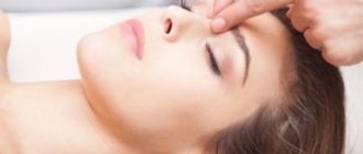
Facial muscle massage is useful. However, due to the need to know the physiological characteristics of the innervation of the face, it must be done by a professional.
Neuritis of the facial nerve should be treated in a hospital setting. Sometimes the doctor allows therapy to be carried out in a day hospital.
Classification
All forms of neuritis of the facial nerve are divided into:
- primary - cold inflammation of the facial nerve (develops against the background of hypothermia);
- secondary - occur against the background of concomitant pathologies (herpes infections, mumps, otitis, odontogenic infections, traumatic brain injuries accompanied by damage to the facial nerve).
For reference. There is also a separate variant of facial neuritis - Bell's palsy. The exact causes of the disease are unknown. Paralysis is seasonal and develops mainly against the background of hypothermia.
Symptoms of the disease
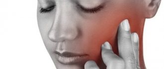
The disease usually develops gradually. The first symptom is a dull, aching pain behind the ear. Less commonly, the pain can be acute.
After a few days, visible facial asymmetry appears. Sometimes symptoms develop faster (a person goes to bed healthy and wakes up with visible facial asymmetry).
On the affected side of the FN it is noted:
- smoothing the nasolabial fold;
- drooping of half the face (eyebrow, corner of the eye and mouth drooping);
- “distortion” of the face.
The patient cannot smile normally (the smile appears asymmetrical), frown his eyebrows, stretch out his lips or show his teeth.
Often, a patient with neuritis of the facial nerve cannot completely close the eye (due to this, drying of the conjunctiva and cornea is noted). When you try to close the eyelids, the eye turns upward (the so-called Bell sign).
Against the background of a slightly open eyelid, the development of lagophthalmos (“hare’s eye”) is observed - a white strip of sclera is constantly visible between the iris and the lower eyelid. Due to the inability to close the eye, constant lacrimation is noted (the patient cries “crocodile tears”). Lacrimation increases sharply during meals.
Also, many patients are concerned about a sharp decrease in the taste sensitivity of the tongue, pain in the jaw and behind the ear.
Increased salivation may occur. Due to the inability to close the mouth normally, there is a constant flow of saliva from the mouth.
Less common is a decrease in the secretion of tears and saliva. Also, some patients have difficulty chewing.
For reference. A common manifestation of the disease is hyperacusis on the side of the nerve inflammation (excessively increased sensitivity to sounds).
Symptoms of secondary neuritis of the facial nerve
With poliomyelitis damage to the nucleus of the FN, severe weakness of the facial muscles is noted.
For reference. Against the background of strokes accompanied by damage to the roots of the FN, as well as the nuclei of the FN and the abducens nerve, the development of convergent strabismus, paresis of the facial muscles and visible asymmetry of the face is noted.
With neuromas, the auditory nerve is also affected, so facial asymmetry is combined with hearing loss and tinnitus .
Against the background of herpetic nerve lesions, severe cutting or burning pain in the ear is noted. Pain may radiate to the face, upper neck and occipital region.
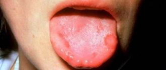
It is also typical for a specific herpes rash to appear on the skin of the auricle, external auditory canal, eyelid skin, lips, as well as mucous membranes lining the pharynx, larynx and tongue.
For reference. Some patients may experience complaints of tinnitus, hearing loss, and constant dizziness . Sometimes there is horizontal nystagmus (fast rhythmic movements of the eyeballs from side to side).
Against the background of mumps (mumps), neuritis develops against the background of general intoxication symptoms and fever. The patient is worried about muscle and joint pain, nausea, weakness, headaches . The appearance of unilateral mumps-specific swelling is also characteristic.
With the development of inflammation of the FN against the background of complicated otitis, facial asymmetry is combined with elevated body temperature and shooting pain in the ear. Trismus (spasm) of the masticatory muscles may also develop.
Classification of facial neuritis
Primary neuritis is distinguished; it occurs as a result of hypothermia, for example. Secondary is also isolated; it occurs as a result of existing inflammation, for example, with otitis media. Separately, some forms of facial neuritis should be noted.
- Hunt's syndrome - facial neuritis in herpes zoster
Damage to the facial nerve is combined with other manifestations of this disease, such as characteristic rashes on the tongue, mucous membrane of the oral cavity and pharynx, as well as in the area of the auricle (see symptoms and treatment of herpes zoster). In this case, the herpes virus affects the ganglion, from which the hearing aid, tonsils and palate receive innervation. The motor branches of the facial nerve are located close to this ganglion. The disease begins with shooting pains in the ear area, followed by facial asymmetry, decreased taste sensitivity in the anterior third of the tongue, dizziness, ringing in the ears and horizontal nystagmus.
- Neuritis due to mumps (mumps)
Can be one-sided or two-sided. Accompanied by fever, signs of intoxication and enlargement of the parotid salivary glands.
- Neuritis due to borreliosis
Bilateral damage to the facial nerve is always observed. Accompanied by a rise in temperature, erythema and widespread neurological symptoms.
- Neuritis with otitis media
Symptoms of neuritis of the facial nerve in this case are accompanied by pain in the ear area, which is acute. An infection from the middle ear contacts the branches of the facial nerve.
- Melkerson-Rosenthal syndrome
This is a hereditary disease, which is quite rare, and is characterized by a paroxysmal course. During an exacerbation, swelling of the face, neuritis of the facial nerve and folding of the tongue are observed.
Why is facial neuritis dangerous?
In any case, inflammation of the nerve leads to the death of some nerve fibers. But the innervation does not completely disappear. The need to control more muscles with less nerve fiber leads to an unpleasant situation called synkinesis.
In fact, synkinesis leads to reflexive conjugal muscle movement. For example, when you blink, the corner of your mouth will rise. Not a very pleasant impression from the outside.
Disruption of the innervation of some small facial muscles can lead to their atrophy. Part of the face will be motionless, which is completely unnecessary.
Due to impaired drooping of the eyelid and constant dryness of the conjunctiva, the development of conjunctivitis, keratitis, and corneal ulcers is possible.
Also, in the absence of timely treatment, inflammation of the FN can be complicated by unilateral contracture of the facial muscles.
For reference. In the vast majority of cases, the mobility of facial muscles is completely restored within 3-6 months with proper treatment and care.
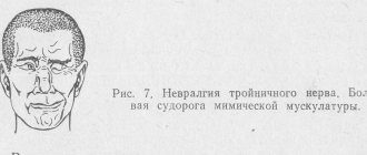
Signs and symptoms of facial nerve neurosis
The main symptom is very severe pain that occurs in any part of the nerve trunk: in the area of the cheekbones, eyebrows, forehead or jaw. As the intensity decreases, a burning sensation occurs in the affected area. The pain goes away during sleep. Paralysis of facial muscles occurs. Most often, the facial muscles are affected on one side. Since the muscles responsible for chewing are also affected, it becomes difficult to chew food, and speech changes. It is not possible to hold liquid food in the mouth. Often a person bites his cheek while eating.
Dry mouth may occur if the endings in the salivary gland are damaged. It becomes difficult to close your eyes, which causes the mucous membrane to dry out. The patient rarely blinks. The tip of the tongue cannot taste food. Hearing decreases, and it becomes especially difficult to distinguish low sounds. Possible sleep disturbances.
The patient's face changes. Due to the fact that one side is paralyzed, symmetry is greatly impaired. The eye on the affected side is open too wide, the nasolabial fold is smoothed out, the corner of the lips is drooping.
Based on the detected symptoms, treatment should be selected by a doctor.
Diagnosis of the disease
If you suspect neuritis of the facial nerve, an examination by a neurologist is required. Moreover, if the disease began with the appearance of sharp facial asymmetry, then first of all it is necessary to exclude stroke and transient cerebrovascular accident.
In particular, if the patient has symptoms uncharacteristic for neuritis of the facial nerve (loss of consciousness, speech impairment, lethargy).
In the classic course of the disease, making a diagnosis is usually not difficult.
For reference. To clarify the cause of the development of the disease, MRI or CT of the brain, electroneurography, electromyography, and examination for viral infections can be performed.
Diagnostic methods
Correct diagnosis is the basis for effective therapy. In most cases, diagnosis is relatively quick and safe. It helps prevent tissue damage such as broken bones, cuts, contusions, bruises, or even acute inflammation. For chronic pain, a correct diagnosis is more difficult. Only the sum of various examinations allows us to obtain correct medical conclusions. Informative tools for the attending physician are a pain questionnaire or diary. If conversation with the patient and physical examination do not give a specific result, additional examinations are carried out. Visual (radiological) and electrophysiological procedures help to correctly identify the disease.
X-ray procedures help identify degenerative, inflammatory, rheumatic, tumor and post-traumatic processes in the body. Single- and multi-slice spiral computed tomography scans are used for inflammatory, neoplastic processes or abnormalities. Other imaging procedures that do not require X-rays include sonography, duplex ultrasound (for example, if there are signs of circulatory problems), and magnetic resonance imaging. MRI is used to visualize soft tissue structures, processes in the bone marrow, blood vessels, processes in the brain and joints.
Skeletal scintigraphy and positron emission tomography are used with great success in medicine to identify the cause of trigeminal neuralgia. Skeletal scintigraphy is prescribed to study inflammatory, degenerative, and metastatic processes. Triphasic skeletal scintigraphy is a routine procedure when Sudek's disease is suspected. Using this research method, the metabolic activity of bones and bone marrow can be determined. PET is used to detect vascular inflammation, dementia, tumors and inflammation.
With the help of electrophysiological studies, muscle damage can be accurately analyzed. Their goal is to determine the type of injury, the nature of the injury, and the extent of muscle damage. If peripheral polyneuropathy or single nerve injury is suspected, neurography, electrography, and electrocardiogram are recommended.
Neurography measures nerve conduction velocity. Electromyography measures the electrical activity of muscles and allows the doctor to determine whether the dysfunction is affecting a muscle or a nerve. Electrocardiography shows the electrical activity of the heart muscle and possibly disturbances in nerve pathways.
Additional methods for obtaining diagnostic information are vibratometry, thermal testing or the iodine-starch reaction. Electrophysiological studies are necessary, for example, for carpal tunnel syndrome, polyneuritis and polyneuropathy.
The microscope is another important tool in the diagnostic chain for determining the cause of neuralgia. Blood and fluid tests in the laboratory help identify indirect signs of more serious diseases - diabetes, multiple sclerosis or Lyme disease. In some cases, the tissue sample is also placed under a magnifying glass. However, a nerve or skin biopsy is only used when all other tests are inconclusive.
Main blood parameters analyzed:
- erythrocyte sedimentation rate (ESR);
- C-reactive protein (plasma protein);
- electrolytes;
- liver enzymes;
- TSH (thyroid gland) values;
- leukocytes.
A blood test is one of the methods for diagnosing facial neuralgia.
Further studies are carried out if diabetes mellitus, alcohol abuse (transaminases, MCV, vitamins) or funicular myelosis (vitamin B12 deficiency) are suspected. There are many other very specific parameters that can be determined by blood.
A lumbar puncture is performed to remove cerebrospinal fluid from the spinal canal. It is a clear liquid that surrounds the brain and spinal cord and can change, for example, during inflammatory processes. Lumbar puncture is important, for example, if inflammatory diseases of the brain are suspected, with multiple sclerosis or Lyme disease.
Another diagnostic option is to remove and examine nerve or skin tissue using a small biopsy. Nerve biopsy should only be considered if an adequate diagnosis cannot be made using other methods. They are allowed if there is a suspicion of treatable polyneuropathies. A biopsy can make a statement about the condition of the nerve fibers in a specific area.
Treatment of neuritis of the facial nerve
All drugs for the treatment of facial neuritis should be prescribed exclusively by a neurologist. Self-treatment is unacceptable and can lead to the development of severe complications.
At the initial stage of the disease, it is recommended to use:
- glucocorticosteroids (prednisolone drugs relieve swelling and reduce the severity of the inflammatory reaction);
- decongestants (diuretics: furosemide, spirolactone);
- vasodilating drugs (nicotinic acid, nicotinate, scopolamine, xanthinol);
- B vitamins.
In the case of the development of secondary neuritis, treatment of the underlying disease that caused inflammation of the facial nerve is also carried out.
To speed up the recovery of facial muscles, complete rest is indicated for the first 7-10 days of illness. From the first days of illness it is allowed to use non-contact heat (sollux).
To prevent the development of conjunctivitis, keratitis and corneal ulcers, artificial tear preparations, as well as moisturizing eye ointments, are used. At night, the eye must be covered with a bandage.
The use of UHF, as well as contact heat (paraffin therapy) is allowed only from 5-6 days.
All physiotherapy procedures are prescribed in courses.
From the second week of illness, massage and exercise therapy can be used carefully. In this case, the load on the muscles should be increased gradually.
For reference. In order to accelerate the restoration of full muscle innervation, from the 14th-15th day of illness, anticholinesterase drugs (neostigmine, galantamine), as well as bendazole, can be used.
According to indications, ultrasound and hydrocortisone administration using phonophoresis are used. Methandienone preparations can also be used to accelerate metabolic processes in nerve tissues.
In rare cases, the use of electrical neurostimulation is indicated.
If there are no signs of FN recovery within 2-3 months, hyaluronidase drugs are prescribed.
Is it possible to use surgery?
Previously, surgery aimed at decompressing the nerve by widening the bone canal was widely used.
Attention. However, this technique can cause nerve damage and hearing loss. In this regard, this technique is not recommended.
Surgery is indicated only if:
- nerve rupture due to trauma (nerve suturing);
- lack of effect from conservative treatment methods for 8-10 months (nerve plastic surgery using autotransplantation is indicated).
Therapy for occipital neuralgia
Treatment of facial neuritis with leeches is gaining increasing popularity. The healing effect is observed due to the properties of leech saliva: it restores the necessary nutrition of tissues, dilates blood vessels, and relieves pain. Thus, the use of hirudotherapy for neuritis gives:
- relieving inflammation;
- pain reduction;
- improvement of vascular function;
- strengthening the immune system;
- removal of edema.
Leeches are placed along the inflamed nerve. 4–6 individuals are used at a time, depending on the affected area. Such treatment must be agreed with your doctor.
Neuralgia of the occipital region causes the greatest discomfort. Severe shooting pains appear in the head. This occurs when surrounding tissue pinches a nerve. Attacks may be accompanied by fainting, vomiting and nausea. Often the pain can be felt in the temples, eyes and forehead. It is important to know the causes of this disease in order to consult a doctor in time.
- Occipital neuralgia is accompanied by headache. Symptoms, the treatment of which is aimed at eliminating the causes of the disease, are characterized by acute, lumbago-like pain that begins and ends unexpectedly.
- When you turn your neck or touch the back of your head, pain occurs.
- The nerve is usually affected on one side of the head. But bilateral inflammation is also possible.
- If you touch the skin on your head, you feel pain and discomfort.
- In bright light, your eyes begin to hurt.
READ MORE: Facial neuritis treatment with folk remedies
In medical practice, two methods of treating this disease are used: conservative and surgical. Drug treatment includes:
- Rest without any physical activity.
- Massage of the nerve affected by the disease.
- Treatment with anticonvulsants: Gabapentin, Finlepsin, Carbamazepine.
- Using muscle relaxants.
- Blockade of nerves in the occipital region. Steroid drugs are used for this procedure. Inflammation is well relieved with Kenalog, Metipred, Dextamethasone, and Hydrocortisone.
If the disease is not treated, occipital neuralgia enters the chronic stage. Symptoms that will be successfully treated, especially with the use of occipital nerve blockade, can be eliminated at an early stage of the disease. Otherwise, surgery will be required. It is carried out in two ways using high quality medical equipment.
How else is occipital neuralgia treated? Symptoms, the treatment of which is often associated with surgery, can be alleviated with the help of folk remedies.
- To prepare the herbal infusion, oregano, peppermint and thyme are used. It is added to the water when a person takes a bath. Herbs are mixed in equal quantities. Place one tablespoon of the mixture in a glass of boiling water, then infuse the solution and filter. Treatment continues for one month, 10 minutes per procedure.
- Compresses made from pickled cucumbers, potatoes and onions relieve headaches. Vegetables are chopped and poured with wine vinegar. They infuse for two hours. Apply a compress to the forehead and back of the head in the morning and evening.
- Drops of beet juice in the ears can quickly relieve headaches. There is no need to cook it. Instead of drops, you can grate the vegetable, wrap the mass in gauze and put it in your ear.
Drug treatment is carried out:
- Painkillers and anti-inflammatory drugs: Diclofenac, Ketoprofen, Ibuprofen, Ortofen. These drugs relieve inflammation and swelling of tissues. But they have a negative effect on the mucous membrane of the intestines and stomach. They are used only as prescribed by a doctor. Physiotherapeutic procedures are prescribed to eliminate pain, but they do not cure the disease. Sciatic nerve neuralgia is treated with various alcohol tinctures, ointments and decoctions. Symptoms, the treatment of which can be effective if the cause of the disease is eliminated, are characterized by impaired sensory function in the area of innervation of the sciatic nerve. Therefore, the use of these funds will only help get rid of the pain syndrome.
- Steroid drugs: Prednisolone, Diprospan, Dexamethasone. These medications are most often prescribed by injection. You cannot use them on your own, as they have side effects.
Remember, ineffective treatment helps to eliminate symptoms for a while. Very soon the pain will return, become even stronger and begin to spread throughout the body. And only comprehensive treatment helps to successfully cope with the disease.