4982
One of the principles of modern dentistry is to preserve living teeth for as long as possible. Removal is resorted to only in extreme cases.
One of the ways to get rid of a source of infection located inside the gum, which is used quite often, is an apexectomy
. In other words, it is called resection of the root apex, because hidden infection is most often located in this area.
In the case of such an intervention, only the infected part of the root, which cannot be cured, as well as a cyst or similar formation that serves as a focus, must be removed. But healthy tissues should remain intact.
With this approach, there is no need to restore the tooth and install artificial roots and crowns. Minimal intervention in most cases gives the maximum positive effect.
When is apex resection indicated?
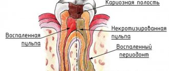
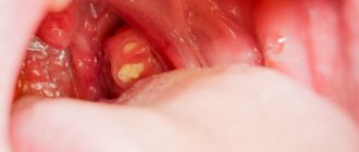
The main indications for such an operation are the presence of a cyst or granuloma in any area of the tooth root.
The formation of a cyst occurs quite unnoticed. At the same time, inflammation and destruction of bone tissue develops. In appearance, the cyst is similar to a sac, which is formed from durable fibrous tissue and filled with purulent masses or liquid. Aggravated forms of this pathology are accompanied by headaches, inflammation of the lymph nodes, periostitis, swelling and swelling of the cheek, and severe pain at the site of its localization.
Often, cystic formations become the cause of the development of chronic sinusitis.
To treat a cyst, public dental clinics use the simplest method - tooth extraction. This is what specialists have used for many years. However, as a result of removal, another significant problem arises - there is no tooth, and chewing food, speech and aesthetics are impaired.
Currently, a microsurgical method that allows you to get rid of cystic formations of a tooth by resection of the apex of its root in addition to various conservative techniques.
These methods involve the introduction of strong medications into the root canal area or inside them, which will help relieve inflammation and prevent further infection.
The duration of such a therapeutic course can be several months.
The most effective way to treat granulomas and dental cysts of all therapeutic methods is to treat the cyst (granuloma) with a laser.
In our clinic, since 2006, we have successfully treated such diseases without surgery or surgical intervention. You can learn about our technique using the SIRONA SLA laser in the section of this site TREATMENT OF DENTAL CYST WITH SIEMENS LASER WITHOUT SURGERY
If treatment of a cystic formation with a laser is ineffective, and this happens in approximately 3% of all clinical cases, microsurgical resection of the apex of the tooth root is prescribed.
Causes of the problem
Fistula (fistula) is not a simple disease. Many factors can influence the formation of a fistula, so the disease can occur in different places and manifest itself in different ways. For example, a fistula on the gum is often a complication of periodontitis. A fistula can form after the removal of a wisdom tooth if the patient did not take good care of the hole and an infection entered the wound. This is also the cause of the appearance of a fistula after the removal of a baby tooth in a child. Often, a fistula occurs after poor root canal filling.
In addition to these main reasons, there are other provoking factors:
- medical errors during tooth extraction, due to which root perforation occurred and the infection remained inside the socket,
- the appearance of a cyst on the root, which over time became inflamed with the formation of suppuration,
- incorrect eruption of the wisdom tooth and its subsequent difficult removal,
- severe caries or advanced inflammation of the nerve (pulpitis), for example, adjacent to the extracted tooth.
A fistula in the maxillary sinus is a serious complication after extraction of an upper jaw tooth. Since the canal formed after the procedure connects the sore spot and the maxillary sinus, inflammation of the latter may occur due to infection.
On a note! Fistula in children is not always caused by tooth decay and inflammation at the root. During teething, your baby's gums are vulnerable. In turn, babies try to lick and suck dirty fingers and other objects. Bacteria enter wounds and can cause inflammation.
Difficulties of conservative treatment of cysts and granulomas when using outdated treatment methods.
As a result of poor-quality filling of dental canals, inflammation in the bone tissue can develop. When depulpation is performed, for example, during the period of treatment of pulpitis, the pulp is removed and the canal is filled to the full depth of the tooth root. There are cases when this procedure was not performed completely, and the empty area became a favorable place for the development of the inflammatory process with the subsequent formation of a cyst. The technique used in Russia in 99% of public dental clinics and 80% of private dentistry is outdated and does not give a positive result of endodontic treatment. The drugs used are obsolete, and the technique does not disinfect the canals 100%.
As a result of such treatment, the dentist does not completely remove the infection from the canal.
The method of laser treatment of dental pulpitis according to the European protocol is recognized throughout the world. You can find it on our website on the LASER TREATMENT OF PULPITIS page. Only such treatment guarantees the absence of complications and excludes the development of granulomas and cysts after canal treatment.
Another situation is when poor-quality filling of tooth canals is associated with the use of the Soviet resorcinol-formalin method of dental treatment. With this technique, popular in the USSR, the tooth canal was not treated, but a mixture of resorcinol and formaldehyde was poured into the tooth, which was supposed to kill and preserve the nerve. However, dentists could not tell by eye whether complete preservation of the nerve had occurred. And most often, the nerve began to rot in the canal a year and a half after using this technique. To treat such a tooth, the dentist must remove the old root filling and perform the canal filling procedure in a new way, using modern technology. However, repeated filling of such areas, especially when the canals are too tortuous, often leads to perforation of the root wall. This is an unpleasant complication that is difficult and expensive to correct. For this reason, based on the data after the CT scan, a decision is made on what will be the best treatment option. Sometimes doctors believe that it is better not to take risks, not to try to clear the resorcinol-formalin channels, but to perform an excision of the apex of the tooth root in order to remove the cystic formation and inflammation.
It is also possible that situations where the canal directly in the part of the apex of the tooth root is not completely sealed, and there is a crown, inlay or pin on the tooth. As you know, conservative therapy involves replacing the old crown, subsequent filling and installation of a new version of the crown. While removing a pin made of high-strength material often causes a root fracture.
In our clinic, we successfully extract stump inlays and pins of any type from the roots of the tooth using an ultrasonic unit SIROSONIC SIEMENS (Germany), however, this is rare equipment and is available in no more than 1% of dental clinics. Therefore, mid-level clinics convince the patient that he is indicated for resection of the apex of the tooth root, since it is the easiest and safest method of eliminating infected tissue.
The same treatment tactics are offered to patients if a foreign body is detected near the tooth roots, and this procedure will also be prescribed. During dental procedures, cases sometimes occur when fragments of the instruments used for this remain in the canals. The consequence of such negligence is usually the development of inflammation, which requires urgent and effective therapy.
Working according to European protocols for endodontic treatment, dentists at the Bionic Dentis clinic in 97% of cases, when a dental cyst or granuloma is detected, carry out therapeutic non-surgical treatment of the dental cyst with a laser.
However, there are those 3% of teeth that cannot be treated with laser. There is also a category of patients who refuse laser tooth treatment through a root canal, for the reason that a crown or bridge is installed on the tooth, and the patients want to keep this crown for financial reasons.
We offer laser resection of the tooth root to such patients.
Using a laser
Modern dentistry and the advent of laser equipment in medicine have made it possible to perform resection of the apex of the tooth root in a more gentle way. The course of the operation and the rehabilitation period are significantly different from the traditional method.
Laser correction consists of the following stages:
- shallow cut. Penetration affects no more than 1 cm of tissue deep;
- carrying out all manipulations to remove the root apex and tumors using an ultrasonic unit, which is less traumatic for the oral cavity;
- laser treatment of inflammation areas;
- closing the wound with synthetic bone tissue and biomembrane.
Among the advantages of using a laser during resection are:
- speed of all manipulations;
- less pain in the postoperative period;
- reducing the likelihood of developing an inflammatory process to a minimum;
- no bleeding.
The only disadvantage of laser correction is its relatively high cost.
The video describes the indications for tooth root resection and the procedure for performing the operation.
When is tooth root resection contraindicated?
It is recommended to refuse this operation if:
- strong mobility of the dentition;
- acute forms of cardiovascular and some infectious diseases;
- exacerbations of periodontitis;
- noticeable damage to the dental crown;
- cracks in the area of the tooth root.
- too short tooth root
How is the apex resection procedure performed at the Bionic Dentis dental clinic?
This operation may require 30-60 minutes.
The fact is that the duration largely depends on the location of the affected area. For example, when performing treatment in the area of the front teeth, less time is required and the entire process is much easier. When foci of inflammation affect the back teeth, surgical intervention may be delayed due to difficult access.
It is important how many roots are affected by the inflammatory process. If it is one root, the operation will be shorter than three roots.
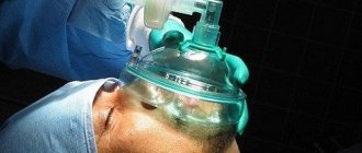
In any case, local anesthesia is performed before the procedure. We use only the German painkiller Ultracain DS Forte (made in Germany), which allows us to perform the operation without discomfort and pain.
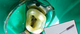
When tooth root resection is planned on unfilled canals, they are first filled. If it is possible to re-treat the canals for at least 2/3 of the root length, they are re-treated.
To fill canals, the Bionic Dentis clinic uses a special technique - 3D canal filling.
Here's what it looks like:
- The canals are expanded and infected tissue is removed from them mechanically using a nickel-titanium M2 instrument. For this purpose, a computerized root canal system from the German company WDV is used.
- After mechanical cleaning, the channels are sterilized with a medicinal preparation, a sodium hypochlorite solution activated by ultrasound. We use the SIEMENS SIROSONIC ultrasound system. Penetrating into the wall of the canal, the disinfectant kills microbes and opens microscopic tubules, clearing them of infection.
- After disinfection, the canal is dried.
- A small amount of epoxy resin, created for use in dentistry, is injected into a dry and clean canal.
- A gutta-percha pin is inserted into the canal and melted under pressure. Melting, gutta-percha fills all the pores in the canal and micro-branches.
- Next, the channel is filled with molten gutta-percha under pressure from an injector.
The use of this technique is standard in Europe. We also follow this method.
Are complications possible?
To avoid undesirable consequences, resection should only be performed by an experienced doctor. Swelling of the gums and minor pain for some time after surgery are normal. But if these symptoms do not go away and intensify after 2-3 days, you need to consult a doctor.
For your information! Like other procedures, resection has its pros and cons. An undoubted advantage is that the removed bone tissue will be completely restored in 3-4 months and will be able to perform its functions. A tooth that has been preserved can last for decades and even serve as a support for dentures. The possibility of relapse is minimized1, soft tissues will heal in 7-10 days. Among the disadvantages are possible complications and relatively high cost.
What other complications are possible: perforation into the maxillary sinus in the upper jaw and hypoesthesia. Perforation is the formation of a hole, in our case connecting the sinuses and the upper jaw. Pathology can be fraught with the formation of a fistula, which will provoke purulent sinusitis. Hypoesthesia is a disturbance of sensitivity. A so-called “numb” tongue or chin may indicate that a nerve was damaged during surgery. If this unpleasant sensation does not go away a couple of hours after the procedure, you need to seek help from a doctor.
What anesthesia is needed for resection of the root apex.
When performing root apex resection on the upper teeth, it is recommended to use infiltration anesthesia. This approach ensures a long-lasting effect of painkillers and penetration deep into the tissue.
Infiltration anesthesia involves injection of anesthetic agents using a needle under the mucous membranes of the gum tissue. This allows the injected medication to gradually travel through the bone, affecting the nerves and tissues of the mouth and periodontal ligaments.
Using infiltration anesthesia, the specialist manages to completely block nerve fibers in all tissues and bleed the periodontium. Therefore, at the injection site you can notice a pronounced change in the color of the gums. To perform high-quality diffusion, a porous bone structure is required. For this reason, infiltration anesthesia is used during treatment of the upper teeth.
At the Bionic Dentis clinic we perform infiltration anesthesia in 2 stages:
- The first stage is to apply a gel to the gums that relieves sensitivity at the injection site.
- The second stage involves submucosal administration of the drug to a place that is “frozen” with topical anesthetic. The patient feels almost nothing during the injection.
The procedure for resection of the apex of the tooth root in the lower jaw is carried out using conduction anesthesia. This anesthetic technique is characterized by the introduction of the drug into areas adjacent to the nerve endings. As a result, the nerve fibers themselves and the tissues close to them are well saturated with this product. Typically, an anesthetic is injected into the surrounding area around the trigeminal nerve.
The procedure itself includes several stages:
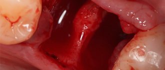
- Providing access to the root apex. To do this, an incision is made on the gum in the shape of an arc and the mucous membrane is peeled off to expose the bone and move the periosteum. After this, in a certain area where the root tip is supposed to be located, it is necessary to make a hole in the bone tissue. When working with bone tissue, we do not use a drill , as this injures the bone tissue and causes tissue burns. We use an innovative European development - the PIEZOSURGERY system . This ultrasonic system for working with bone tissue and teeth is the best option to perform root resection without harming surrounding tissues. With all these actions, patients will not feel any pain.
- Resection procedure. The hole created by ultrasound using the PIEZOSURGERY system will be used to locate the root tip and directly carry out the cutting procedure. Then, using a special device, the dentist will remove the affected fragments along with the cystic formation and the upper part of the root. When an empty niche remains at the excision site, it is filled with synthetic material, which can accelerate the recovery processes in bone and other tissues. We use only fully synthetic bone tissue in our practice and do not use cadaveric bone tissue or tissue of animal origin due to possible rejection.
- After instrumental cleaning of the cystic cavity, the SIRONA SIEMENS laser is switched on. Using it, our dentists process the cavity, achieving 100% sterilization and clean it of pathogenic microorganisms. The laser allows you to avoid the recurrence of dental cysts.
- The innovative resection technique at the Bionic Dentis clinic is carried out using Active tissue regeneration proteins , which we obtain from the patient’s blood. 10 minutes before the operation, blood is drawn from a vein, the blood is processed, and tissue regeneration proteins are released from it. These proteins are introduced into bone tissue. Under their influence, rapid healing and restoration of the bone occurs.
- After installing synthetic bone tissue and active regeneration proteins into the bone cavity, the defect is closed with a bioresorbable membrane to prevent leaching of the implanted components.
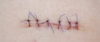
- Suturing the resection site. After such an event, a wound remains on the mucous and bone membranes, which must be carefully sutured. For this purpose, specialized suture material is used.
Complications after surgery
Tooth root resection is a serious surgical intervention that requires the specialist to have certain knowledge and extensive practical experience in dental surgery. Only then can you be sure of a positive outcome of the procedure and the absence of complications.
Resection can only be performed by a certified dental surgeon.
When performing a conventional tooth root resection, there are types of complications:
- injury to blood vessels;
- damage to the alveolar nerves;
- perforation of the nasal cavity;
- suppuration of a postoperative wound;
- reappearance of the cyst if the affected area was not properly cleared of inflammation.
Sometimes the development of complications is caused by unfavorable anatomical factors, for example, the close location of the upper dentition from the bottom of the maxillary sinus. Although extreme caution during surgery and larger incisions can be used to avoid this complication.
As a result of nerve injury, facial parasthesia may form. Such injuries require physiotherapeutic treatment using UHF and electrophoresis with potassium iodide. Dibazole and vitamin therapy are also used.
When performing ultrasound resection using active PRF regeneration proteins, such complications do not occur.
At the Bionic Dentis dental clinic, when using the apicotomy technique using ultrasound, laser and active regeneration proteins, the level of complications is reduced to 0.5%. We work according to the European safety standard for our patients and carry out multi-level quality control of our work.
Possible risks during tooth resection
Like any surgical intervention, apiectomy is associated with the risk of complications:
- risk of opening into the nasal cavity. It is possible only with a horizontal incision in the gum above the upper incisor. As a result, over time the tooth begins to “whistle”;
- When operating on lower molars, there is a possibility of touching the mental nerve. This is fraught with disorders of the surface sensitivity of tissues (from mild tingling to complete numbness);
- there is a certain risk of damage to the maxillary sinus. The consequence is the danger of purulent formations such as sinusitis;
- “standard” complications: swelling or edema near the operated area, sharp pain when pressing.
It is important to understand that the consequences of tooth root resection can “dormant” for a long time and appear after a considerable period of time. Therefore, patients who have undergone an apiectomy must be regularly monitored by a dentist.
Advantages of ultrasonic root resection PIEZOSURGERY:
- Vessels and nerves are not damaged, since the ultrasound system acts only on the bone and does not injure soft tissues.
- Bone tissue is not rejected, since it is completely synthetic and not cadaveric or animal.
- There is no allergy to the implanted synthetic bone.
- There is no burn or injury to bone tissue, since the bone is not cut out using a cutter or drill.
- There is no unpleasant smell of burnt bone during surgery.
- Ultrasound will completely remove infected, altered tissue from the root apex and will not leave an infection.
- There are no relapses - the cyst is removed forever with a 100% guarantee.
- Laser bone sterilization is the key to complete cleansing of bacteria.
Features of postoperative rehabilitation
After such surgery, the patient should refrain from any physical activity for 24 hours. Eating food is allowed only after 3 hours. Subsequently, care must be taken to ensure that the oral cavity is not exposed to thermal irritants, to limit the consumption of spicy and salty foods, as well as the use of overly aggressive means for rinsing and brushing teeth.
Swelling may persist for 1-2 days and moderate pain may be felt. To prevent the development of a purulent and inflammatory process, antibacterial agents and antiseptic solutions are prescribed, which must be used to thoroughly rinse the mouth. After 6 months you should undergo an x-ray examination.
Treatment of fistula
You cannot self-medicate; you need to consult a dentist. The doctor must assess the extent of the disease, if necessary, open the fistula and remove the pus. If fillings, crowns, etc. interfere with this process, the doctor can remove them.
Antibiotics are used as an aid. But you can’t do this without a trip to the dentist. A good specialist will study the problem and prescribe the correct course of antibiotics. Usually, tablets are taken for 5 to 10 days, depending on the size of the fistula.
Traditional medicine is often used. This is an integral part of complex treatment. The consequences of using traditional medicine on the body can be unpredictable. Therefore, it is better not to self-medicate without the approval of a specialist. You can't guess the body's reaction. Many medicines are made from herbs, so infusions can be used as an antiseptic or to relieve pain.
You can rinse your mouth with decoctions of eucalyptus, millennial, and calendula. Such procedures can be done even when there is no disease, but simply for prevention. Recipes for preparing decoctions should only be obtained from a doctor.
Reviews of tooth root resection using the PIEZOSURGERY ultrasonic system
According to many patients who underwent the procedure of excision of the root apex, they continued to have swelling on their face for some time and felt moderate pain. For such a surgical procedure, these symptoms are considered quite normal. Sometimes they persist for up to 5 days, but usually disappear after 3 days.
As a result of resection, the length of the tooth root is shortened, which can affect its subsequent stability and functioning. However, this procedure allows you to avoid the development of serious complications that require long-term treatment and drastic interventions. But the most important thing is to preserve the remaining teeth, preventing them from becoming infected and forming various dental problems.
Patient reviews of ultrasonic resection of the apex of the tooth root are positive in 99% of cases. Patients note the absence of an unpleasant smell of burnt bone during surgery, absence of pain and minimal swelling in the postoperative period.
| № | Procedure/manipulation | Price, rub.) |
| 1 | Treatment of cyst/granuloma with Sirona SIEMENS laser on a tooth with 1 channel (I visit) | 65 000 |
| 2 | Treatment of cyst/granuloma with Sirona SIEMENS laser on a tooth with 2 channels (I visit) | 70 000 |
| 3 | Treatment of cyst/granuloma with Sirona SIEMENS laser on a tooth with 3 channels (I visit) | 75 000 |
| 4 | Treatment of cyst/granuloma with Sirona SIEMENS laser on a tooth with 4 channels (I visit) | 80 000 |
| 5 | Treatment of cyst/granuloma with Sirona SIEMENS laser on a tooth with 1-4 channels (II visit) | 25 000 |
Ozerov Petr Vladimirovich
Chief physician. Dentist, implantologist, orthopedist, surgeon. Laser dentistry specialist
More details
Andreev Dmitry Lvovich
Dentist orthopedist-implantologist.
More details
Frolov Andrey Konstantinovich
Orthopedic dentist
More details
The Bionic Dentis dental clinic uses the best currently known method of root apex resection.
We use:
- Ultrasound system PIEZOSURGERY (Italy).
- Dental laser SIRONA SIEMENS (Germany).
- Synthetic bone NanoBone (Germany), which is not rejected and does not cause an allergic reaction.
- The use of active regeneration proteins iPRF allows you to significantly speed up healing and eliminate relapses.
- Our dental surgeon is certified, which is confirmed by a diploma, and has 15 years of experience.
By choosing our clinic for resection, you will receive all the best that modern world dentistry has in this area.