The facial nerve is a cranial nerve that is located between the medulla oblongata and the pons. Its paths run along the facial muscles, which it innervates. The facial nerve contains another nerve, the intermediate nerve. This nerve is directly connected to the innervation of the lacrimal gland and stapedius muscle, and is also responsible for certain parts of the taste sensitivity of the tongue.
The facial nerve is formed from cell processes, is motor, however, as part of the intermediate nerve, it performs mixed functions. Both nerves run at the base of the brain, connecting with the vestibulocochlear nerve. Next, three nerves move into the internal auditory canal through the auditory opening of the pyramid. In the auditory canal, reunited, the intermediate and facial nerves enter the facial nerve canal. Next, the formation of the genu of the facial nerve occurs from the bend of the canal, and the genu, having formed into a node, gives sensitivity to the composition of the intermediate nerve.
Before entering the thickness of the parotid gland, the facial nerve branches into separate branches: the posterior auricular nerve (2 branches - the anterior auricular branch and the posterior - occipital branch), stylohyoid branch, digastric branch, lingual branch.
And the intermediate nerve, being inside the temporal bone, gives rise to the following branches: the greater petrosal nerve, the connecting branch with the tympanic plexus, the stapedial nerve, the connecting branch with the vagus nerve, the chorda tympani (terminal branch).
Another branching of the facial nerve occurs already in the thickness of the parotid gland and gives rise to two main branches - a powerful upper and smaller lower branch, which in turn also branch. This branching is radial: up, forward and down to the facial muscles. As a result, the radial branching forms the parotid plexus.
The task of the facial nerve is the motor functions of the face, but its structure contains taste and secretory fibers related to the composition of the intermediate nerve. This suggests that the fibrous structure of the facial nerve is born from several nuclei. One motor nucleus, formed from separate cell groups, is responsible for motor functions in the nerve. These groups innervate different facial muscles. One of the cell groups provides bilateral cortical innervation to the eyelids and forehead. The facial nerve facilitates the work of muscles in performing a synergistic act of both areas of the face: muscles can contract simultaneously or separately, creating different facial expressions for different functions (eating, emotions, etc.).
Peripheral facial paralysis
When the motor function of the facial nerve is damaged, peripheral paralysis occurs. The disease manifests itself as facial asymmetry, in which characteristic signs are observed: the absence of muscle movements on the face and their sharp movement during facial expressions. The affected facial area remains motionless; when trying to wrinkle the skin on the forehead in the affected part of the face, it does not lead to the desired result and the skin folds do not gather. The patient cannot close his eye and during such an attempt the eyeball rolls upward, exposing the sclera.
However, with paresis of the orbicularis muscle, if the lesion is moderate, then the patient can close both the left and right eyes, but do this only symmetrically. Closing just one healthy eye does not present any difficulty or obstacle. During rest, when the patient sleeps, the eye muscle relaxes, which helps improve its closure. When trying to inflate the cheek on the affected facial area, air passes through the affected part of the mouth (corner), expressing the sail sign. With paralysis, the corner of the mouth takes a downward direction, and the fold between the lip and nose is smoothed. Since muscle tone is reduced, when you try to lift the affected corner of the mouth with your own hands, it lifts without any changes in shape. Baring is not done properly - the teeth remain covered by the lips.
As a result, the picture of the disease is as follows:
- pronounced asymmetry of the mouth is a symptom of a racket, as it resembles its shape
- paralyzed facial muscles make it difficult to eat
- bites of the oral mucosa occur on the affected part
- voluntary release of drool and liquid food from the corner of the mouth
- difficulty speaking
- difficulties with some functions (attempting to whistle, blowing out a candle)
Damage to the facial nerve in the pyramid of the temporal bone has several causes
- regarding the tympanic: peripheral paralysis of the facial nerve, there is an absence of taste buds on the front of the tongue (2/3); the disease is characterized by dry mouth, due to dysfunction of the secretion of the sublingual and submandibular salivary glands
- regarding the stapedius nerve: the symptoms are the same as above; in addition, hyperacusis occurs
- relatively large petrosal nerve: symptoms are the same as for the relatively tympanic nerve; sometimes nerve deafness is observed, in its absence hyperacusis occurs; xerophthalmia occurs
The possibility of developing the following syndromes: internal auditory canal syndrome, which is called Lyanitz syndrome; Pontine lateral cistern syndrome, otherwise called cerebellopontine angle syndrome.
Other injuries in peripheral facial palsy are damage to the facial nerve in the cranial cavity and damage to the facial nerve nucleus.
Innervation of the facial muscles: intraparotid plexus of the facial nerve
Medicine has been dealing with colds of the facial nerve for a long time and today. This is mainly a seasonal illness associated with cold weather.
Diseases of the facial nerve usually manifest as problems with the movement of facial expressions. The symptoms of the disease may be similar to other ailments, which may be chronic and seem harmless in the early stages. Therefore, if you have alarming symptoms, you should not hesitate to go to the doctor.
The first signs may be mild pain in the ears. Then it is better to immediately contact an ENT doctor. If it does not go away and the face becomes distorted (the skin may also freeze in the mask), then most likely the facial nerve is damaged or inflamed.
A type of disease is paralysis of the motor muscles of the face. In this case, the person has visible facial asymmetry, one side resembles a mask, the eye on it does not close.
Paralysis usually develops due to injury to the tympanic portion of the facial nerve canal. This often happens during ear surgery. The disease also has associated problems:
- Getting dust and dirt onto the mucous membrane (with the eye open).
- The likelihood of conjunctivitis.
- With this type of disease, the patient cannot wrinkle his forehead himself.
Peripheral paralysis occurs when basic motor function is affected. Then complete asymmetry of the face occurs due to paralysis of the facial muscles. The movements of the speech apparatus are also impaired.
In addition to paralysis, there are paresis of the facial nerve. They are not always noticeable. With paresis, muscle movement functions are only partially impaired. The disease manifests itself most often when a person is talking. Paresis is classified into:
- Easy. Changes in facial expressions are minor.
- Heavy. The face turns into a mask. He is close to paralysis.
The cause of paresis and paralysis can be both compression of the fibers and nerve damage.
Symptoms of paresis and paralysis are as follows:
- difficulties with swallowing, speech;
- drooling, lacrimation;
- asymmetry of facial muscles;
- pain;
- eye twitching;
Signs may also include:
- bulging of the eyeball or turning it to the side;
- difficulty in pulling out the lips;
- hearing impairment, greater sensitivity;
- lacrimation.
The phase of the disease is also important. In its acute form, the symptoms appear most clearly.
Unlike paresis, neuritis is characterized by inflammation. The inflammatory process can occur in any part of the face.
There are secondary and primary neuritis. The first develops due to a cold. The second is a complication from other diseases. For example, the disease often develops with chronic otitis media, and neuritis of the facial nerve is also associated with otorhinolaryngology.
There may be many causes of the disease. It is important which part of the nerve is affected. Neuritis can also develop due to diseases:
- diabetes;
- middle ear disease;
- stroke;
- ischemia;
- infection;
- oncology;
- multiple sclerosis;
- cerebral circulatory disorders and others;
The main reason is hypothermia.
1. Temporal branches - the anterior and superior auricular muscles, as well as the muscles that surround the eye;
2. Zygomatic branches - the zygomaticus major muscle and the orbicularis oculi muscle;
3. Buccal branches – muscles of the same name and facial muscles located above the oral fissure;
4. Marginal mandibular branch - facial muscles located below the oral fissure;
5. Cervical branch - part of the superficial loop of the neck (ansa cervicalis superficialis).
The above branches diverge in the form of rays, forming the so-called pes anserinus major - a large crow's foot.
Central facial palsy
Central facial palsy occurs due to pathological manifestations in the cerebral cortex. Also, the cause of paralysis lies in the corticonuclear pathways that run to the facial nerve system. Central facial paralysis occurs opposite the pathological focus, usually in the lower region of the face.
Thanks to the connection of the facial nerve with the extrapyramidal system, the facial muscles perform involuntary movements, which are expressed in the form of tics and facial spasms. The disease may be accompanied by isolated supranuclear palsy, as well as attacks of epilepsy.
Types of facial muscles and their functions
The main functions of these muscles are already clear from the name. Chewing muscles are necessary for chewing food, facial muscles are necessary for expressing emotions. A cosmetologist works with facial muscles, so it is most important for him to know the structure of this group.
The topography of the deep region of the face is associated not only with the facial nerve. Its interweaving with the intermediate means that the front one shares responsibilities with it.
The facial nerve is responsible for innervation of almost all facial muscles. It provides motor functions of the face. Nerve fibers innervate:
- facial expressions;
- subcutaneous muscle of the neck;
- stylohyoid muscle;
- occipital
Parasympathetic autonomic fibers supply nerves to the following glands:
- tear;
- sublingual;
- submandibular;
- nasal mucosa;
- hard and soft palate.
VII pair - facial nerves
The facial nerve (n. facialis) is mixed, has motor, sensory and parasympathetic fibers (Fig. 528).
528. Branches of the facial nerve. 1 - rr. temporales; 2 - rr. zygomatici; 3 - rr. buccales; 4 - rr. marginalis mandibulae; 5 - r. colli; 6 - pi. parotideus; 7 - n. facialis
The motor part of the facial nerve begins from the nucleus located in the dorsal part of the medullary pons, surrounded by the reticular formation, on the border with the medulla oblongata posterior and outward from the superior olive. The intracerebral part of the nerve root rises upward and goes around the nucleus of the abducens nerve. This bend represents the intracerebral genu of the facial nerve. The facial nerve exits the ventral surface of the brain between the posterior edge of the pons and the olive of the medulla oblongata and enters the internal auditory canal (porus acusticus internus), and then into the canal of the facial nerve of the pyramid of the temporal bone. Initially, the nerve lies horizontally, reaching the large stony foramen (hiatus canalis n. petrosi majoris), near which the nerve turns back and laterally at an angle of 90°. This first bend of the nerve is called the genu (geniculum n. facialis). Having passed 6-8 mm above the tympanic cavity, the facial nerve forms a second bend and changes its horizontal position to a vertical one. The vertical part of the nerve passes behind the tympanic cavity and through the stylomastoid foramen (for. stylomastoideum) enters the retromaxillary space, in which the parotid salivary gland lies. In its thickness, the facial nerve is divided into 5-10 branches, diverging radially to the facial muscles. The branches of the nerve form small and sometimes large loops of the parotid nerve plexus.
A number of branches arise from the motor fibers of the facial nerve.
1. The stapedius nerve (n. stapedius) is very short and thin, extending from the second flexure of the facial nerve. Penetrates into the tympanic cavity, ending in the stapes muscle (m. stapedius).
2. The branch for innervation of the muscle that lifts the soft palate arises in the facial canal. Motor fibers, together with parasympathetic fibers, exit through the canaliculus chordae tympani into the petrotympanic fissure at the base of the skull, where they enter the gangl. oticum. The nerve innervates m. levator veli palatini.
3. The connecting branch with the glossopharyngeal nerve (r. communicans cum n. glossopharyngeo) is separated from the nerve near the stylomastoid foramen and along the m. stylopharyngeus reaches the wall of the pharynx, connecting with the branches of the glossopharyngeal nerve.
4. The posterior auricular nerve (n. auricularis posterior) departs from the facial nerve on the outer base of the skull near the stylomastoid foramen, goes back upward, bending around the mastoid process in front. Innervates the occipital belly of the supracranial muscle, the posterior and superior auricular muscles.
5. The digastric branch (r. digastricus) is thin, extends below the previous nerve, innervates the posterior abdomen of the m. digastricus and m. stylohyoideus.
6. Temporal branches (rr. temporales) emerge from the parotid plexus. Among them, the anterior branches are conventionally distinguished (they innervate the upper part of the orbicularis oculi muscle and the corrugator muscle), the middle ones - the frontal muscle, the posterior ones - the anterior and partially the superior auricular muscles.
7. Zygomatic branches (rr. zygomatici), numbering 2-5, innervate the lower part of the orbicularis oculi muscle and the zygomatic muscle.
8. Buccal branches (rr. buccales), numbering 2-4, innervate the buccal, orbicularis oris muscles, the levator anguli oris muscles and the upper lip.
9. The marginal branch of the mandible (r. marginalis mandibulae) is located along the edge of the lower jaw and innervates the laughter muscle, the mental muscle, the depressors of the angle of the mouth and the lower lip.
10. The cervical branch (r. colli) passes near the angle of the lower jaw to the neck and innervates m. platysma.
The sensitive part of the facial nerve consists of two parts: the first is the fibers of the taste analyzer, arising from the receptors of the taste fields of the tongue, the second is the fibers of general sensitivity.
In the first part, sensitive unipolar cells are located in the ganglion ganglion (gangl. geniculi), located in the genu of the facial canal. The node has dimensions 1×0.3 mm. The taste buds are located on the front 2/3 of the tongue in the taste pores. The fibers of the taste nerve enter n. lingualis and leave it at the upper edge of the medial pterygoid muscle, penetrating the chorda tympani. Sensitive fibers of the chorda tympani enter through the petrotympanic fissure into the tympanic cavity and pass in its submucosal layer between the long leg of the incus and the handle of the malleus. From the tympanic cavity through the petrotympanic fissure they enter the facial canal. Exiting through the porus acusticus internus at the base of the skull, the fibers penetrate the brain and switch in the sensitive nucleus (nucl. tr. solitarii).
The second part of the nerve contains general sensory fibers that contact receptors located in the skin of the inner surface of the auricle. Their sensory cells are located in the gangl. geniculi.
3. Parasympathetic (secretory) fibers of the facial nerve are directed from the superior salivary nucleus (nucl. salivatorius superior), located in the dorsal part of the medullary pons. The root fibers of this nerve exit the base of the brain next to the motor fibers of the facial nerve and, together with them, enter the facial canal. Preganglionic parasympathetic fibers are divided into two portions and leave the facial canal (Fig. 529).
529. Diagram of autonomic and sensory nodes with nerve fibers located in the head (according to Muller). The blue line is parasympathetic fibers from the midbrain and boulevard regions, the red line is sympathetic preganglionic fibers; red dashed - sympathetic postganglionic fibers. 1 - n. oculomotorius; 2 - n. trigeminus; 3 - n. facialis; 4-n. glossopharyngeus; 5 - gangl. sublinguale; 6 - gangl. oticum; 7 - gangl. sphenopalatinum; 8 - gangl. ciliare
The first portion is separated in the area of the knee and, through the entrance to the canal of the greater stony nerve (hiatus canalis n. petrosi majoris), enters the cavity of the middle cranial fossa called the greater stony nerve (n. petrosus major) (Fig. 529). This nerve passes through the connective tissue of the foramen lacerum of the skull and enters the pterygoid canal (canalis pterygoideus) of the sphenoid bone. Before entering this canal, the great petrosal nerve is joined by the deep petrosal nerve (n. petrosus profundus), composed of postganglionic sympathetic fibers from the cells of the internal carotid plexus (plexus caroticus internus). The pterygoid nerve exits into the pterygopalatine fossa, where parasympathetic fibers switch to neuron II and form the pterygopalatine ganglion (gangl. pterygopalatinum) (Fig. 525).
The following fibers come to the node: parasympathetic - through n. petrosus major, which have contacts in the node with the next neuron; sympathetic - through n. petrosus profundus, which pass the node and, as part of its branches, reach the vessels and mucous membrane of the nasal cavity and nasopharynx; sensory fibers form branches: rr. orbitales, nasales posteriores superiores, palatini. Parasympathetic postganglionic fibers also begin from the pterygopalatine ganglion, which pass as part of the nn. pterygopalatini, maxillaris, zygomaticus. In the orbit they leave the zygomatic nerve, forming an anastomosis with n. lacrimalis. In its composition they reach the lacrimal gland.
The second portion of preganglionic parasympathetic fibers continues its path initially along the facial canal, and then passes into the canaliculus chordae tympani, located in the same bundle with sensory (taste) fibers called chorda tympani. The drum string is connected to n. lingualis. Its parasympathetic fibers exit from the lingual nerve to the submandibular and sublingual salivary glands. Near the submandibular gland they form a gangl. submandibulereis, in the sublingual - gangl. sublingualis. Postganglionic parasympathetic fibers emerge from the nodes for secretory innervation of the submandibular and sublingual salivary glands and mucous glands of the tongue.
Embryogenesis. The motor nucleus is formed in the 4th week of embryonic development near the bottom of the IV ventricle in the column of tegmental cells of the medulla oblongata and comes into contact with the derivatives of the II branchial arch. During development, the nucleus of the facial nerve shifts in the ventrolateral direction and its fibers become curved. Axons come into contact with visceral myotomes, where facial muscles are formed.
Phylogenesis. In fish and amphibians, the facial nerve departs from the medulla oblongata by several roots, having a node into which the lateral and facial nerves themselves flow. The lateral nerve innervates seismosensory organs, which disappear in terrestrial animals, which causes a reduction of this nerve.
The facial nerve itself in aquatic and terrestrial animals has sensory and motor branches. Sensory fibers begin from the taste buds of the oral mucosa and lateral line. In terrestrial animals, the sensitive part of the lateral line disappears, and the head part passes through the tympanic cavity, maintaining connection with the taste buds of the tongue, and is called chorda tympani. Motor fibers innervate the muscles of the suspension and operculum in fish, the premaxillary muscle, the muscle that depresses the mandible, and the subcutaneous cervical muscles in terrestrial animals. Mammals have well-developed facial muscles, which are also innervated by a special branch of the facial nerve, which in humans, due to the development of facial muscles, has received predominant development.
Arteries of the face
The largest percentage of blood supply to the face and neck comes from vessels that arise from the external carotid artery. The largest arteries are listed below:
- facial;
- supraorbital;
- supratrochlear;
- infraorbital;
- chin.
Branches of the facial artery guarantee most of the blood supply to the face. It branches off from the external carotid artery at the level of the mandible. From here it goes to the corner of the mouth, and then approaches the corner of the palpebral fissure, closer to the nose. At the level of the mouth, branches branch off from the facial artery, carrying blood to the lips. When the artery approaches the canthus, it is already called the angular artery. Here it communicates with the dorsal nasal artery. The latter, in turn, arises from the supratrochlear artery, a branch of the ophthalmic artery.
The supraorbital artery supplies blood to the superciliary ridges. The infraorbital vessel, true to its name, carries blood to the area of the face under the eyeball.
The mental artery provides blood supply to the lower lip and, in fact, the chin.
Topographic anatomy of vessels and nerves of the face
⇐ PreviousPage 6 of 15Next ⇒
Facial vessels The main source of blood supply to the face is the external carotid artery. From the neck area, the facial artery comes to the face, which is projected onto the skin from the middle of the body of the lower jaw to the inner corner of the eye. Gives large branches: the arteries of the upper and lower lips and the final branch - the angular artery, anastomoses with the orbital artery through the nasal arteries.
The second large artery - the maxillary artery (a. xillaris) - departs from the external carotid artery in the thickness of the parotid gland at the level of the neck of the articular process of the lower jaw, goes into the deep region of the face, lies on the outer surface of the external pterygoid muscle and lies first in the temporo- pterygoid tissue space, then - in the interpterygoid space.
A. maxillaris is the largest branch of the external carotid artery, it gives off 19-20 branches and supplies blood to the entire deep area of the face with the masticatory muscles and the dentofacial apparatus. The artery is inaccessible for ligation, therefore, if necessary, they resort to ligation of the external carotid artery in the neck in the carotid triangle. In the deep area of the face near the artery, it is customary to distinguish three sections:
1) Mandibular (pars mandibularis) - behind the neck of the articular process. The largest branch is the inferior alveolar artery (a. alveolaris inferior);
2) Pterygoid (pars pterygoidea) - between the temporal muscle and the external pterygoid. Branches:
a) middle meningeal artery (a. meningea media);
b) deep temporal artery;
c) masticatory artery;
d) superior alveolar artery;
e) buccal arteries;
e) pterygoid arteries.
3) Pterygopalatine (pars pterygopalatine) - in the pterygopalatine fossa. Branches: infraorbital, pharyngeal, palatine, etc.
The venous system of the face is divided into two layers. The first layer of veins is formed by the facial vein system, v. facialis, the origins of which are the angular vein, supraorbital, external nasal, tube veins, nose, as well as the retromandibular vein, v. retromandibularis, located in the thickness of the parotid gland. In the region of the root of the nose, the facial vein has wide anastomoses with the superior orbital veins and through them with the vein-sinuses of the dura mater. Infection can enter the sinus veins due to carbuncles and boils of the upper lip, nose, with the development of thrombophlebitis (sinus thrombosis) and inflammation of the membranes of the brain.
The deep venous network of the face is represented by the pterygoid venous plexus (plexus pterygoideus). It drains into the postmaxillary vein. Thus, both systems are interconnected. It should be noted that the pterygoid plexus, located in the intermaxillary space, is connected with the vein-sinuses of the dura mater. The postmaxillary and facial veins merge posteriorly from the angle of the mandible into the common vein of the face, which flows into the internal jugular vein.
Nerves of the face. The facial innervation is carried out by the facial, trigeminal, glossopharyngeal nerves, and cervical plexus.
The facial nerve (7th pair of cranial nerves) primarily supplies motor innervation to the facial muscles. The nerve leaves the pyramid of the temporal bone through the stylomastoid foramen and forms the posterior auricular nerve 1 cm below.
The main trunk of the facial nerve enters the thickness of the gland and here it is divided into superior and inferior branches, from which five groups of branches arise. The branches extend radially from a point 1 cm downward from the auditory canal. Inflammatory processes in the gland can cause paralysis and paresis of the facial nerve. Incisions on the face are made only taking into account the course of the branches of the facial nerve. The nerve lies relatively shallow, and there is a high risk of damage to its branches, which also leads to paralysis of the facial nerve or its individual branches.
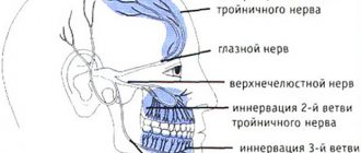
The trigeminal nerve (5th pair of cranial nerves) is mixed (sensory-motor) in structure and function. After leaving the brain stem, the nerve forms the semilunar gasserian ganglion. The node is located on the anterior surface at the apex of the pyramid of the temporal bone and lies in the cavity formed by the dura mater. Three main branches of the trigeminal nerve depart from the anterior edge of the ganglion: I) orbital; 2) maxillary; 3) mandibular.
According to its topographic and anatomical structure, the trigeminal nerve is one of the most complex. Its branches pass through hard-to-reach anatomical areas and enter into complex relationships with blood vessels. At the same time, since the nerve carries sensitive pain innervation for the dentofacial apparatus, anesthesia of the branches of the nerve is necessary during operations on the face. Therefore, we will consider the places where the large branches of the nerve exit to the face.
It should immediately be noted that the skin of the face receives pain innervation from the trigeminal nerve.
The first branch innervates the skin of the frontal and orbital areas.
The second branch of the trigeminal nerve provides pain innervation to the infraorbital region, nose, upper lip, teeth, and upper jaw. It leaves the skull through a round hole in the pterygopalatine fossa and gives off the main branches. The infraorbital nerve exits through the inferior orbital fissure, enters the orbit, lies in the infraorbital groove and exits through the infraorbital foramen. It is located 0.5 cm below the middle of the edge of the orbit, forms a “crow’s foot”, from which the labial and nasal branches extend to the lower eyelid. Along the way, the nerve gives off the superior posterior, middle and anterior alveolar nerves; they enter the upper jaw in the area of the tubercle. These nerves connect in the canaliculi of the alveolar process of the upper jaw and form the superior dental plexus.
In addition, in the pterygopalatine fossa, the pterygopalatine branches and branches of the maxillary nerve (n. petrosus major and n. facialis) form the autonomic pterygopalatine ganglion, from which the palatine nerves depart: large (exits through the greater palatine foramen), middle and posterior (enters through the lesser palatine hole), innervating the gum, soft and hard palate.
The posterior nasal nerves, a large branch of which, the nasopalatine nerve, exits through the incisive foramen and innervates the anterior part of the palate.
The mandibular nerve exits through the foramen ovale. The mixed nerve carries motor innervation for the masticatory muscles: temporal, masseter, and pterygoid muscles. Its largest branches are: buccal, auriculotemporal, inferior alveolar and lingual nerves. The inferior alveolar nerve runs down the inner surface of the external pterygoid muscle, then between the pterygoid muscles it enters the mandibular foramen and exits into the mandibular canal along with the artery. Provides pain innervation to the teeth of the lower jaw, its final branch is the n. mentales (mental). This nerve exits through the mental foramen. The lingual nerve goes to the tongue from below.
The mental nerve innervates the skin of the lower lip, the gums in the area of the canines and premolars, and the skin of the chin. The mental foramen is located midway between the lower edge of the jaw and the alveolar process.
Projection anatomy of vessels and nerves of the facial part of the head:
1. The facial artery (a. facialis) is projected from the intersection of the anterior edge of the masticatory muscle with the lower edge of the lower jaw in an ascending direction to the inner corner of the eye.
2. The mandibular foramen (foramen mandibulare) is projected from the side of the oral cavity onto the buccal mucosa in the middle of the distance between the anterior and posterior edges of the mandibular branch, 2.5-3 cm upward from its lower edge.
3. The infraorbital foramen (foramen infraorbitalis) is projected 0.5-0.8 cm downward from the middle of the lower orbital margin.
4. The mental foramen (foramen mentalis) is projected in the middle of the height of the body of the lower jaw between the first and second small molars.
5. The trunk of the facial nerve (truncus n.facialls) corresponds to a horizontal line drawn through the base of the earlobe.
Incisions for purulent parotitis
Indications. Cellulitis and abscess of the parotid gland.
Technique. The patient is placed on his back, his head is turned to the side. Three radial incisions 5-6 cm long are made. The incisions begin at the tragus of the ear: the upper one - along the lower edge of the zygomatic arch, the middle one - in the direction of the corner of the mouth, reaching the anterior edge of the masticatory muscle (m. masseter), the lower one - in the direction from the middle of the distance between the angle of the lower jaw and the chin, also reaching to the anterior edge of m. masseter
The direction of the cuts coincides with the course of the branches of the facial nerve (Fig. 83).
The skin with subcutaneous fatty tissue is dissected. Use hooks to widen the wound. The parotid-masticatory fascia is dissected using a scalpel along a grooved probe. Then the capsule and the superficial layer of the substance of the parotid salivary gland are dissected. The main danger with incisions is damage to the branches of the facial nerve that penetrate radially through the thickness of the parotid salivary gland.
Nerve branches cannot be crossed. It should be borne in mind that the stenon duct is projected along the line connecting the lower edge of the external auditory canal with the corner of the mouth or the wing of the nose; within these limits, the incision should be made with extreme caution to avoid injury to the excretory duct of the parotid salivary gland. Gauze strips (tampons) are inserted into the incisions.
When the abscesses are localized in the deep parts of the gland (mandibular fossa), an incision is made according to Voino-Yasenetsky. A 3 cm long incision is made with the head thrown back, from the earlobe downwards between the posterior edge of the ascending ramus of the mandible and the anterior edge of the sternocleidomastial muscle. The incision should be 1-1.5 cm behind the edge of the mandible so as not to damage the inferior branch of the facial nerve, which remains in front of it.
| Rice. 83. Scheme of incisions on the face and their relationship to the branches of the facial nerve |
The edges of the wound are stretched with sharp hooks and a blunt instrument (forceps), passing to a depth of 2.5 cm towards the styloid process and the posterior wall of the pharynx, penetrating through the tissue of the parotid gland (see Fig. 83).
Test tasks (choose the correct answer)
1. The transverse sinus corresponds to the anatomical formation of the bones of the skull:
1) external occipital protuberance;
2) mastoid process;
3) upper nuchal line;
4) lower nuchal line.
2. The arteries of the soft tissues of the head have the following directions:
1) axial;
Topographic anatomy of the neck. Neck fascia and fibrous spaces. Neurovascular bundles of the neck. Neck organs
Boundaries and external landmarks. The upper border of the neck area is drawn along the edge of the base of the lower jaw, through the apexes of the mastoid processes and posteriorly along the upper nuchal line. The lower border is drawn along the jugular notch of the sternum, along the upper edges of the clavicles, through the humeral processes of the scapula (acromion) to the spinous process of the 7th cervical vertebra.
To facilitate orientation in the complex topography of the neck area, and above all in the numerous vessels and nerves, various external landmarks are used, which can be divided into five groups: bone, cartilage, muscle, vascular and skin folds. Landmarks allow you to divide the neck into sections and areas, and also help plan surgical approaches to the neck.
The midline divides the neck into right and left halves. The frontal plane, drawn through the transverse processes of the cervical vertebrae, divides the neck into anterior, visceral and posterior muscular (neck) sections. The transverse plane, drawn through the hyoid bone, divides the anterior part of the neck into the suprahyoid and infrahyoid regions.
The muscles of the anterior neck form a special coordinate system in the form of triangles (Fig. 84).
The boundaries of the triangles are drawn along the contours of large muscles. The sternocleidomastoid (sternocleidomastoid) muscle divides each half of the anterior neck into an inner and outer (lateral) triangle. Within the internal triangle, the submandibular triangle is distinguished, bounded by the bellies of the digastric muscle. The unpaired mental triangle is distinguished between the anterior bellies of the digastric muscle. In addition, the internal triangle contains the carotid and scapulotracheal triangles. In the outer triangle, the scapular-trapezoid and scapular-clavicular triangles are distinguished. Triangles help navigate the complex anatomy of the neck. Each triangle is distinguished by its unique layer-by-layer anatomy and arrangement of neurovascular elements.
| Rice. 84. Triangles of the anterior surface of the neck (expanded view with the head tilted back): 1 - hyoid bone; 2 - sternocleidomastoid bone (m. sternocleidomastoidea); 3 - digastric and 4 - omohyoid muscles; 5—submandibular (trigonum submandibular); b - chin (trigonum mentale); 7—sleepy (trigonum caroticum); 8—scapular-tracheal (trigonum omotracheale); 9 — scapular-trapezoidal (trigonum omotrapezoideum); 10—scapular-clavicular triangles (trigonum omoclaviculare) |
Layers. In the layer-by-layer anatomy of the neck area, the issue of fascia and cellular spaces as anatomical elements that determine the course of purulent-inflammatory processes should be highlighted.
The fascia of the neck is an anatomical element that makes the neck a single whole. The most widespread and practically acceptable is the classification of neck fascia according to V.N. Shevkunenko (Fig. 85), according to which five fascia are distinguished on the neck (Table 12). Between the sheets of fascia there are fatty tissue and lymphoid tissue, so the fascia determines the location of phlegmon in the neck (mainly adenophlegmon) and the direction of purulent leaks.
| Rice. 85. Neck fascia on a cross section: 1 - superficial layer of the neck’s own fascia; 2—prevertebral fascia; 3 - platysma; 4 - sternocleidomastoid muscle; 5—deep layer of own fascia; b—parietal leaf of the splanchnic fascia; 7—visceral leaf of the splanchnic fascia; 8—esophagus; 9— sheath of the main neurovascular bundle; 10— long neck muscles; 11 - anterior scalene muscle; 12 - posterior scalene muscle; 13 - trapezius muscle; 14 - superficial fascia |
| Table 12 Fascia and cellular spaces of the neck Fascia | Cellular spaces |
| Superficial | Subcutaneous tissue |
| Superficial layer of proper fascia | Sac of the submandibular salivary gland Sac of the sternocleidomastoid muscle |
| Deep layer of own fascia | Blind pouches of Grubber Suprasternal cellular space |
| Internal fascia (parietal and visceral layers) | Gap of the neurovascular bundle Pre-organ space Posterior organ space |
| Prevertebral fascia | Deep prevertebral space Cellular spaces of the lateral triangle of the neck (between the 3rd and 5th and 2nd and 5th fascia) |
Neurovascular bundles of the neck. There are two large neurovascular bundles in the neck: main and subclavian.
The main neurovascular bundle of the neck consists of the common carotid artery, internal jugular vein, and vagus nerve. It is located in the neck in the area of the sternocleidomastoid (sternocleidomastoid) muscle and the carotid triangle. Thus, the main socistonervous bundle along the carotid artery has two sections: 1st section in the area of the sternocleidomastoid muscle, 2nd section in the carotid triangle. In the area of the sternocleidomastoid muscle, the neurovascular bundle lies quite deep, covered by the muscle, the 2nd and 3rd fascia. The sheath of the bundle is formed by the parietal leaf of the 4th fascia and, in accordance with Pirogov’s laws, has a prismatic shape, with spurs the sheath is fixed to the transverse processes of the cervical vertebrae.
The relative position of the elements of the neurovascular bundle here is as follows: a vein lies in front and outward of the artery, the vagus nerve is located between the vein and artery and posteriorly.
Above, the main neurovascular bundle is located in the carotid triangle (Fig. 86), which is bounded above by the posterior leg of the digastric muscle, in front by the upper belly of the omohyoid muscle, and behind by the anterior edge of the sternocleidomastoid muscle. The neurovascular bundle is not covered by muscle and third fascia. With the head thrown back, the pulsation of the carotid artery is clearly visible on the neck, and with palpation the pulse here can be determined even with a significant decrease in blood pressure. The relative position of the elements of the neurovascular bundle remains the same, the venous elements lie more superficially, and the common facial vein flows into the internal jugular vein here. The common carotid artery in the carotid triangle at the level of the upper edge of the thyroid cartilage (according to Pirogov) is divided into internal and external branches. It is practically important to know their differences. An anatomically reliable sign of the external carotid artery is the presence of lateral branches in the carotid triangle, of which the superior thyroid, lingual and facial arteries are constant. Ligation of the external carotid artery to stop bleeding throughout injuries to the maxillofacial area is performed immediately after the separation of the superior thyroid artery. The internal carotid artery in the neck does not give branches. The internal carotid artery is usually divided into three sections:
1) from the bifurcation of the common carotid artery to the hypoglossal nerve;
2) from the hypoglossal nerve to the entry into the carotid artery canal and 3) intracranial. To perform surgical interventions, the internal carotid artery is accessible only in the first section.
| Rice. 86. Carotid triangle (right side): 1 - sternocleidomastillary muscle; 2 - omohyoid muscle; 3—posterior belly of the digastric muscle; 4 - common carotid artery; 5 - external carotid artery; 6— internal carotid artery; 7—facial artery; 8—lingual artery; 9—superior thyroid artery; 10—hypoglossal nerve; 11—vagus nerve; 12—sympathetic trunk |
An anatomical feature of the carotid triangle is the presence of large nerve trunks. As part of the main neurovascular bundle, the vagus nerve (10th pair of cranial nerves) runs here. Forming an arch, the external carotid artery is crossed by the hypoglossal nerve (12th pair of cranial nerves), here it gives off a descending branch lying on the anterior surface of the common carotid artery, which further anastomoses with the cervical plexus (cervical loop). In the bifurcation of the common carotid artery lies the carotid glomerulus, the so-called intercarotid paraganglia, the receptor body (glomus caroticus). Behind the internal carotid artery lies the superior node of the sympathetic trunk. The location in a narrow space of large vessels, cranial nerves, receptor formations, and the sympathetic trunk makes it possible to distinguish the carotid triangle as a reflexogenic zone of the neck.
Sympathetic trunk. The cervical sympathetic trunk has 3-4 nodes. The upper node is located at the level of the 2nd and 3rd cervical vertebrae, lies on the 5th fascia and the longus colli muscle. The middle node is unstable, it is located at the intersection of the common carotid and inferior thyroid arteries, at the level of the 6th cervical vertebra, and lies in the thickness of the 5th fascia. The intermediate node lies on the surface of the vertebral artery before entering the transverse processes, at the level of the upper edge of the 7th cervical vertebra. The inferior, or stellate, node is located behind the subclavian artery, at the level of the lower edge of the 7th cervical vertebra.
The close proximity of the main neurovascular bundle to the sympathetic trunk and the presence of anastomoses with the vagus nerve explains the effect of vagosympathetic blockade according to Vishnevsky. In some cases, vagosympathetic blockade can cause acute reflex cardiac arrest, which is associated with the departure of the superior cervical cardiac nerve from the superior sympathetic ganglion, and the depressor nerve from the vagus nerve to the heart, the so-called Zion nerve.
The subclavian neurovascular bundle is formed by the subclavian artery, subclavian vein and brachial plexus. Along the course of the subclavian artery and according to its relationship with the anterior scalene muscle, three sections are distinguished. The subclavian neurovascular bundle is located in the internal and external triangles of the neck. In the internal triangle of the neck, elements of the subclavian neurovascular bundle occupy the deep intermuscular spaces of the neck.
Deep intermuscular spaces of the neck. On the neck in the inner triangle in the deep layers of the sternocleidomastial region, the following deep intermuscular spaces are distinguished: I) prescalene fissure; 2) scalene-vertebral triangle; 3) interscalene gap.
The first intermuscular space - the prescalene fissure (spatium antescalenum) is limited in front and outside by the sternocleidomastoid muscle, behind - by the anterior scalene muscle, from the inside - by the sternohyoid and sternothyroid muscles. In the spatium antescalenum there is the lower part of the main neurovascular bundle (a. carotis communis, v. jugularis interna, n. vagus), the phrenic nerve and the venous angle of Pirogov - the confluence of the internal jugular vein and the subclavian vein. On the surface of the body, the venous angle is projected onto the sternoclavicular joint. All large veins of the lower half of the neck (external jugular, vertebral, etc.) flow into the venous angle. The thoracic lymphatic duct flows into the left venous angle. The right lymphatic duct flows into the right venous angle. The thoracic lymphatic duct (HLD) is an unpaired formation. It is formed in the retroperitoneal space at the level of the 2nd lumbar vertebra. Two variants of the final section of the GLP at the point of its confluence with the venous angle are described: scattered and main.
The terminal section of the subclavian vein is located in the prescalene fissure. The vein crosses the clavicle at the border of the inner and middle third of the clavicle and lies on the first rib. The subclavian vein starts from the lower border of the first rib and is a continuation of the axillary vein. The topography of the right and left subclavian veins is almost the same. The subclavian vein can be divided into two sections: behind the clavicle and at the exit from under the clavicle in the trigonum clavipectorale. The subclavian vein passes between the anterosuperior surface of the first rib and the posterior surface of the clavicle. The length of the subclavian vein is 3-4 cm, diameter 1-1.5 cm or more. The subclavian vein lies anterior to the anterior scalene muscle. The vein is distinguished by its constant location, its walls are fixed in the space between the first rib and the collarbone, the periosteum of these formations and the spurs of the fifth fascia. In this regard, the subclavian vein does not spasm, its walls never collapse. This makes it possible to perform puncture and catheterization of the subclavian vein during severe hypovolemia (shock, massive blood loss). The high volumetric velocity of blood flow in the subclavian vein prevents the formation of blood clots and fibrin deposition on the catheter. At the lower edge of the middle third of the clavicle, the subclavian artery and vein are separated by the anterior scalene muscle. The artery is removed from the vein, which avoids mistakenly hitting the artery instead of the vein. At the same time, the artery separates the vein from the trunks of the brachial plexus. Above the clavicle, the vein is located closer to the dome of the pleura; below the clavicle, it is separated from the pleura by the first rib.
Immediately behind the sternoclavicular joint, the subclavian vein connects with the internal jugular vein, the brachiocephalic veins are formed on the right and left, which enter the mediastinum and, having united, form the superior vena cava. Thus, along the entire front, the subclavian vein is covered by the clavicle. The subclavian vein reaches its highest point at the level of the middle of the clavicle, where it rises to its upper edge. In front, the subclavian vein is crossed by the phrenic nerve; in addition, on the left, above the apex of the lung, the thoracic lymphatic duct passes into the venous angle formed by the confluence of the internal jugular and subclavian veins.
Features of the subclavian vein in young children. In newborns and small children, due to the high position of the chest (the jugular notch of the sternum is projected onto the 1st thoracic vertebra), the neck is relatively short. Its shape is cylindrical. The subclavian vein is thin-walled, tightly adjacent to the 1st rib and clavicle directly posterior to the costosubclavian ligament. The final segment of the subclavian vein at the venous angle lies directly on the dome of the pleura, covering it in front. In newborns, the diameter of the vein ranges from 3 to 5 mm, in children under 5 years old - from 3 to 7 mm, over 5 years old - from 6 to 11 mm. The subclavian vein is covered in front by the clavicle and only in young children can it protrude slightly above the clavicle. The subclavian vein is accompanied throughout its entire length by loose fiber, which is especially well developed in children. In children of the first five years of life, the subclavian vein is projected onto the middle of the clavicle; at older ages, the point of projection of the vein shifts medially and is located on the border of the middle and inner third of the clavicle.
| 10 Rice. 87. Topography of the subclavian artery (left side): 1 - subclavian artery; 2—vertebral artery; 3 - thyrocervical trunk; 4 - inferior thyroid artery; 5 - suprascapular artery; 6— internal mammary artery; 7—transverse artery of the neck; 8 - phrenic nerve; 9 - anterior scalene muscle; 10—cervical vertebra; 11—brachial plexus; 12 - dome of pleura |
| And |
| The third intermuscular space is the interscalene gap (spatium interscalenum), the space between the anterior and |
The second intermuscular space, the scalenovertebral triangle (trigonum scalenovertebrale), is located posterior to the prescalene fissure. The outer edge of the triangle is formed by the anterior scalene muscle, the inner edge by the longus capitis muscle, the base by the dome of the pleura, and the apex by the transverse process of the 6th cervical vertebra. The 1st section of the subclavian artery lies in the triangle. The importance of this department is very great, since three important branches pass here: the vertebral, thyrocervical trunk, and internal thoracic artery. The anatomical features of the position of the vertebral artery allow relatively free manipulation only in a small area from its mouth to its entry into the bone canal of the cervical vertebrae, i.e. in the scalene-vertebral triangle - its first section. The second section is located in the bone canal, the third - at the exit from the atlas with the formation of a siphon, and the fourth - intracranial. The scalene-vertebral triangle is the second reflexogenic zone of the neck, since behind the subclavian artery lies the lower node of the sympathetic trunk, in front of the vagus nerve, and outside on the anterior scalene muscle is the phrenic nerve (Fig. 87).
middle scalene muscles. Here lie the second section of the subclavian artery with the outgoing costocervical trunk and the bundles of the brachial plexus.
The third section of the subclavian artery is located in the outer triangle of the neck, here the transverse artery of the neck departs from the artery, all elements of the subclavian neurovascular bundle join together to pass into the axillary fossa of the upper limb. Inwardly from the artery lies a vein, posteriorly, above and outwardly, 1 cm from the artery, there are bundles of the brachial plexus. The lateral part of the subclavian vein is located anterior and inferior to the subclavian artery. Both of these vessels cross the upper surface of the 1st rib. Behind the subclavian artery is the dome of the pleura, rising above the sternal end of the clavicle.
⇐ Previous6Next ⇒
Recommended pages:
Clinical lesions
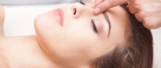
Inflammatory processes of various etiologies affecting the tissues of the trigeminal nerve lead to the development of a disease called neuralgia. Based on its location, it is also called “facial neuralgia.” It is characterized by a sudden paroxysm of sharp pain piercing different parts of the face.
This is how the trigeminal nerve is damaged.
The causes of this pathology are not fully understood, but many factors are known that can provoke the development of neuralgia.
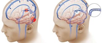
If a malfunction occurs or the facial nerve canal is pinched, this can lead to paralysis of the motor facial muscles. Facial asymmetry is visually diagnosed. The part of the face that is relaxed and motionless creates a mask effect, the eye does not close on the affected side, and lacrimation increases. It occurs due to irritation of the eye mucosa by air and dust, therefore leading to inflammation and conjunctivitis.
Wrinkles on the forehead and nasolabial area are straightened. The corners of the mouth “look” down; the victim cannot wrinkle his forehead on his own. Paralysis of the orbicularis oculi muscle and the non-adjacent part of the eyelid to the eyeball lead to disruption of the formation of the capillary gap. Because of this, problems with tear production occur.
If for some reason motor function is affected, then we can talk about peripheral paralysis. The clinical manifestations are as follows: complete asymmetry of the face, paralysis of facial muscles, limited intake of liquids, impaired speech apparatus. If damage to the nerve occurs when it is located in the pyramidal bone, then the following is observed: absence of taste symptoms, deafness and all of the above symptoms are observed.
Neuritis
A neurological disease characterized by inflammation. Neuritis can be located in the central part of the face and in the peripheral part. Symptoms depend on which part of the nerve is involved. As a rule, there are no erroneous diagnoses in differentiation and formulation. The development of the disease can be due to hypothermia, the so-called primary neuritis, and secondary, manifested due to some other diseases.
The clinical picture is described by an acute onset. The pain radiates behind the ear, and after a few days, facial asymmetry is noticeable. Symptoms may vary, depending on the affected part. If the nucleus of the facial nerve is affected, then the person suffers from facial muscle weakness. The process of infringement located in the area of the pons of the brain leads to strabismus and paralysis of almost all facial muscles. If infringement occurs at the exit, this can lead to impairment and short-term hearing loss.
Neuritis can be concomitant, for example, with chronic otitis media. And it arises due to the ongoing process of inflammation in the middle ear. Therefore, facial paresis occurs with accompanying “shooting” in the ear. When accompanied by mumps, general intoxication of the body occurs - fever, chills, body aches.
The treatment regimen for inflammation and injuries should be comprehensive and timely. Drug therapy necessarily includes:
- glucocorticosteroid drugs;
- diuretics that remove fluid from the capillary network;
- drugs that promote vasodilation;
- vitamin therapy, usually group B.
Further, complex treatment of this nerve includes exclusion and treatment of the underlying cause. Since neuralgia is the result of an illness or a secondary disease. Usually, nervous diseases are accompanied by quite painful sensations; analgesics are prescribed to reduce or stop them. For more effective and rapid treatment, the facial muscles should remain completely at rest.
Surgical intervention is performed in rare cases, if neuralgia is congenital or the nerve is severely damaged due to mechanical trauma. This operation involves stitching together torn or incorrectly fused ends. Another case that provokes surgical intervention is the ineffectiveness of drug therapy for 6-8 months.