Panoramic photograph of teeth (orthopantomogram)
When diagnosing and further treatment, the question for the dentist is: choose a classic x-ray or take a panoramic photograph of the teeth (orthopantomogram)?
A classic x-ray is taken to take pictures that show in detail the condition of one or more teeth. Dentists often take several of these x-rays to look at the problem area from all angles. When we ask a doctor about the differences from a traditional x-ray, we will receive the following answers from doctors. A dental panoramic photograph creates a single image of the entire oral cavity: the upper and lower jaws, the temporomandibular joints, all teeth, the nose and sinuses. When a panoramic photograph of the teeth is taken, the image provides a good view of the entire curved structure of the jaw, making it easier to analyze each part. The price of a panoramic dental photograph, although higher than a classic x-ray, allows you to assess the condition of the oral cavity in one procedure. A panoramic photograph of teeth on film or digitally can significantly help in diagnosis.
Online consultation with a doctor: why use a panoramic dental image?
Asking a doctor about the specifics of an orthopantomogram will allow you to get the following answers from doctors. Because a dental panoramic photograph shows the entire mouth in one image, it does not provide detail. But, as a question to a dentist shows, if you take a panoramic x-ray of your teeth, this type of x-ray shows the main problems, such as abnormalities and bone fractures, cysts, pulpitis, caries, affected teeth, infections and tumors. Doctors' answers are clear: a dentist who suspects any of these problems may choose to take a panoramic photo of the teeth. The price of a panoramic photograph of teeth will, of course, be higher than a targeted photograph of one tooth, but if you take into account the number of photographs that will be needed to diagnose the problem, then a panoramic photograph of teeth on film or digital will ultimately be cheaper. Questions to the doctor will reveal that the dentist may also use this method when planning procedures such as braces, implants and dentures, as well as orthodontic treatment, bite correction and tooth extraction.
Asking your dentist will reveal that dental x-rays help dentists and dental surgeons identify dental problems that would otherwise be difficult or impossible to detect. These include:
- Fillings, caries and wisdom teeth on an x-ray.
- Tooth decay, especially under a filling or between two teeth
- Dental infections, including abscesses at the root of the tooth or below the gum line
- Affected wisdom teeth
- Loss of bone in the jaw from gum disease or a missing tooth
- Hyperdontia and hypodontia (conditions in which there are genetically too many or too few teeth)
- Tumors
If a doctor is told about the general benefits of an orthomantomogram for people seeking dental treatment, then the doctors' answers will be clear. Panoramic dental imaging allows dentists to accurately assess your oral health and provide a cost estimate for your treatment plan. An online consultation with a doctor will clarify that the price of a panoramic photograph of the teeth is not commensurate with the benefits of such a diagnosis.
Modern diagnostic methods - panoramic and targeted dental imaging
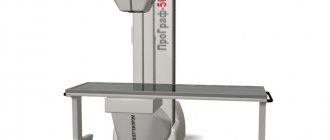
Our clinic has installed new generation diagnostic equipment - a computer tomography (CT) machine from SIRONA, ORTHOPHOS XG3 (Germany), which allows you to take a 3D image of the tooth for a comprehensive study, which means to establish an accurate diagnosis and prescribe the most effective course of procedures and manipulations. Dental diagnostics using three-dimensional computed tomography allows for a detailed examination in the following cases:
- Dental injuries;
- Abnormal development of the dental system;
- To identify tooth defects, as well as to determine the exact location of impacted teeth;
- To identify chronic diseases and foci of infections;
- To identify neoplasms in the soft tissues and bones of the jaws;
- When undergoing a dental implantation procedure (does it hurt?);
- When removing teeth;
- For dental prosthetics;
- To obtain a complete picture of the condition of the patient’s dental system and draw up an accurate dental treatment plan;
- To determine the exact location of the mandibular canal.
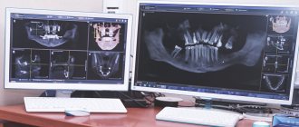
The results of the tomographic examination represent a 3D model of the patient’s dental system, which displays data in three planes at once:
- Sagittal
- Frontal
- Axial
A 3D study allows you to obtain clear information about the structure and structure of bone tissue, the individual characteristics of the location of teeth and their roots, the presence of inflammatory processes and much other information necessary for an accurate diagnosis. A high-quality photograph of a tooth makes it possible to visualize information not only of a general nature, but also for literally each tooth and area of the jaw. The clinic’s specialists also recommend taking a targeted photograph of the tooth for a more detailed diagnosis of a specific problem area. Usually it can be done after a consultation, or immediately when you have been prescribed a panoramic photograph of your teeth.
The results of dental x-rays are recorded on electronic media, which makes it possible to use them for consultation and treatment by specialists from different fields of dentistry. This allows for a comprehensive examination and successful treatment of the most complex cases. It is very important to take dental x-rays during diagnosis and treatment.
Dental diagnostics using computed tomography is one of the safest examination methods, since the dose and time of radiation are minimized - the examination lasts no more than 10 seconds.
The use of computed tomography - the advantages of a 3D tooth image
The computed tomography equipment installed in our clinic allows you to take an image of the tooth and conduct high-precision research in the following areas of dentistry:
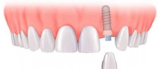
- Surgical dentistry: diagnosis of cysts and granulomas, bone grafting, implantation
- Orthopedic dentistry: modeling of dental structures
- Endodontics: study of dental canals without nerves
- Orthodontics: determining the condition of teeth and bone tissue, the shape of the dentition for subsequent correction of the bite, you need to know which braces are best to install? Sign up for a consultation with our specialists, and they will tell you how to install braces.
- Periodontology: identification of sclerotic changes in the periodontium, determination of the level of bone tissue resorption in the presence of progressive periodontitis
Computed tomography makes it possible not only to conduct an accurate diagnosis, but also to create a three-dimensional model of the patient’s dental system, which allows you to create a positioning template and plan the further course of the operation.
12 reasons why you should choose Eurodent
- We provide a full range of dental services.
- Our clinics operate 24/7.
- The most modern and high-quality materials are used in the treatment process
- Qualitative diagnostics using targeted or panoramic images of teeth
- High-precision diagnostics using computed tomography eliminates errors when making a diagnosis.
- Application of the latest treatment methods. Our specialists undergo training and internships in leading clinics in Europe.
- Examination and treatment of children of all ages.
- The Antihepatitis and AntiAIDS programs are actively working in our clinics.
- Our specialists provide free consultations.
- Affordable prices with a high level of service. We provide a guarantee for all our services.
- There is a flexible system of discounts for our regular clients, and we also provide the opportunity for treatment in installments.
- When starting any treatment, it is mandatory to take a photo of the tooth (panoramic or targeted) for better and more correct treatment.
We do everything to make our treatment as effective and comfortable as possible for our patients!
How to take an x-ray?
Unlike traditional X-rays, a panoramic dental photograph on film or digital is extraoral. This means that both the device and the film are located outside of your mouth. A panoramic dental X-ray machine projects a beam through the mouth onto a film or sensor that is positioned opposite the X-ray tube.
The online doctor's consultation advises that when preparing for a dental x-ray you may be asked to:
- Remove any jewelry, glasses, or other metal objects that could become an obstacle.
- Wear a lead apron to protect the rest of your body.
- Bite a special thin block to open your mouth a little.
Asking your dentist will clarify that in order to get a clear image, it is important that you remain absolutely still. The procedure usually takes place while you are standing. But if you feel more comfortable sitting, the device can be adjusted.
Capturing an image takes about 10-20 seconds. The computerized x-ray or panoramic film of the teeth is sent directly to the radiologist so he can immediately review and evaluate it.
Your dentist may recommend that you take a panoramic dental photo every 6 to 24 months, depending on your oral health. The price of a panoramic dental photograph will be repaid by timely and accurate diagnosis.
Expert commentary
Alexandrova Margarita SergeevnaPrimary doctor |
A panoramic photograph of teeth on film or digitally allows you to get a complete picture of the condition of the oral cavity in one procedure.
An online consultation with a doctor will clarify that the basic design of a panoramic X-ray machine includes an imaging tube mounted on a horizontal arm and aimed at one side of your face. Asking the doctor what is on the opposite horizontal bracket, located on the other side, will yield the following answers from doctors: there is an x-ray film or sensor located there. As a rule, the patient's head rests on special supports on the side, at the chin and on the forehead. During the image, the patient's mouth remains open, and the moving part of the X-ray machine rotates around the head, starting from one side of the jaw and ending on the other.
Advantages
- The price of a panoramic photograph of the teeth is significantly lower than individual targeted photographs of the teeth individually.
- A panoramic photograph of teeth on film or digitally, taken with modern equipment, is of very high quality.
- Taking a panoramic photo of your teeth gives your dentist a complete view of your entire mouth in one photo.
- A panoramic photograph of your teeth can be performed in a short time during 1 visit.
- According to the latest data from an online doctor’s consultation, the radiation exposure from one panoramic photograph of the teeth is only 0.02 millisieverts, which is absolutely safe.
Where to get a dental x-ray done in Astrakhan inexpensively and with high quality
In terms of importance, detail and informational usefulness, dental x-rays as a method of research in dentistry have no equal. In fact, no relatively serious intervention can be done without a tooth image. X-rays give an idea of the severity of the disease and help make an accurate diagnosis. This is especially important in the early stages, when the pathology does not yet manifest itself visually. Moreover, during the treatment process it is often necessary to take a picture in order to assess its correctness and make adjustments if any are required. All this allows the specialists of the regional dental center to undertake the treatment of any pathologies and ailments and do this with a guaranteed and long-term result.
X-ray in Astrakhan - safe and fast diagnostics
An X-ray of teeth is a safe procedure, with virtually no contraindications (you just need to warn the doctor about pregnancy
). At City Clinic No. 4 in Astrakhan, the study is carried out by competent and experienced specialists, with maximum speed, so as not to delay patients. The office is equipped with modern, highly sensitive, low-level radiation equipment, and all patient safety and radiological control standards are observed in the strictest possible manner.
You can take an X-ray of a tooth in Astrakhan in our clinic for an appointment with a local doctor, as well as to confirm the correctness of treatment in another dental center. Images of primary, molar, filled, pulpless teeth, after root resection, treatment of periodontitis or extraction - all this is performed by our knowledgeable radiologists. Without an x-ray, it is impossible to identify the initial stage of a disease of hard tissues, pulp, or to recognize a cyst or crack in a tooth, an abscess or granuloma, or inflammation in the canal. It helps to clarify the location of incorrectly growing teeth (including wisdom teeth). With the latter, problems very often arise due to improper growth and eruption - before removal, it is necessary to take a photo to determine the tactics of the removal operation.
Panoramic photograph of teeth – the highest information content
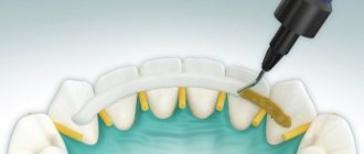
Along with the periapical (that is, 1-3 adjacent teeth), a panoramic photograph of the teeth is also performed in our X-ray room. It is done before all serious dental and surgical interventions, prosthetics, implantation, installation of braces to identify the slightest pathologies and developmental anomalies. A panoramic photograph (experts call it an orthopantomogram) shows the upper and lower jaw, the condition of the roots and basal tissues, the joint, and the nasal sinuses. This is the most complete study that provides an accurate picture for selecting therapy.
Price of dental photographs in clinic No. 4
The affordable price of dental photographs in the regional center attracts patients from other medical institutions to us. And if you take into account the quality, accuracy and modern level of equipment, the popularity of this service becomes clear. The specific cost of each type of image is indicated on the website, but in any case, this is a service that will not exceed the budget planned for a visit to the dentist.
Is panoramic dental photography safe?
A question to your dentist will reveal that, when performed in accordance with the precautions taken, panoramic dental photography on film or digital is considered an extremely safe procedure. Although a dental panoramic image produces a small amount of radiation exposure, it does not accumulate in the body after the procedure. Doctors' answers are clear: the extent of exposure to panoramic dental imaging is not considered risky, even for infants and young children, who are more sensitive to radiation. Panoramic X-rays are of external origin. Therefore, this method is often preferred for radiography of the teeth of children and adolescents. The price of a panoramic dental x-ray also matters in order not to undergo numerous procedures of classical x-rays, but to take a single image.
Online Doctor Consultation reports that panoramic dental imaging is a widely used imaging modality in dental practice and can be a valuable diagnostic tool. However, panoramic images are difficult to interpret and require professionalism from the dentist. In addition to showing the jaws with multiple overlaps and distortions, the panoramic dental photograph depicts numerous anatomical structures outside the jaws that can create additional problems. Successfully interpreting a dental panoramic film or digital film begins with an understanding of the normal anatomy of the head and neck and experience in reading such films.