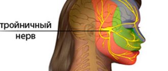
What is the danger of x-rays and its consequences?
It has been found that ionizing radiation has an adverse effect on DNA chains.
This leads to a number of disruptions in the functioning of organs and systems. Radiation-sensitive organs are more at risk:
- thyroid;
- Bone marrow;
- mammary gland.
If X-rays are used adequately, there are no serious consequences from the procedure. Scanning is carried out only when necessary, without exceeding recommended doses. The picture below shows approximate radiation doses from an X-ray machine, not the dose received by a person.
The exact dose must be asked from the x-ray technician if there is an exposure meter on the device. Also, these indicators depend on the manufacturer, type of device (film, digital), body weight, structure and density of the organ being studied.
Radiation exposure standards for X-ray examinations
Every year, on average, a person receives a total dose of radiation exposure (from natural sources) equal to two to three mSv (millisievert). The norm is within the limits of the permissible background radiation and is 0.20 μSv/hour (which corresponds to 20 μR/h - microroentgen per hour). The upper limit is 0.50 µSV/h (microsievert per hour), but exposure to even a few µSv/h is acceptable over just a few hours.
Radiation accumulates in the body, so the maximum accumulated amount of ionizing radiation over a lifetime should not exceed 100-700 millisieverts. The significant range allowed by regulations is due to different levels of radiation. Thus, residents of high mountainous areas are accustomed to a higher natural background and tolerate slightly increased doses of radiation without negative health consequences.
How an X-ray is taken
The radiation doses that a patient receives during various types of medical procedures are presented in the following table:
| Medical procedure | Radiation dose | The period during which a patient receives a similar dose of radiation in the natural environment |
| Chest X-ray | 0.1 mSv | ten days |
| Fluorography (digital) | 0.03-0.06 mSv | three to five days |
| Fluorography (film) | 0.1-0.2 mSv | two weeks - a month |
| Mammography | 0.7 mSv | three months |
| CT scan of the abdomen and pelvis | 10 mSv | three years |
| Whole body computed tomography | 10 mSv | three years |
| X-ray of the stomach and small intestine | 8 mSv | three years |
| X-ray of the large intestine | 6 mSv | two years |
| X-ray of the spine | 1.5 mSv | six months |
| CT head | 2 mSv | eight months |
| CT scan of the spine | 6 mSv | two years |
| Bone Density Determination | 0.001 mSv | less than a day |
| X-ray of the sinuses | 0.5-1 mSv | two to four months |
| CT scan of the chest | 7 mSv | two years |
| Dental X-ray (depending on equipment) | from 0.015 to 0.3 mSv | one to three days |
For comparison, a single dose of radiation received by an airplane passenger in one hour is 10 μSv, which corresponds to a day of exposure from natural sources.
Allowable passing frequency
The permissible frequency of x-ray examinations is determined by the attending physician, depending on the disease and the characteristic features of the body. Only one diagnostic test is carried out during the day; if a repeat analysis is necessary, you will need to wait several days.
How often can an x-ray be taken? If diagnostics are carried out using old-style X-ray machines, it is worth remembering some recommendations:
- X-ray of teeth. If a lateral X-ray is performed, examination is allowed up to 5 times a year. With direct X-ray and simultaneous scanning of the brain - no more than once a year.
- Nasal scan. It is recommended no more than once a year.
- X-ray of the skull. The procedure is carried out no more than once a year to avoid negative effects on brain tissue.
- X-ray of the spine. It is recommended no more than once a year.
Re-passing is recommended after at least 1 month. Fluorography performed once a year does not pose any danger; it is a generally recommended procedure.
If a repeat scan is necessary, doctors advise contacting specialized centers that have modern X-ray equipment. This equipment reduces radiation exposure tenfold.
Taking X-rays using new-style machines up to 5-6 times a year.
Types of research
In recent years, dental radiography has been used increasingly. This is due to the development of devices that allow you to obtain instant and accurate images. Thus, treatment is faster and patients experience minimal discomfort.
Diagnosis can be carried out using old and new technology. Depending on the equipment used, there are four types of dental radiography:
- bitewing: to detect caries and tartar;
- targeted: to determine the internal condition of the tooth and gums;
- panoramic: to obtain a more accurate picture of the general condition of the jaw structure;
- digital: to obtain a clear image of both an individual tooth and the entire dental structure.
Read also: How to relieve severe toothache
The newest type of dental diagnostics is 3D X-ray. This research method allows you to obtain a panoramic or three-dimensional image,
which is displayed on the computer screen.
As a result of image processing, the doctor receives the most accurate picture.
In order to avoid having to undergo repeated diagnostics and to ensure that the image is of high quality, the examination must be carried out according to certain rules, which must be followed not only by a medical specialist, but also by the patient himself.
What is the permissible dose and how to reduce the radiation load
In order to control ionizing radiation, after each procedure, data on the load received is entered into the patient’s medical record.
To minimize radiation and reduce its negative impact on the body, use:
- protective glass plates;
- X-ray protective screen;
- aprons based on lead material.
According to regulatory documents on radiation safety, the permissible radiation dose for one person is no more than 5 mSV per year. For children and patients during pregnancy (lactation), the dose is halved.
Applications and properties of X-rays
X-rays are used mainly in medicine during diagnostic examinations, as well as to monitor the dynamics of the course of the disease. There are two types of X-ray radiation: bremsstrahlung and characteristic.
How dangerous is the procedure? The radiation that a patient receives during a preventive examination once a year is within the acceptable range and does not harm health.
X-rays are often not recommended, as the likelihood of irreversible changes in the quality of the blood or the development of leukemia, cancer, and cataracts increases. Characterized by premature aging and a whole list of complications of varying severity.
X-rays during pregnancy
If a bone fracture is suspected, radiography is performed with simultaneous screening of the pelvic area, mammary glands and abdomen.
Dental x-rays are considered safe because they are performed quickly and with minimal radiation dosage. If possible, you should refuse to undergo scanning on old-style devices.
In modern diagnostic centers, radiography is performed using new generation equipment, which greatly reduces the negative impact on the human body.
Radiography in childhood
How many times a year can children be x-rayed? According to the recommendations of the World Health Organization (WHO) for children under 15 years of age, undergoing a diagnostic test should be excluded. Prescribing a study is only advisable if there is a clear medical need. For example: an x-ray of the face area is indicated if a nasal fracture or bone crack is suspected.
Regarding radiography in pediatrics, as a preventive method, diagnosis is categorically not recommended. Prescribing the procedure to children requires strict indications.
How often can x-rays be taken?
How many times can an X-ray be taken to avoid excessive radiation exposure? The frequency with which an x-ray beam can affect a patient’s body is allowed by medical standards, one procedure per year. However, figures suggest that theoretically, X-rays of the spine, jaw, skull and other organs can be taken at least ten times a year without negative consequences for the body.
The possible risk to the patient is assessed and taken into account when ordering further diagnostic or control tests in cases where the person has already received X-ray exposure equal to 50 mSv during the last year.
In general, X-rays can be taken as long as necessary to make a correct diagnosis or monitor the dynamics. The required (or acceptable) period for the procedure depends on several factors and is determined solely by the attending physician.
In addition, the safe frequency of the procedure (x-ray of the sinuses, abdominal cavity, lungs, mammography or fluorography) varies for different groups of patients:
- relatively healthy people can be exposed to x-rays for preventive purposes once a year (the period is counted from the last procedure);
- the permissible number of procedures for persons who are not at risk (experienced smokers, workers in hazardous industries) is one or two per year;
- employees of the service sector, public catering and children's institutions are required to undergo an X-ray examination twice a year;
- For patients for whom X-rays are a necessary measure (for example, patients with complex pneumonia), the procedure can be performed even several times a week.
In the latter case, the risk from exposure to X-rays is not comparable with the complications and consequences of an untreated disease or an incorrectly prescribed course of treatment.
The feasibility of conducting a study depends on several factors. Many (multiple) x-rays are indicated for some diseases, but the need for diagnosis or monitoring of therapy does not always outweigh the potential harm caused by radiation. The attending physician takes into account the date of the previous procedure, the medical need for the x-ray, and the total radiation dose the patient received over the past year.
The doctor taking the x-ray is exposed to minimal danger if safety precautions are followed. Radiologists “for their harmfulness” receive additional days of vacation, bonuses and the right to early retirement.
X-ray for sinusitis, pneumonia, in dentistry
Individual questions from patients who often have to stand in front of an X-ray machine are caused by x-rays of the nose, lungs or teeth. How often can an X-ray of the lungs be taken, for example, in case of pneumonia, or how many procedures are acceptable in dental treatment? Images of the sinuses, lungs and teeth are taken not only for diagnostic purposes, but also to monitor the success of therapy, therefore the permissible number of studies is determined solely by the attending physician.
X-ray during pregnancy
How often can x-rays be taken during pregnancy? The answer is not so clear. It is advisable for pregnant women to avoid the procedure altogether, but in some cases it is simply necessary to undergo an x-ray. If the life or health of the expectant mother is at risk, doctors will not worry too much about the well-being of the fetus. The main goal in this case is to save the woman.
A separate topic is dental x-rays during pregnancy. Going to the dentist is usually a forced event.
How often can dental x-rays be taken during pregnancy and is it even possible? Dentists note that the study will not have a negative impact on the health of the fetus, but the procedure is undesirable in the first trimester.
As for the general risks, x-rays taken in the first trimester can lead to spontaneous abortion, which the woman may not be aware of. Taking an X-ray in the middle of pregnancy is fraught with developmental disorders of the organs and systems of the fetus, serious pathologies; an examination in the third trimester can appear in the first year of the child’s life or provoke complications during labor.
X-ray in childhood
How often can children be x-rayed? According to WHO recommendations, X-ray examinations in childhood should be done only if medically necessary. An X-ray of a child’s nose, for example, is indicated if a fracture is suspected; serious damage to the hip joints, jaw, skull, and birth injuries are also sufficient indications.
Is it possible to do an X-ray on a child for preventive purposes or not? Preventive examinations are strictly not recommended for children under 14 years of age; children are subject to X-rays only when indicated.
Alternative diagnostic methods
Medicine may offer other examination options. Today, the main diagnostic methods are CT and MRI, but are they a full replacement for X-rays?
CT (computed tomography)
The CT machine is based on the use of x-rays, in fact, it is a modern replacement for standard x-rays, but this method is more harmful. A classic X-ray is a single image, while a CT scan creates many “slices.”
The body part in question is metaphorically “cut into slices.” More advanced machines are even capable of creating a three-dimensional image of the organ or tissue being examined.
MRI (Magnetic resonance imaging)
MRI is a highly informative research method, but its purpose is not always reasonable. Thus, computed tomography is used to examine parts of the body (chest, abdomen, spine.). MRI is rather used to study a specific organ (brain, heart, pancreas, bone marrow).
If you need to undergo a high-quality diagnosis of bone tissue, then you need to turn to an x-ray or CT scan. If diagnostics of soft tissues and organs is required, then MRI is the best way to identify pathological processes.
Even with modern digital equipment, x-rays remain an indispensable method for identifying pathologies, in particular bone tissue. To minimize the risks of the negative impact of the procedure, you should undergo it according to the doctor’s indications, and not prescribe it yourself.
How often can dental x-rays be taken?
This question interests many patients. A doctor of the highest category, Svetlana Vasilievna Voitova, talks about dental x-rays.
X-ray of teeth
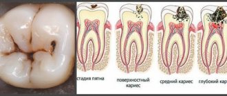
X-ray examination of teeth is a very effective way to diagnose most dental diseases. A dental (dental) photograph allows you to detect hidden caries, determine how deeply the hard tissues of the tooth are affected, timely notice inflammation in the periodontium, cracks and fractures of teeth, control the quality of obturation (filling) of root canals after their treatment, identify overhanging edges of fillings, etc. .
Modern dentistry is impossible without X-rays - they make dental treatment high-quality and reliable. Today, computer radiovisiographs are very widespread, replacing conventional X-ray machines. These radioimaging devices significantly save time, are easy to use and, most importantly, provide much less radiation exposure to the body, while allowing the dentist to obtain more information.
Depending on the relative position of the film and the object of study (teeth and surrounding tissues), a distinction is made between an intra-oral radiograph (the film is inserted into the oral cavity) and an extra-oral one (the film is located outside). An internal oral radiograph, depending on the position of the film in the oral cavity, is divided into contact (the film is adjacent to the area under study) and occlusal (the film is held by closed teeth and is located at some distance from the area under study). The structure of teeth and surrounding tissues is most clearly obtained on an intraoral contact radiograph, as well as periapecal and occlusal photographs.
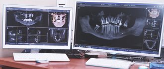
In dental clinics, to examine the dental system, they mainly use overview images - orthopantomograms and spot - targeted images (1-2 teeth).
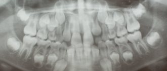
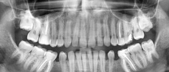
Orthopantomogram
An orthopantomogram is a large image that allows you to see not only both dentitions, but also the condition of the jaw bones, periodontium, TMJ (temporomandibular joint) and maxillary sinuses.
Read also: How to treat sores in the mouth
Such an image is necessary for the general treatment plan; it allows for complex surgical procedures - implantation, flap operations, removal of eighth teeth and much more; for orthodontic treatment, that is, to correct incorrect position of teeth and bite in general.
You cannot do without it when planning the restoration of missing teeth - orthopedic treatment.
Sight shot
However, an orthopantomogram only gives a general idea of the teeth. If the problem is related to a specific tooth, then a targeted or spot image is required.
With it you can see the tooth and periodontium more clearly. This is especially important for making a diagnosis. A targeted image allows not only to identify the pathological process, but also to determine its stage and extent of prevalence.
Often, targeted images are used during therapeutic dental treatment - in this case they are called control images, since they allow monitoring the effectiveness and quality of treatment.
Other types of dental x-rays
In addition to these two most popular types of dental x-rays, others are sometimes used.
For example, iteraproximal radiography is used to obtain images in which the image of the marginal areas is not distorted. With the help of such radiography, it is possible to detect hidden caries that is not noticeable upon direct examination.
There is also occlusal radiography, which allows you to examine a certain area of the jaw, while spot radiography will not be enough, and an orthopantomogram will be unnecessary.
X-ray examination techniques:
*The standard technique is that the X-ray film is wrapped in opaque paper and placed behind the tooth. The X-ray tube is brought directly to the tooth being examined, after which an image is taken. The X-ray dose in this case is small.
*radiovisiography. With this method of x-ray examination, a small sensitive matrix is located near the tooth being studied, then an image is taken, and the resulting image is displayed on a high-contrast, high-resolution monitor. Since the matrix is extremely sensitive, the radiation power is reduced tens of times compared to a conventional x-ray.
*orthopantomogram is a type of dental examination in which an X-ray image of all teeth and jaws is obtained in one image. This research method is used when it is necessary to determine a large amount of information about the condition of the teeth and jaw bones. In addition, the method is indicated in cases of dental prosthetics, to determine the anatomical and topographic relationship of the teeth, prosthesis and jaw. An orthopantomogram is recommended when several teeth are to be removed, and the possibility of further implantation is also assessed.
*computed tomography is a method of non-invasive diagnosis of diseases based on X-ray radiation. CT is one of the most common diagnostic methods in dentistry.
The peculiarity of X-rays is that they react differently to different anatomical structures, so that the result of the study accurately reflects the structure of the patient's teeth and jaw. This result is output to the computer through the detector - and a computer tomogram is obtained. In this case, the image on the tomogram turns out to be three-dimensional, thanks to this property it is possible to select the treatment as accurately as possible and select the type and size of the implant; the computed tomography procedure allows for a complete examination of the entire dental system in just 20-30 seconds; it practically does not irradiate a person due to modern digital technologies and a short exposure time. Due to the almost complete absence of radiation, tomography can be used to examine even children and pregnant women.
With the help of computed tomography, before surgical treatment, you can easily analyze the general condition of the bone tissue, its height, width and even density. CT also allows you to determine the distance from the maxillary sinus to the mandibular canal and accurately say if there are additional bone septa or inflammatory processes in the maxillary sinus.
The maximum permissible radiation dose is 150 mSv per year ; it is received only by people who need regular x-ray monitoring, or for health reasons (accident, severe injury, internal bleeding). If you do only routine diagnostic examinations - fluorography, mammography, x-rays at the dentist - you will gain only about 15 mSv per year (1 millisievert = 114 mRoentgen).
Comparative table of radiation doses:
Fluorography (1 projection) 0.6–0.8 mSv
Digital fluorogram (1 projection) 0.03–0.05 mSv
Mammography 0.2–0.3 mSv
Dental X-ray 0.15–0.35 mSv
Determination of complete dental status (10 images) 1.1–1.8 mSv
Orthopantomogram (panoramic photograph of both jaws) 0.006–0.02 mSv