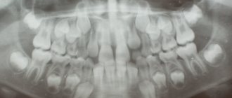
A panoramic photograph of teeth or an orthopantomogram is an integral part of diagnosis and quality dental treatment. The effectiveness of medical services largely depends on the research methods used. In dentistry, this cannot be done without radiography, and more specifically, an orthopantomogram.
Nowadays, modern safe devices are used, which, even with frequent exposure to X-rays, cannot cause harm.
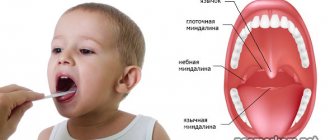
Panoramic photographs of teeth can be taken during pregnancy, during the examination of baby teeth and in a nursing mother. But this opinion is not shared by all experts. Some doctors agree to perform an orthopantomogram or MRI during pregnancy only if absolutely necessary.
Visiography is considered the safest research method in dentistry during pregnancy. As for adults and children, dental MRI and panoramic X-ray can be done without problems.
Is the danger real?
In modern medicine, doctors' opinions about the harmful effects of dental x-rays vary. Some of them are inclined to believe that X-ray examinations during pregnancy are unacceptable.
According to European doctors, based on statistics, a certain percentage of women after such an examination gave birth to low-birth-weight children. However, there is no evidence that underweight children were a consequence of dental x-rays.
Most experts deny the danger of X-ray diagnostics if it is not used in the first weeks, when the child’s organs are forming and his cells are especially vulnerable to radiation exposure.
At later stages of fetal development, the use of x-rays in dental treatment is simply necessary. After all, pathological microorganisms can negatively affect the development of a child.
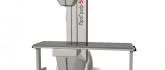
However, undergoing an X-ray examination at a public dental clinic is somewhat risky for a pregnant woman. In these clinics, the equipment is mostly old, which can produce increased loads.
Modern equipment is much safer. It emits gamma radiation in minute dosages, which allows the patient to take 5 pictures in one visit.
The radiovisiograph is safe in that its beam has a narrow focus, that is, its object is exclusively the tooth, and other tissues remain outside its field of action.
Thus, the area where the fetus is located will not be affected. And the doses of the visiograph are not higher than those of normal background radiation in a modern urban environment.
How to do a dental x-ray
Toothache is unbearable. Expectant mothers are especially concerned about the sensations of the fetus. It is believed that he feels the same as his mother. As a rule, it is not recommended to take an X-ray of one tooth in the early stages of pregnancy, but thanks to the development of modern technologies, the appearance of a visiograph has become possible - a special device used in dental clinics. The small size of the detector and radiation source made it possible to significantly reduce the radiation dose. In this case, dental x-rays during pregnancy do not have serious consequences.
To protect the bodies of women and children, it is necessary to wear a lead apron. It will not allow X-rays to penetrate itself. The visiograph is brought close to the tooth being studied, so the radiation passes through it pointwise, without reaching other organs and tissues. The pictures are taken immediately in digital form, which significantly improves the detail of the image. Therefore, there is no need to take several pictures, one is enough. Also, thanks to low radiation, you can take up to 10 pictures in one trip to the dentist. This allows you to ensure that the treatment is performed correctly.
A dental x-ray can be done during pregnancy if the doctor, knowing all the circumstances, recommends this procedure. Don't be afraid of radiation. As observations of women who survived the explosion of a nuclear bomb in Japan showed, they were able to give birth to full-fledged and healthy children. An equally difficult question is whether it is possible to treat teeth using anesthesia; it can also affect the child’s health.
Process principles
The main thing in x-ray diagnostics is to carry it out without errors. Although it is believed that the procedure does not contain any risk, it is prescribed only taking into account the indications .
In addition, the duration of pregnancy cannot be ignored. All diagnostic safety measures must be strictly observed.
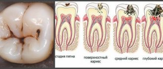
This procedure is not always required. If the picture of caries is clear, then the dentist does not need to x-ray the tooth for treatment.
An X-ray is necessary if the carious process has deeply affected the tooth.
What is the purpose of dental physiotherapy and the stages of the procedure. Come here if you are interested in how to correctly read a plot for toothache.
At this address https://dr-zubov.ru/lechenie/zuby/izbavleniya-ot-boli-chesnoka.html find out how you can use garlic against toothache.
The inevitability of holding
Main indications for x-ray examination:
- extensive infection (several teeth);
- externally invisible pathologies of the tooth (root or crown);
- periodontal disease;
- purulent tissue damage in the form of a cyst;
- complication caused by the growth of wisdom teeth;
- tooth injury;
- Finding out the possibility of saving the tooth.
An X-ray image gives the dentist information about the position of the roots, canals, and a picture of the pulp chamber. Without this data, treatment is unlikely to be effective.
It is better, of course, to plan your pregnancy and get your oral cavity in order before it occurs. In this case, new caries that overtakes a woman already in position will be superficial, and x-rays will not be needed.
Suitable timing
The first trimester of pregnancy is least suitable for x-ray examination. It is in the first trimester that conditions are important for the normal formation of the child’s body, and there should be no negative impact on the fetus.
Statistics show that during this period most spontaneous abortions and deviations from the norm in fetal development occur.
The first weeks of pregnancy are the riskiest for this diagnosis. The procedure can only be carried out if it is not possible to postpone it to a later date.
In life such cases are very rare. It is usually possible to delay treatment.
It may be that a woman, not knowing about her pregnancy, underwent an X-ray examination at the dentist. In this case, she needs to undergo examination to identify possible genetic abnormalities of the fetus.
The 2nd trimester is most suitable for carrying out the described diagnosis, when the formation of the child’s organs is completed, and the negative external influence on him will be minimal.
Therefore, all a woman’s health problems that require medical intervention are best addressed in the second trimester. And the 3rd trimester - the risk will increase again.
Precautionary measures
In any trimester, even the most optimal for treatment, X-ray safety measures must be observed:
- Lead collars and aprons for patients. Lead plates enclosed in a fabric cover protect the patient from gamma radiation.
- Maintain the distance between the beam tube and the patient. Further down the tube means less radiation.
- It is important to maintain the time : 5 seconds after irradiation, the patient should not remove the lead apron, since during this time the gamma rays undergo a decay process.
- No more than 5 pictures per visit , take a break of several minutes between pictures.
All these rules of x-ray diagnostics protect the woman’s fetus from the negative effects of gamma radiation.
Features of the event
To minimize possible negative consequences for the child, the procedure must be carried out taking into account the woman’s position, and adhere to the optimal timing and methods of conducting the study.
Making the analysis safe is the main task of both the patient and the doctor.
The inevitability of holding
Therapy without deep and high-quality diagnostics can be fraught with a lot of unpleasant surprises, especially when it comes to dental treatment.
Most problems are hidden from external view and can only be identified as accurately as possible using modern equipment.
The procedure is inevitable in the following clinical manifestations:
- pulpitis with the need to fill the root canals - for high-quality treatment it is necessary to know its size and shape;
- with atypical growth of eighth units;
- with the development of cystic formations , granulomas and other neoplasms of various origins;
- if there is a suspicion of inflammatory processes in fragments of periodontal surrounding organs and tissues;
- in case of mechanical trauma to the root part or its partial fracture.
Optimal timing
In the first trimester of pregnancy, the fetus develops all the most important systems and organs of the human body, therefore, a period of up to 12 weeks is the most dangerous in terms of the development of various pathological processes.
This period is extremely dangerous, and X-rays should be taken only for special indications, when the harm to the mother’s health is higher than the risk of complications in the fetus.
The 2nd trimester is a more favorable time. The woman’s health improves significantly, her immunity stabilizes. The fetus already has strong placental protection, and all organs have received a genetic seed and are developing safely.
The period from 4 to 6 months is the optimal time for such studies.
The third trimester is a potential danger of any impact on the mother’s body. The tone of the uterus increases, and any, even psychological, impact can lead to premature birth, therefore, if possible, you need to wait a little before taking an x-ray.
Precautionary measures
The main safety measure during manipulation is the use of only modern equipment with minimal radiation exposure.
Every woman, before going to the clinic, should familiarize herself with the basic rules that will help minimize possible risks from the procedure:
- do not use a film device - the ideal solution in this case is a computer visiograph. Its impact is of a point nature, and the direction of the flows is local. This negative load is tens of times lower than that of a film X-ray scanner;
- Before starting the manipulation, the stomach must be covered with a protective apron made of thick lead. The same goes for the chest. Lead is an excellent obstacle to the penetrating action of beam and radiation flows. Without such a special apron, x-rays can pose a potential danger to both mother and fetus;
- if the period is short enough, it is imperative that you notify a specialist about the presence of pregnancy and indicate a specific period in weeks. It is important!
Let's find out together why teeth hurt at night and what can be done. Visit here to learn more about the causes of tooth decay.
At this address https://dentist-pro.ru/lechenie/zuby/v-chem-sut-premedikacii-v-stomatologii.html read why premedication is prescribed in dentistry.
Possible abnormalities in fetal development
X-ray diagnostics carried out in the first three months (12 weeks) of bearing a child can harm the development of such organs as the spinal column and bronchi.
Untimely x-rays can cause cardiac problems in a child, improper development of the maxillofacial region, and cranial underdevelopment.
Irradiation can cause further manifestations in the child:
- dystrophy;
- anemia;
- leukemia;
- incurable diarrhea;
- oncological disease.
A fetus exposed to radiation may simply stop developing. A miscarriage is also possible.
But for all the above-mentioned developmental abnormalities in the fetus to occur, the radiation dose must reach 3 msv. And the dosages of a modern visiograph do not exceed 0.02 msv, the doses of outdated devices do not exceed 0.3 msv.
Factors that provoke the development of necrosis of hard dental tissues and methods for eliminating pathology. In this article, read about the reasons for the development of wedge-shaped dental defects.
Here we will discuss the cost of laser dental treatment.
Consequences of radiation exposure of potential parents
As it turned out, the fetus can suffer from dosed radiation of 3 msv. But radiation, according to scientists, harms not only the embryo. Gamma rays also harm the egg.
Therefore, when planning a pregnancy, a woman should postpone conception until the next cycle if for some reason she had to take such a dose of radiation.
A man involved in conception must avoid such radiation exposure for 72 days . It is during this period that the sperm is renewed.
A number of scientists suggest that the radiation received by a man accumulates in sperm. Such sperm, having fertilized the egg, can negatively affect the development of the fetus.
Is it possible to take x-rays during early pregnancy?
The use of X-rays in the early stages of pregnancy is regulated by SanPiN standards, and there are no categorical instructions that this study cannot be done. As a rule, radiologists do not advise women in the first month of gestation to come into contact with ionizing radiation, since the risks to the fetus are too great.
If you cannot do without an x-ray in the first weeks of pregnancy, and this can happen due to injury, disease of internal organs, etc., consider the ratio of potential harm and benefit for the expectant mother and child. If the patient’s condition is life-threatening without timely diagnosis, the doctor will undoubtedly allow her to have an x-ray. It is indicated to be carried out in the early stages in an apron with lead inserts, which will protect the pelvic organs from radiation.
Good to know! If the study area is located “peripherally,” that is, away from the pelvis, the risk to the fetus is reduced.
What if I took an x-ray without knowing that I was pregnant?
Of particular concern to expectant mothers are situations where they had to take an x-ray without knowing about pregnancy. As a rule, this happens in situations where the doctor prescribes an examination before a missed period, that is, in the first weeks of gestation. Since in the first week of pregnancy there is the greatest risk of damage to the embryo, the woman and the gynecologist will have to decide whether to save the fetus or decide to have an abortion. The following points will help you make your final decision:
- The dose of ionizing radiation received during the procedure. If the total radiation exposure of the procedure was more than 100 mSv, the likelihood of damage to the fetus is extremely high. In this case, the doctor may recommend termination of the pregnancy.
- X-ray exposure area. If an examination of the pelvic organs, sacral spine, urinary tract or fallopian tubes was carried out, pregnancy after an x-ray will develop with pathologies in 90% of cases. The doctor will recommend termination. If other parts of the body were examined and the abdomen was protected with a lead apron, the risk of damage to the fetus is minimal.
In any case, when a woman finds out that she is pregnant after an x-ray, a thorough examination is necessary. In gynecological practice, a lot of screening studies have been developed aimed at identifying fetal pathologies in the early stages of pregnancy. If their results are positive, the issue of abortion will become especially acute.
In what cases is the risk justified?
Medicine knows many situations when the doctor recommends taking an x-ray during pregnancy, even if there is a danger to the fetus. For what reason may an expectant mother be prescribed an x-ray:
- Skeletal injuries - bruise of the spine, chest, neck, fractures and dislocations of the limbs. If the doctor suspects the presence of urgent injuries that may complicate the course of pregnancy, x-rays will be performed at any stage.
- Pathological processes of the joints of the pelvic region - signs of symphysitis (divergence of the pubic bones), rupture of the ligamentous apparatus and others. Such conditions can directly affect the course of pregnancy and delivery. The results obtained during the X-ray will determine which method of birth will be chosen - natural birth or cesarean section.
- Inflammatory processes, acute injuries of the maxillofacial system, accompanied by signs of brain damage. In this situation, there is a risk to the life of not only the fetus, but also the mother, and timely diagnosis will help save the lives of both of them.
X-rays are used when symptoms do not clearly indicate a specific diagnosis. In this case, only an X-ray image will help the doctor decide on treatment tactics. If it is possible to postpone X-ray diagnostics, it is postponed to the 3rd trimester, when the danger to the fetus is minimal.
Important! If the diagnosis does not affect the pelvic area, it is protected from ionizing radiation with a special lead apron. This minimizes risks for the baby at any stage of pregnancy.
Other methods
Today, no method can completely replace X-ray diagnostics. If necessary, you can use the equipment offered by the market.
Of all the market offers, the best option is a digital visiograph. It produces a very low radiation load. There is no need to wait for the snapshot; it is issued immediately and saved in the database.
The disadvantage of a visiograph is that the pictures it takes can only capture three teeth.
An orthopantomograph can provide a complete image of the jaws. Its radiation doses are quite low, and the images are characterized by excellent digital quality.
When the pathology is simple, X-ray diagnostics can be replaced by an apex locator. The device gives a complete picture of the dental canal without x-rays.
How to prevent exposure
X-ray radiation will bypass a woman carrying a child if she does not develop caries and consults a doctor in a timely manner.
It's even better if she takes proper care of her oral cavity. Additional funds are used for care during pregnancy, as teeth and gums weaken during this period.
A woman planning a pregnancy must first take care of the health of her oral cavity and undergo a preventive dental examination.
It is better to heal incipient caries immediately. An orthopantomogram can serve as diagnostic equipment for all teeth.
A proper diet, including essential vitamins and minerals, which have a beneficial effect on strengthening tooth enamel and gums, will contribute to the preservation of teeth.
Confectionery products should be avoided. High carbohydrate foods like these stimulate the growth of bacteria in the mouth.
The video presents the opinion of one of the experts.
Other Imaging Techniques
| Method | Peculiarities |
| Radiography | The radiation dose is less than with CT. Can be used to diagnose traumatic bone changes and pneumonia. |
| MRI | There is no radiation exposure, the risk of teratogenic effects on the fetus is much lower, however, MRI is also not recommended in the first trimester. |
| Ultrasound | The method does not involve ionizing radiation and is an acceptable alternative in the evaluation of, for example, kidney stones. Ultrasound has some limitations, such as not assessing structures that are deep or hidden behind the bones. |
It is recommended to do a computed tomography scan during pregnancy, unless it is planned to terminate it, only under strict indications, as prescribed by a doctor, and with the use of protective equipment.
The most significant contraindication to CT scanning is pregnancy. Of course, scientists have not yet established exactly the effect of x-ray radiation on the state of the nascent organism. But the fact remains that the fetus cannot expect any benefit from such rays. Unfortunately, science cannot yet predict the possible consequences for the mother and child. Therefore, doctors try not to refer women to CT scans during pregnancy.
In addition to the fetal reaction to ionizing radiation, it is worth thinking about the reaction of the expectant mother. Scientists do not yet know how the female body can react during hormonal changes in the body. Indeed, during this special time for a woman, changes occur in the functioning of the digestion, circulatory system, metabolism, and nervous system. Experts have not yet established the possible consequences of the X-ray field on the condition of the expectant mother.